A
A
A
A
Anatomic study of the double-bundle of the anterior cruciate
natomic study of the double-bundle of the anterior cruciate
natomic study of the double-bundle of the anterior cruciate
natomic study of the double-bundle of the anterior cruciate
natomic study of the double-bundle of the anterior cruciate
ligament with the knee in 90
ligament with the knee in 90
ligament with the knee in 90
ligament with the knee in 90
ligament with the knee in 90ººººº flexion
flexion
flexion
flexion
flexion
Estudo anatômico das duas bandas do ligamento cruzado anterior com o joelho
Estudo anatômico das duas bandas do ligamento cruzado anterior com o joelho
Estudo anatômico das duas bandas do ligamento cruzado anterior com o joelho
Estudo anatômico das duas bandas do ligamento cruzado anterior com o joelho
Estudo anatômico das duas bandas do ligamento cruzado anterior com o joelho
em 90º de flexão
em 90º de flexão
em 90º de flexão
em 90º de flexão
em 90º de flexão
E
DMARS
TIEVEN-F
ILHO1; E
DUARDOT
OSTAG
ARSCHAGEN2; M
ARION
AMBA3; J
OÃOL
UIZV
IEIRADAS
ILVA3; O
SVALDOM
ALAFAIA,ECBC-PR
4;
L
UIZA
NTÔNIOM
UNHOZDAC
UNHA5A B S T R A C T
A B S T R A C T
A B S T R A C T
A B S T R A C T
A B S T R A C T
Objective Objective Objective Objective
Objective: To anatomically evaluate the femoral origin and tibial insertion of the anteromedial and posterolateral bands of the anterior cruciate ligament. MethodsMethodsMethodsMethods: We studied eight cadaver knees as for the following: in the femur, distance from theMethods center of the anteromedial band to the deep cartilage and the ceiling; also in the femur, distance from the center of the posterolateral band to the deep cartilage, to the inferior cartilage and to the superficial cartilage. In the tibia, we measured the distances between the anterior tibial bone edge to the anterior region of the anteromedial band, to the center of the anteromedial band and to the center of the posterolateral band. We also measured the distance between the center of the posterolateral band to the tibial posterolateral bone and the total length of the anteroposterior tibial insertion of the anterior cruciate ligament. Results
Results Results Results
Results: In the femur, the distance from the center of the anteromedial band to the deep cartilage was 6.3 ± 1.4 mm, and 11.2 ± 2 mm to the ceiling. Also in the femur, the distance from the center of the posterolateral band to the deep cartilage was 9 ± 4 mm, to the superficial cartilage 7.6 ± 1.8 mm, and to the inferior cartilage 4.2 ± 0.9 mm. In the tibia, the distance from the anterior tibial bone edge to the anterior region of the anteromedial band was 11.9 ± 2.8 mm, to the center of the anteromedial band 18.8 ± 2.6 mm, and to the center of the posterolateral band 26.5 ± 2.3 mm. The distance from the center of the posterolateral band to the tibial posterior bone edge was 19.6 ± 4 mm and the total length of the anteroposterior tibial insertion of the anterior cruciate ligament was 19.4 ± 1.8 mm. ConclusionConclusionConclusionConclusion: The center of the tibial insertion of the anteromedial bandConclusion is approximately 20mm distant from the anterior edge of the tibia, while the center of the posterolateral band is approximately 30mm. The distance between the center of the origin of the anteromedial band to the deep cartilage is 6mm, and to the posterior lateral 10mm.
Key words Key words Key words Key words
Key words: Anatomy. Anterior cruciate ligament. Knee joint. Tendon injuries. Cadaver.
Work conducted at the Department of Anatomy, Division of Biological Sciences, Universidade Federal do Paraná – UFPR, Curitiba, Paraná State – PR, Brazil.
1. Volunteer Physician, Universidade Federal do Paraná; 2. Post-Graduate, Sports Traumatology, Universidade Federal do Paraná; 3. Physician, Universidade Federal do Paraná; 4. Professor, Department of Surgery, Universidade Federal do Paraná; 5. Professor, Department of Orthopedics Universidade Federal do Paraná.
INTRODUCTION
INTRODUCTION
INTRODUCTION
INTRODUCTION
INTRODUCTION
T
he incidence of anterior cruciate ligament (ACL)
reconstruction is 100,000 cases per year in the United
States of America
1. Every year there are more cases of
injury due to the growing number of practitioners in sports
activities
2-4.
The reconstruction techniques, focusing on the
repair of the anteromedial band, are well established with
good results, also with regards to the return to high
performance physical activities
5,6.
Despite the wide use of the reconstruction with
a single band, the ACL consists of two bands: the
anteromedial (AM) and posterolateral (PL). Proponents of
the reconstruction of both bands claim that there is
residu-al instability in the conventionresidu-al reconstruction, which
cau-ses degenerative changes
7-12. Based on the fact that ACL
reconstruction is not effective in preventing the progression
to osteoarthritis in the operated knee, questions arose about
whether the conventional AM band reconstruction is able
of keeping the joint healthy, especially in women
13-15.
One of the most discussed questions about the
surgical technique of reconstruction is the placement of
the tunnels. The papers describe ACL anatomy in
anatomical position
16(extension), whereas surgical position
is at 90° flexion
17. This complicates the interpretation of
the anatomy by the surgeon during the operation.
proposed for analysis at 90° knee flexion. The new
nomenclature suggests that, when the knee is at 90°, the
anatomic relationships should be called superficial / deep,
superior / inferior
17.
This applies to the femur, as in the case of the
tibia there is no change in the anatomical relationship
between the two situations.
The objective of this study is to assess the
anatomic relationships of the two macroscopic bands of
the ACL in its femoral origin in surgical position and in the
tibial insertion.
METHODS
METHODS
METHODS
METHODS
METHODS
This work was conducted by the Department of
Anatomy, Division of Biological Sciences, Federal University
of Paraná. Twenty different cadaver knees were dissected
in order to study the anatomy in the surgical position,
according to the new nomenclature
17(Figures 1 A and B),
and measurements of anatomic structures and relationships
in the ACL femoral origin and tibial insertion were performed.
As inclusion criteria, we used only cadaver knees
with intact cruciate ligaments (anterior and posterior),
without prior arthrotomy. Eight knees met the criteria. The
dissection was anterior, with medial parapatellar approach
till proper exposure of the cruciate ligaments was achieved
18.
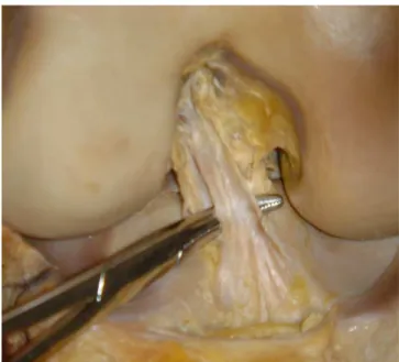
All knees were preserved in formaldehyde (Figure 2).
For comparison of measurements we used
40x12mm needles in marking the specific points and a
metallic caliper. The study was conducted with the knee
flexed at 90 degrees for measurement of femoral data.
The measurements of the following distances were
performed in the femur (Figure 3): 1) from the center of
the anteromedial band to the deep cartilage (AM-CP); 2)
from the center of the anteromedial band to the intercondyle
ceiling (AM-teto); 3) from the center of the posterolateral
band to the deep cartilage (PL-CP); 4) from the center of
the posterolateral band to the superficial cartilage (PL-CS);
and 5) from the center of the posterolateral band to the
inferior cartilage (PL CI).
The measurements of the following distances were
performed in the tibia (Figure 4): 1) from the anterior edge
to the anterior portion of the anteromedial band
(AM-AT-A); 2) from the anterior edge to the center of the
anteromedial band (AT- AM); 3) from the anterior edge to
the center of the posterolateral band (TA-PL); 4) from the
posterior edge to the center of the posterolateral band (PL
PT); and 5) anteroposterior diameter of the ACL (AP).
RESULTS
RESULTS
RESULTS
RESULTS
RESULTS
The measures, means and standard deviations
of the femoral origin of the ACL of the two bands are shown
in table 1 and the ones from the tibia in table 2.
In the femur the measurements were: AM-CP,
6.3 ± 1.4 mm; AM teto, 11.2 ± 2 mm; PL-CP, 9 ± 4 mm;
PL-CI, 4.2 ± 0.9 mm; PL-CS 7.6 ± 1.8 mm. In the tibia we
found: AT-AM-A, 11.9 ± 2.8 mm; AT-AM, 18.8 ± 2.6
mm; AT-PL, 26.5 ± 2.3 mm; PL-PT, 19.6 ± 4mm; AP,
19.4 ± 1.8 mm.
DISCUSSION
DISCUSSION
DISCUSSION
DISCUSSION
DISCUSSION
The restoration of the anatomy of the knee to
the nearest normal, with the positioning of the tunnels in
the most anatomically possible position, is crucial to the
success of ACL reconstruction
19-23.
Figure 1 Figure 1 Figure 1 Figure 1
Figure 1 - A) anatomical position and anatomical description, B) surgical position and description proposed by Zantop17
Figure 2 Figure 2 Figure 2 Figure 2
Most articles study the anatomy of the ACL as a
whole, not analyzing the bands separately, which constitutes
a limiting factor in studying the placement of tunnels with
dual band reconstruction.
We found no anatomical description with the
femur at 90° flexion, which is the surgical, though
non-anatomical, position. Despite the anatomical analysis of
the flexed knee does not have a direct influence on the
measures, it facilitates the interpretation of surgical data.
The distance between the center of the femoral
origin of the posterolateral (PL) band to the inferior cartilage
was 4.2 ± 0.9 mm. Petersen et al.
24found a value of 4 to
5mm. This shows that the origin of the PL band almost
touches the articular cartilage.
Regarding the measure of the distance between
the center of the PL band to the region of the deep femoral
cartilage, we obtained 9 ± 4 mm. Yasuda et al.
25reported
values of 5 to 8 mm for this lenght. These numbers help
as a location parameter of the origin of this band in the
knee. It is known that the PL band is more superficial than
the anteromedial (AM), because the distance from the AM
band to the deep cartilage was 6.3 ± 1.4 mm.
Figure 3 Figure 3Figure 3 Figure 3
Figure 3 - References for measurements of the femur: 1) center of anteromedial band to deep cartilage (AM-CP); 2) center of anteromedial band to the intercondyle ceiling (AM-teto); 3) center of posterolateral band to deep cartilage (PL-CP); 4) center of the posterolateral band to superficial cartilage (PL-CS); and 5) center of posterolateral band to inferior cartilage (PL-CI)
Figure 4 Figure 4Figure 4
Figure 4Figure 4 - References for measurements of the tibia: 1) anterior tibia edge to anterior portion of anteromedial band (AM-AT-A); 2) anterior tibia edge to center of anteromedial band (AM-AT); 3) anterior tibia edge to center of posterolateral band (TA-PL); 4) posterior tibia edge to center of posterolateral band (PL-PT); and 5) antero-posterior diameter of ACL (AP).
Table 1 -Table 1 -Table 1 Table 1
-Table 1 - Anatomical measures of the femoral origin of ACL (mm).
AM - CP AM - CP AM - CP AM - CP
AM - CP AM – tetoAM – tetoAM – tetoAM – tetoAM – teto PL – CPPL – CPPL – CPPL – CPPL – CP PL - CSPL - CSPL - CSPL - CSPL - CS PL – CIPL – CIPL – CIPL – CIPL – CI
Average 6.3 11.2 9 7.6 4.2
Standard deviation 1.4 2 4 1.8 0.9
Legend: AM-CP: center of anteromedial band to deep cartilage; AM-teto: center of anteromedial band to the ceiling of the intercondyle; PL-CP: center of posterolateral band to deep cartilage; PL-CS: center of posterolateral band to superficial cartilage; PL-CI: center of posterolateral band to inferior femur cartilage.
Table 2 -Table 2 -Table 2 Table 2
-Table 2 - Anatomical measures of the femoral insertion of ACL (mm).
A T - A M - A A T - A M - A A T - A M - A A T - A M - A
A T - A M - A A T - A MA T - A MA T - A MA T - A MA T - A M AT-PLAT-PLAT-PLAT-PLAT-PL PL-PTPL-PTPL-PTPL-PTPL-PT A PA PA PA PA P
Average 11.9 18.8 26.5 19.6 19.4
Standard deviation 2.8 2.6 2.3 4.1 1.8
Another parameter measured was the distance
from the superficial cartilage to the center of the PL band
and we found 7.6 ± 1.8 mm. There are no values in the
literature to compare with the determination of this
measure. In an arthroscopic view these numbers mean that
the superficial edge of the ACL would touch an imaginary
line that cuts the condyle in half.
The anatomical visualization of the ACL on the
tibia is not changed between the surgical position and the
classical anatomical one; so it is said that the name of the
ACL bands is due to its tibial position. But this is only one
way to avoid confusion in the interpretation of the surgical
anatomy and does not correspond to reality.
According to Petersen et al.
24, the insertion of
the ACL begins 10 to 14 mm posterior to the anterior border
of the tibia and the diameter of the sagittal plane of the
ligament varied from 15 to 19 mm. These numbers have
clinical relevance in double band reconstructions, as in the
realization of the double tibial tunnel it is essential that the
size of the tunnels is adapted to the size of the knee. Small
knees require more delicate drills to prevent communication
between the tunnels, preserving a bone bridge between
them. In this work the distance from the anterior edge of
the tibia to the anterior portion of the AM band was 11.9 ±
3 mm, similar to that found by Petersen et al.
24, whereas
the antero-posterior diameter was 19.4 ± 2 mm, greater
than the one found by them.
The distance obtained from the center of the AM
band to the anterior edge of the tibia is an important finding
because it can be used as a parameter for the conventional
ACL reconstruction; it is known that an error in this position
may bring bad results to the ACL reconstruction
26. In this
work the values of this measure was 18.8 ± 3 mm, and
Petersen et al.
24had 13-17mm.
The distance from the center of the PL band to
the anterior tibial edge serves as parameter for dual-band
reconstructions. We obtained 26.5 ± 2 mm in this study,
whilst Petersen et al found 20 25mm
24.
It is interesting to note that the tibial insertion is
long in the antero-posterior plane (19.4 ± 2 mm), the center
of the PL band being closer to the posterior edge of the
tibia (19.6 ± 4mm) than to the anterior one (26.5 ± 2mm).
In conclusion, the distance from the center of
origin of the anteromedial band to the deep cartilage of
the femur is approximately 6 mm, and to the posterolateral
band 10 mm. The center of the tibial insertion of the
anteromedial band is approximately 20 mm from the
ante-rior edge of the tibia, and the posterolateral band 30 mm.
The distance between the center of the origin of the
posterolateral band to the inferior cartilage is about 5 mm.
R E S U M O
R E S U M O
R E S U M O
R E S U M O
R E S U M O
Objetivo Objetivo Objetivo Objetivo
Objetivo: Avaliar anatomicamente a origem femoral e inserção tibial das bandas ântero-medial e póstero-lateral do ligamento cruzado anterior. MétodosMétodosMétodosMétodosMétodos: Estudados oito joelhos de cadáveres, foram feitas as seguintes medidas no fêmur: distância do centro da banda ântero-medial à cartilagem profunda e a ao teto. Ainda no fêmur, do centro da banda póstero-lateral à cartilagem profunda, a cartilagem inferior e à cartilagem superficial. Na tíbia, foi aferido do bordo ósseo tibial anterior à região anterior da banda ântero-medial, ao centro da banda ântero-medial e ao centro da banda póstero-lateral. Também foi medido o centro da banda póstero-lateral ao bordo ósseo posterior da tíbia e o comprimento ântero-posterior total da inserção tibial do ligamento cruzado anterior. ResultadosResultadosResultadosResultadosResultados: No fêmur, a distância do centro da banda ântero-medial à cartilagem profunda foi de 6,3 ±1,4mm e ao teto 11,2 ±2mm. Ainda no fêmur, a medida do centro da banda póstero-lateral à cartilagem profunda 9 ±4mm, à cartilagem superficial 7,6 ±1,8mm e a cartilagem inferior 4,2 ±0,9mm. Na tíbia, a distância do bordo ósseo tibial anterior à região anterior da banda ântero-medial foi de 11,9 ±2,8mm, ao centro da banda ântero-medial 18,8 ±2,6mm e ao centro da banda póstero-lateral 26,5 ±2,3mm. A medida do centro da banda póstero-lateral ao bordo ósseo posterior da tíbia foi 19,6 ±4mm e o comprimento ântero-posterior total da inserção tibial do ligamento cruzado anterior 19,4 ±1,8mm. ConclusãoConclusãoConclusãoConclusãoConclusão: O centro da inserção tibial da banda ântero-medial encontra-se a aproximadamente 20mm da extremidade anterior da tíbia, enquanto o centro da póstero-lateral se encontra a 30mm. A distância entre o centro da origem da banda ântero-medial até a cartilagem profunda é 6mm e da póstero-lateral 10mm.
Descritores Descritores Descritores Descritores
Descritores: Anatomia. Ligamento cruzado anterior. Articulação do joelho. Traumatismos dos tendões. Cadáver.
REFERENCES
REFERENCES
REFERENCES
REFERENCES
REFERENCES
1. Griffin LY, Agel J, Albohm MJ, Arendt EA, Dick RW, Garrett WE, et al. Noncontact anterior cruciate ligament injuries: risk factors and prevention strategies. J Am Acad Orthop Surg 2000; 8(3):141-50. 2. Stern HP, Bradley RH, Prince MT, Stroh SE. Young children in recreational sports. Participation motivation. Clin Pediatr 1990; 29(2):89-94.
3. Chen AL, Mears SC, Hawkins RJ. Orthopaedic care of the aging athlete. J Am Acad Orthop Surg 2005; 13(6):407-16.
4. Filho ES, Sampaio EB, Namba M, Silva JL, Albano M, Rocha LE, et al. Is it possible tp predict the lenght of knee flexor tendons by anthropometry ? Rev Col Bras Cir 2010; 37(4):274-8.
5. Cooley VJ, Deffner KT, Rosenberg TD. Quadrupled semitendinosus anterior cruciate ligament reconstruction: 5-year results in patients without meniscus loss. Arthroscopy 2001; 17(8):795-800. 6. Freedman KB, D’Amato MJ, Nedeff DD, Kaz A, Bach BR Jr.
7. Järvelä T, Paakkala T, Kannus P, Järvinen M. The incidence of patellofemoral osteoarthritis and associated findings 7 years after anterior cruciate ligament reconstruction with a bone-patellar tendon-bone autograft. Am J Sports Med 2001; 29(1):18-24. 8. Ristanis S, Giakas G, Papageorgiou CD, Moraiti T, Stergiou N,
Georgoulis AD. The effects of anterior cruciate ligament reconstruction on tibial rotation during pivoting after descending stairs. Knee Surg Sports Traumatol Arthrosc 2003; 11(6):360-5. 9. Tashman S, Collon D, Anderson K, Kolowich P, Anderst W. Abnormal
rotational knee motion during running after anterior cruciate ligament reconstruction. Am J Sports Med 2004; 32(4):975-83. 10. Ristanis S, Stergiou N, Patras K, Vasiliadis HS, Giakas G, Georgoulis
AD. Excessive tibial rotation during high-demand activities is not restored by anterior cruciate ligament reconstruction. Arthroscopy 2005; 21(11):1323-9.
11. Fithian DC, Paxton EW, Stone ML, Luetzow WF, Csintalan RP, Phelan D, et al. Prospective trial of a treatment algorithm for the management of the anterior cruciate ligament-injured knee. Am J Sports Med 2005; 33(3):335-46.
12. Roe J, Pinczewski LA, Russell VJ, Salmon LJ, Kawamata T, Chew M. A 7-year follow-up of patellar tendon and hamstring tendon grafts for arthroscopic anterior cruciate ligament reconstruction: differences and similarities. Am J Sports Med 2005; 33(9):1337-45.
13. Fithian DC, Paxton LW, Goltz DH. Fate of the anterior cruciate ligament-injured knee. Orthop Clin North Am 2002; 33(4):621-36. 14. Fink C, Hoser C, Hackl W, Navarro RA, Benedetto KP. Long-term outcome of operative or nonoperative treatment of anterior cruciate ligament rupture—is sports activity a determining variable ? Int J Sports Med 2001; 22(4):304-9.
15. Lohmander LS, Ostenberg A, Englund M, Roos H. High prevalence of knee osteoarthritis, pain, and functional limitations in female soccer players twelve years after anterior cruciate ligament injury. Arthritis Rheum 2004; 50(10):3145-52.
16. Girgis FG, Marshall JL, Monajem A. The cruciate ligaments of the knee joint. Anatomical, functional and experimental analysis. Clin Orthop Relat Res 1975; (106):216-31.
17. Zantop T, Petersen W, Fu FH. Anatomy of the anterior cruciate ligament. Operat Tech Orthop 2005; 15(1):20-8.
18. Abbott LC, Carpenter WF. Surgical approaches to the knee joint. J Bone Joint Surg Am 1945; 27:277-310.
19. Steiner ME, Battaglia TC, Heming JF, Rand JD, Festa A, Baria M. Independent drilling outperforms conventional transtibial drilling in anterior cruciate ligament reconstruction. Am J Sports Med 2009; 37(10):1912-9.
20. Buoncristiani AM, Tjoumakaris FP, Starman JS, Ferretti M, Fu FH. Anatomic double-bundle anterior cruciate ligament reconstruction. Arthroscopy 2006; 22(9):1000-6.
21. Abebe ES, Moorman CT 3rd, Dziedzic TS, Spritzer CE, Cothran RL, Taylor DC, et al. Femoral tunnel placement during anterior cruciate ligament reconstruction: an in vivo imaging analysis comparing transtibial and 2-incision tibial tunnel-independent techniques. Am J Sports Med 2009; 37(10):1904-11.
22. Scanlan SF, Blazek K, Chaudhari AM, Safran MR, Andriacchi TP. Graft orientation influences the knee flexion moment during walking in patients with anterior cruciate ligament reconstruction. Am J Sports Med 2009; 37(11):2173-8.
23. Stieven Filho E, Malafaia O, Ribas-Filho JM, Diniz OE, Borges PC, Albano M, et al. Biomechanic analysis of the sewed tendons for the reconstruction of the anterior cruciate ligament. Rev Col Bras Cir 2010; 37(1):52-7.
24. Petersen W, Zantop T. Anatomy of the anterior cruciate ligament with regard to its two bundles. Clin Orthop Relat Res 2007; 454:35-47.
25. Yasuda K, Kondo E, Ichiyama H, Tanabe Y, Tohyama H. Surgical and biomechanical concepts of anatomic anterior cruciate ligament reconstruction Operat Tech Orthop 2005; 15(2):96-102. 26. Jackson DW, Gasser SI. Tibial tunnel placement in ACL
reconstruction. Arthroscopy 1994; 10(2):124-31.
Received on 30/09/2010
Accepted for publication 02/12/2010 Conflict of interest: none
Source of funding: none
How to cite this article: How to cite this article:How to cite this article: How to cite this article:How to cite this article:
Stieven Filho E, Garschagen ET, Namba M, Silva JLV, Malafaia O, Cunha LAM. Anatomic study of the double-bundle of the anterior cruciate ligament with the knee in 90º flexion. Rev Col Bras Cir. [peri-ódico na Internet] 2011; 38(5). Disponível em URL: http://www.scielo.br/ rcbc
Address correspondence to: Address correspondence to:Address correspondence to: Address correspondence to:Address correspondence to: Edmar Stieven-Son