29
REVISTA BRASILEIRADE OTORRINOLARINGOLOGIA 71 (1) PART 1 JANUARY/ FEBRUARY 2005 http:/ / w w w .rborl.org.br / e-mail: [email protected]
Neck dissection in squamous
cell carcinoma of the tongue
Summary
Ali Amar , Otávio Alber to Cur ioni, Ser gio Altino Fr anzi, Daniel Knabben Or telado, Abr ão Rapopor t1
1 Department of Head and Neck Surgery and Otorhinolaryngology, Hospital Heliópolis, Hosphel, Sao Paulo, Brazil.
Address correspondence to: Prof. Dr. Abrão Rapoport – Rua Iramaia, nº 136 Jd. Europa São Paulo SP 01450-020. Article submited on September 16, 2004. Article accepted on January 21, 2005.
A
im: The purpose of this study w as to assess the prognosis of patients with tonsillar squamous cell carcinoma with different stages of lymph node involvement and to determine the best elective neck dissection for those cases. Study design: Case series. Mater ial and Method: 51 patients with tonsillar tumors were treated between 1992 and 2001. The incidence of different tumor-node-metastasis stages was evaluated according to primary tumor extension. Results: cN0 patients had metastases in stages I and II only. Among pN+ subjects with stage I metastases, 6/ 7 had primary tumor extending to oral cavity. Conclusion: Supraomohyoid neck dissection (stages I, II and III) is the elective treatment of choice when tonsillar primary tumor extends to oral cavity. When primary tumors are limited to the oropharynx, selective neck dissection of stages II and III proved to be more adequate.Key w ords: metastasis, lymph node, tonsil.
« «
Rev Bras Otorrinolaringol. V.71, n.1, 29-31, jan./feb. 2005
O RIG IN AL ARTIC LE
30
REVISTA BRASILEIRADE OTORRINOLARINGOLOGIA 71 (1) PART 1 JANUARY/ FEBRUARY 2005 http:/ / w w w .rborl.org.br / e-mail: [email protected] INTRODUCTION
The proposed elective extended neck dissection in surgical approaches of oropharyngeal tumors remains unclear. Among selected standard neck dissections, the supraomohyoid dissection (stages I, II and III) or jugular dissection (stages II, III and IV) consist of the treatments
of choice1. Generally, oropharyngeal tumors present stage
II metastases and, less frequently, they produce stage III metastases, w hile incidence of lymph node involvement
in other stages is low2. The present study aimed at assessing
lymph node stages involved in tonsillar squamous cell car-cinomas, taking into account primary tumor extension, in an attempt to best determine the most appropriate elective neck dissection.
MATERIAL AND METHOD
Medical records of 51 patients with tonsillar squamous cell carcinoma treated at the Head and Neck Surgery Service of Hospital Heliópolis in the period of January 1992 and December 2001 were revised. Ages ranged from 37 to 83 years, mean age of 55. Regarding gender, there were 45 men and 6 w omen. All patients w ere initially treated by surgery, of w hich 49 cases included neck dissection. Traditional radical neck or radical modified dissections were performed in 40 patients, out of w hich 2 w ere bilaterally dissected; supraomohyoid dissection w as performed in 7 subjects and comprehensive supraomohyoid dissection (stages I to IV) w as performed in 2 patients. One subject was treated with lymphonodectomy and 1 was not treated in the neck. Concerning disease staging, patients w ere classified as T1 (3), T2 (20), T3 (24) and T4 (4). Postoperative radiotherapy with mean dosage of 61Gy (45 to 71 Gy) was applied in 44 patients, out of which 5 had previously been irradiated. Incidence of metastases was assessed based on different lymph node staging, according to American Head
and Neck Society classification1. Additionally, primary tumor
w ith extension to adjacent anatomical subsites and its correlation w ith lymph node involvement w ere assessed. Absolute numerical outcomes were expressed without the need of developing alternative hypotheses to justify the application of statistical tests, as therapeutic paradigm was uniform.
RESULTS
Seventeen patients were staged cN0, out of which 6 were false-negatives. Among 34 cN+ patients, 28 had lymph node metastases according to histological exam. Stage II metastases were observed in 32/ 34 pN+ cases. Distribution of metastases according to lymph node stages is shown on Table 1. Stage Ib metastases were found in 7 cases, among w hich 6 had primary lesion extended to oral cavity (3 to
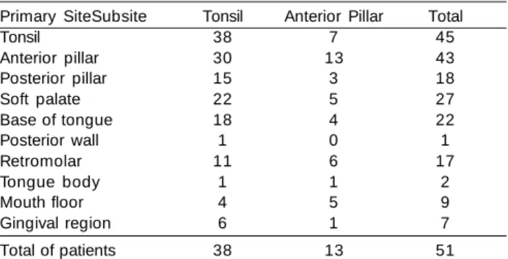
retromolar region and 3 to tongue body). There w ere no stage Ia metastases among these patients. Out of 17 N0 subjects, 2 presented stage Ib metastases only. Regarding neoplasia extension to oral cavity, out of 51 studied patients, 38 (74%) had tonsil-limited lesions and 13 (26%) presented extraoral extension (Table 2).
DISCUSSION
Selective dissections w ere performed to reduce morbidity related to radical dissection and they proved to be appropriate not only to stage N0 necks, but also to treat N+ neck s of sel ected cases, w i th or w i thout
postoperative radiotherapy3. Reduced dissection extension
w as stimulated by positive outcomes in controlling regio-nal disease and improvement of overall survival. Although cervical metastases were related to significantly low survival, poor outcomes mainly occur due to therapy failures on p ri mary si te and di stantl y, w hi l e i sol ated regi onal recurrences are early detected and treated at patients’
follow-up 4,5.
Selective dissections of oropharyngeal lesions may consist of lateral or jugular (stages II, III and IV) approach
Table 1. Distribution of metastases in different lymph node
stages.
N0p N+ N+ p N+ Total
Ia 0/6 0/28 0/34
Ib 4/6 3/28 7/34
IIa 5/6 27/28 32/34
IIb 2/6 3/28 5/34
III 0/6 9/28 9/34
IV 0/6 4/28 4/34
V 0/6 4/28 4/34
Table 2. Anatomical subsites involved in tonsillar tumors* Primary SiteSubsite Tonsil Anterior Pillar Total
Tonsil 38 7 45
Anterior pillar 30 13 43
Posterior pillar 15 3 18
Soft palate 22 5 27
Base of tongue 18 4 22
Posterior wall 1 0 1
Retromolar 11 6 17
Tongue body 1 1 2
Mouth floor 4 5 9
Gingival region 6 1 7
Total of patients 38 13 51
31
REVISTA BRASILEIRADE OTORRINOLARINGOLOGIA 71 (1) PART 1 JANUARY/ FEBRUARY 2005 http:/ / w w w .rborl.org.br / e-mail: [email protected]
or of supraomohyoid dissection (stages I, II and III). As metastases mainly occur at stages II and III, they are both acceptable alternatives. Supraomohyoid dissection has been advocated, as stage I is the only involved and lateral dissection incorrectly stages these patients, while stage IV
metastases are not commonly found alone6. In our sample,
most patients w ith stage I metastases presented tumor extension to oral cavity w ith involvement of tongue body or retromolar region. In patients w ith tumor limited to oropharynx, dissection in stages II and III yielded adequate cN0 neck staging. Although this is not a standard procedure, its indication is based on regional lymphatic drainage and
frequent metastases distribution 2,7,8,9. Reduction of elective
dissection may lead to investigation of sentinel lymph node, an established concept that involves w ider technical knowledge.
Differently from mouth lesions, oropharyngeal lesions present higher incidence of stage IIb metastases, demanding elective dissection, despite the morbidity related to management of the accessory nerve, considering that stage IIb recurrences are hardly rescued.
Supraomohyoid dissection (stages I, II and III) has proved to be an appropriate elective neck treatment for cases of tonsillar tumors with extension to oral cavity. In N0 cases limited to oropharynx, neck dissection of stages II and III patients is the most appropriate indication.
REFERENCES
1. Robbi ns KT, Cl ayman G, Levi ne PA et al . Neck di ssecti on classification update: revisions proposed by the American Head and Neck Society and the American Academy of Otolaryngology-Head and Neck Surgery. Arch Otolaryngol Otolaryngology-Head Neck Surg 2002; 128:751-8.
2. Lindberg R. Distribution of cervical lymph nodes metastases from squamous cell carcinoma of the upper respiratory and digestive tracts. Cancer 1972; 29:1446-9.
3. Muzaffar K. Therapeutic selective neck dissection: a 25-year review . Laryngoscope 2003; 113:1460-5.
4. Myers EN, Fagan JJ. Treatment of the N+ neck in squamous cell carcinoma of the upper aerodigestive tract. Otolaryngol Clin North Am 1998; 31:671-86.
5. Amar A, Franzi AS, Rapoport A. Evolution of patients w ith squamous cell carcinoma of upper aerodigestive tract. Sao Paulo Med J 2003; 121:155-8.
6. Vartanian JG, Pontes E, Agra IM et al. Distribution of metastatic lymph nodes in oropharyngeal carcinoma and its implications forr the elective treatment of the neck. Arch Otolaryngol Head Neck Surg 2003; 129:729-32.
7. Werner JA, Dünne AA, Myers JN. Functional anatomy of the lymphatic drainage system of the upper aerodigestive tract and its role in metastasis of squamous cell carcinoma. Head Neck Surg 2003; 25:322-32.
8. Rouvière H. Anatomie des lymphatiques de l’homme. Paris: Masson et C Editeurs; 1932.