Prognostic factors in patients with breast cancer
metastasis in the femur treated surgically
Guilherme Grisi Mouraria, Silvia Raquel Frick Matte, Carlos Hideo Hanasilo, Maria Julia Rosso de Carvalho, Mauricio Etchebehere
University of Campinas, Unicamp, Medical School, Department of Orthopedics, Campinas, SP, Brazil
BACKGROUND:Breast carcinoma is a common malignancy in the developed world. Late diagnosis of the disease is associated with bone metastasis. The femur is commonly affected. Prognostic factors of mortality in patients with bone metastases originating from cancers in general have been reported. However, there is no specific report of prognostic factors in relation to breast cancer metastasis in the surgically treated femur. The determination of prognostic factors in patients with bone metastasis can assist in therapeutic decisions. OBJECTIVE:To determine clinical and orthopedic factors related to mortality in patients with breast cancer and metastases to the femur treated surgically.
METHOD: This was a retrospective study and included 41 patients undergoing surgical treatment of femoral metastases. We analyzed the following variables: (i) number and location of bone metastases, (ii) visceral metastases, (iii) presence of pathological fracture, (iv) fixation method, and (v) laboratory tests. These factors were correlated with mortality using Cox Multivariate Logistic regression and Kaplan-Meier curves.
RESULTS:There was a high prevalence of multiple metastases associated with pathological fractures at the time of surgery. Mortality was high and early. Subtrochanteric location, the presence of fractures, anemia, and alterations in renal function were associated with higher mortality. The fixation method, the number of bone metastases, and the presence of metastasis in other organs did not affect mortality.
CONCLUSIONS:Breast cancer with metastasis to the femur is an advanced disease with early mortality. Clinical and orthopedic factors should be considered. Surgery is recommended when lesions occur, regardless of the type of implant used.
KEYWORDS: Breast Cancer; Femur Metastasis; Prognosis; Mortality.
Mouraria GG, Matte SRF, Hanasilo CH, de Carvalho MJR, Etchebehere M. Prognostic factors in patients with breast cancer metastasis in the femur treated surgically. MEDICALEXPRESS. 2014 Oct;1(5):221-226.
Received for publication onMay 10 2014;First review completed onMay 21 2014;Accepted for publication onJune 22, 2014
E-mail: [email protected]
B INTRODUCTION
Breast carcinoma is a common disease in the developed world. In the presence of metastases, the bone is affected in more than 50% of patients1. In the appendicular skeleton, the femur is most commonly affected2,3.
Although metastases to the axial skeleton are more frequent, metastases to the lower limbs cause greater loss of function. Bone metastases in the lower limb, even in the absence of fractures, lead to pain, functional limitations, and reduced quality of life for patients, due to strict bed rest and its complications, such as deep vein thrombosis, pneumonia, and decubitus sores4. The presence of fractures leads to a further increase in morbidity and mortality in these patients5. In practical terms, there is no specific clinical treatment for metastatic lesions in the femur4,6.
Prognostic factors for mortality in cancer patients in general and for bone metastases have been reported. The presence of
multiple bone metastasis, visceral metastasis, low hemo-globin, presence of fractures, and prosthetic replacement therapy are associated with higher mortality in this population7 – 10. However, we are aware of no report of specific prognostic factors related to metastases derived from breast cancer that occur in the femur. Thus, the aim of this study was to determine factors related to clinical orthopedic issues and mortality in this cohort of patients.
B SUBJECT AND METHODS
We conducted a retrospective cohort study involving 41 women diagnosed with breast cancer metastasis in the femur who underwent surgery between 1994 and 2007. We analyzed the following orthopedic variables: (i) the number of bone metastases, (ii) the presence of pathological fractures, (iii) the fixation method used in the treatment of the metastasis (synthesis or prostheses/endoprostheses), and (iv) the topography of the metastasis in the femur. The number and location of bone metastases was determined by scintigraphy and RX. In order to establish the topography of
DOI:10.5935/MedicalExpress.2014.05.02
Patient follow-up ranged from 8 to 63 months (average, 37 months). One patient was lost to follow-up. The mean age was 53.8 years. The average number of bone metastases, including the femur, was 10.8. The proximal femur proved to be the most common location of bone metastases and fractures. Most of the patients (30) had pathological fracture at the time of surgery.
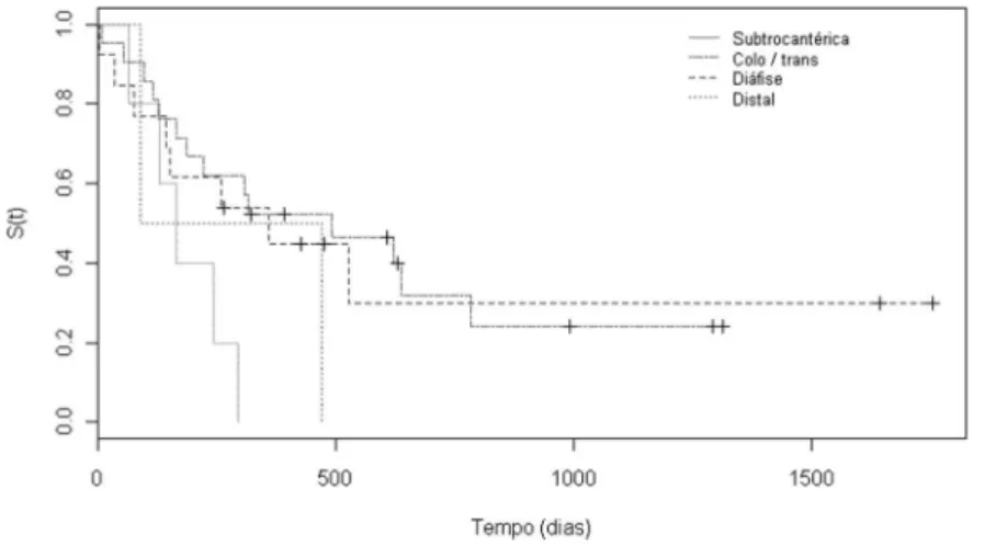
number of bone metastases were not, as shown in Table 1. For statistical purposes, the subtrochanteric region was used as a reference. The presence of metastasis in this region increased mortality by 3- to 3.3-fold in comparison to those located in the proximal femur and diaphysis, respectively (Table 1). Figure 2 shows the distribution of mortality according to the location of the femoral lesion.
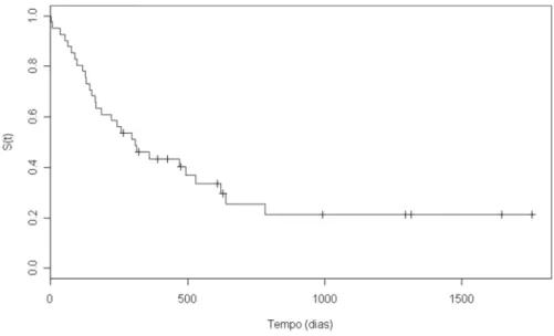
Figure 1 -Kaplan-Meyer curve for overall mortality. The median survival was 8.1 months, with 70% of deaths occurring during the first year after surgery.
Table 1 -Mortality estimates obtained by Cox multivariate logistic regression
95% confidence interval
Variables regression coefficient hazard ratio (HR) lower upper p value
proximal(1)
21.088 2.969(2) 1.010 4.929 0.050
Topography of lesions diaphysis 21.20 3.322(2) 1.362 5.820 0.050
distal 20.443 1.557(2)
20.462 3.517 0.610
Hemoglobin 20.233 1.263(3) 0.697 3.223 0.002
Urea 0.026 1.026 0.934 1.968 0.090
Creatinine 1.295 3.650(4) 1.690 5.610 0.002
Presence of fracture 1.14 3.130 1.170 5.060 0.016
Prosthetic replacement/osteosynthesis 20.251 0.900 21.006 2.806 0.996
Metastasis in other organs 0.419 0.400 21.560 2.306 1.520
Number of bone metastasis 0.030 0.910 21.010 2.900 0.987
Clinical data showed that a 1 mg/dL increase in serum Creatinine was associated with about a 3.6-fold increased risk of death (Table 1). Creatinine values above 1.2 mg/dL also increased mortality (Fig. 3A). Urea levels did not appear to affect mortality.
A drop in the hemoglobin value of 1 mg/dL increased the risk of death by approximately 1.2 fold (Table 1). In patients with Hb,11 mg/dL, mortality was significantly increased (Fig. 3B).
The presence of fractures was associated with increased mortality, especially in the first 12 months postoperatively (Table 1). However, after 24 months, there was no statistical difference in mortality (Fig. 4).
Mortality was not influenced by the number of bone metastases or the presence of multiple lesions (Table 1, Fig. 5B). Moreover, the presence of metastasis in other organs was not associated with increased mortality (Table 1, Fig. 5A). The type of procedure performed (osteosynthesis or arthroplasty) did not affect mortality (Table 1, Fig. 6).
B DISCUSSION
Metastasis of breast cancer to the femur requires, in most cases, surgical intervention because conservative treatments and radiotherapy are not effective in preventing pathological fractures. The study population had advanced neoplasia, because the presence of bone metastasis indicates dissemi-nated disease in stage IV. The average age here was 56 years, lower than that reported in the literature1.
The average number of bone metastases at the time of treatment of a femoral lesion was high, so there was a tendency to also find metastasis elsewhere, an observation consistent with the literature9. In this series, femoral metastasis was associated with lesions in other organs in 58% of cases, an indicator of advanced disease. Thus, the staging of the disease is important because there is a high likelihood of finding other bone lesions and/or metastases in other organs at the time of diagnosis of a femoral lesion. These factors are important in preoperative planning and overall treatment of the disease.
In terms of topographic distribution, the femoral lesions were predominantly located in the proximal region, which is consistent with published reports11. The proximal location of the lesion results in a higher probability of fracture due to
Figure 2 -The Kaplan Meyer curves for distribution of mortality according to the location of the femoral lesion. Subtrochanteric lesions are significantly worse than all other femoral locations.
mechanical forces acting on the hip3. Additionally, a proximal lesion more often needs prosthetic replacement, reflecting an increase in the duration of the surgery and its complications, thereby increasing mortality12. However, in this study, the subtrochanteric region resulted in worse prognosis, in contrast to reported data13. This difference may be due to the fact that other reports considered this region to be part of the proximal femur. Thus, differentiation between lesions in the subtrochanteric proximal, intertrochanteric, and neck regions is important because of different associated mortalities. Thus, a subtrochanteric location, which is associated with reduced survival, may be treated with palliative procedures and, eventually, minimally invasive procedures to improve the quality of life.
In our study, the type of surgery (osteosynthesis or prosthetic replacement) had no effect on mortality. This divergence from the literature14,15 may be due to the surgeon’s level of expertise with arthroplasty. In the literature, fixation was associated with a greater chance of implant failure and revision surgery. These techniques should therefore be used in patients with reduced life expectancies. Prosthetic replacements should be reserved for patients with a better prognosis due to the reduced possibility of implant failure8.
Fractures were associated with high mortality in the first 12 postoperative months (Fig. 5), in agreement with the literature 2 Fractures caused increased mortality due to restriction to bed rest and associated complications. Prophylactic treatment of bone lesions can result in shorter hospital stay and reduced blood loss during surgery, leading to lower mortality5. Fractures associated with bone metastasis of breast cancer have higher mortality than do other cancers, such as those of the lung or kidney9,15.
Clinically, renal function parameters and hemoglobin levels were, on average, normal. Renal insufficiency and anemia are important in recovery and postoperative mortality2,17. Anemia leads to slower wound healing, with an increased possibility of infection. An Hb level,11 mg/ dL is associated with poor prognosis and higher mortality17. In this study, Hb,11 mg/dL (Fig. 4) and Cr.1.2 g/dL (Fig. 3) were independent predictors of mortality, which is consistent with the literature17.
Figure 4 -Kaplan Meyer distribution of mortality according to the presence or absence of femoral fractures. The presence of fractures significantly affects mortality during the first year after surgery, but not after that.
Visceral metastases occurred in 58% of the patients. The lung was most commonly affected, and liver and brain metastases were next. Distant metastasis is a relevant comorbidity because it leads to organ failure and frequent hospitalizations. Thus, it is associated with poor prognosis and higher mortality2,12. However, in this cohort, there was no statistical relationship between the presence of visceral metastasis and higher mortality. There may be a bias in this result, as only patients who underwent surgery were included in our sample; we excluded patients with visceral metastases and other metastasis who did not undergo femoral surgery. Further studies are necessary to examine this.
Overall, 29 patients (70%) died after a minimum two months of follow-up. The average time between surgery and death was 8.1 months. This high mortality and early deaths reflects the typical behavior of an aggressive disease.
In conclusion, surgeons should assess these patients globally (orthopedic and clinical factors) before a surgical procedure is decided upon, because in most cases, a palliative surgical treatment should be considered. The surgical treatment of metastasis in the femur should be
performed as soon as possible and allow rapid rehabilitation so that the patient can enjoy the last months of survival with the best possible quality of life.
B ACKNOWLEDGEMENTS
The authors declare that they have no conflict of interest. There was no external funding source for this study.
B RESUMO
INTRODUCA˜ O:O carcinoma de mama e´ um tumor maligno comum no mundo desenvolvido. O diagno´stico tardio da doenca esta´ associada com meta´stases o´sseas. O feˆmur e´ comumente afetado. Fatores progno´sticos de mortalidade em pacientes com meta´stases o´sseas provenientes de caˆnceres em geral teˆm sido relatados. No entanto, na˜o ha´ nenhum relato´rio especı´fico de fatores progno´sticos em relaca˜o a` meta´stase do caˆncer de mama no feˆmur tratados cirurgicamente. A determinaca˜o de fatores progno´sticos em pacientes com meta´stases o´sseas pode auxiliar a tomada de deciso˜es terapeˆuticas.
OBJETIVO:Determinar os fatores clı´nicos e ortope´dicos relacionados com a mortalidade em pacientes com caˆncer de mama e meta´stases ao feˆmur tratados cirurgicamente.
Incidence of bone metastases and skeletal-related events in breast cancer patients: A population-based cohort study in Denmark. BMC Cancer. 2011;11(Jan 24):29.
2. Narazaki DK, Alvarenga Neto CC, Caiero MT, Camargo OP. Prognostic factors in pathologic fractures secondary to metastatic tumors. Clinics. 2006;61(4):313-20.
3. Keene JS, Sellinger DS, Mcbeath AA, Engber WD. Metastatic breast cancer in femur. A search for the lesion at risk of fracture. Clin Orthop Relat Res. 1986;203:282-8.
4. Dutka J, Sosin P. Time of survival and quality of life of the patients operatively treated due to pathological fractures due to bone metastase. Ortop Traumatol Rehabil. 2003;5(3):276-83.
5. Saad F, Lipton A, Cook R, Chen YM, Smith M, Coleman R. Pathologic fractures correlate with reduced survival in patients with malignant bone disease. Cancer. 2007;110(8):1860-7.
13. Jacofsky DJ, Haidukewych GJ. Management of pathologic fractures of the proximal femur: state of art. J Orthop Trauma. 2004;18(7):459-69. 14. Moholkar K, Mohan R, Grigoris P. The long Gamma Nail stabilization of
existing and impending pathological fractures of the femur: an analysis of 48 cases. Acta Orthop Belg. 2004;70(5):429-34.
15. Weber KL, O’Connor MI. Operative of long bone meta´stase: focus on the femur. Clin Orthop Relat Res. 2003; (415 Suppl):S276-8.
16. Chrobok A1, Spindel J, Mrozek T, Miszczyk L, Koczy B, Tomasik P, et al. Partial long stem resection Austin – Moore Hip endoprotesis in the treatment of metastases to the proximal femur. Ortop Traumatol Rehabil. 2005;7(6):600-3.