Introduction
Musculoskeletal disorders are considered the most prevalent pathologies in developed countries1. Among these, low back pain (LBP), which is deined as pain and discomfort, localized below the costal margin and above the inferior gluteal folds, with or without leg pain2 can be highlighted. The most com-mon form of LBP is nonspeciic and occurs when the cause of anatomopathological pain cannot be determined3. In Brazil, the LBP prevalence is higher than 50% in adults and 13.1% to 19.8% in adolescents4,5.
The etiology of LBP is multifactorial and may be associ-ated with factors such as age, sex, smoking, alcoholism, body weight, social class, level of schooling, physical activity and work activities. The literature has highlighted the inluence of the imbalance between the function of the extensor and lexor muscles of the trunk in increasing the probability of developing disorders that affect and disrupt the stability of the lumbar spine6,7. There is still no consensus in the literature whether the lack of muscle pre-activation is a cause or manifestation of LBP8, but it is known that the lack of resistance of the trunk muscles may be associated with episodes of this disorder9. Muscles that have functional and morphological alterations can be investigated by surface electromyography (EMG)10. Its use can provide informa-tion about the amount of muscular activity that some exercise or positioning requires, thus facilitating the choice of the most appropriate treatment for each individual11.
Currently, the use of a lumbar pain classiication system, based on the signs and symptoms helps in choosing the most appropriate intervention. Among the classiications for subgrouping patients, the System of Treatment-based Classiication (TBC) is able to increase the eficacy of conservative interventions for patients
with low back pain12. Therapeutic exercises are still considered the most effective resources for treating chronic LBP, although in clinical practice there are varieties of applied exercises13. The clinical guideline published by the American College of Physicians (2017) recommended that the irst suggestion for patients with chronic low back pain should be nonpharmaco-logic treatment including exercises and motor control exercise in addition to other therapies14.
The trunk stabilization exercise has been recommended by most guidelines as a treatment for individuals with LBP who have a coordination deicit of motion15. The Pilates Method is an exercise program that is often prescribed for these individuals, since they are used in the activation and strengthening of the stabilizing muscles of the trunk, such as multiidus and abdominal musculature16. Pereira, Queiroz, Loss, Amorim, Sacco17 showed that Pilates is an effective way to allow lumbopelvic stabilizer activation even in the irst session in healthy and chronic low back pain individuals17. However, only low to moderate-quality evidence showed that Pilates resulted in effects on pain when compared to other physical activity14,18. Few high quality evidence clinical trials compared the effects of Pilates to other interventions19. Moreover, studies evaluating the improvement of muscle activation of lumbar multiidus (LM) and transversus abdominis/internal oblique muscles (TrA/IO) in individuals with LBP were not found, speciically after an exercise protocol based on the Pilates Method. Since Pilates acts in the contrac-tion of this muscle, it is believed that the method can be used in individuals with lumbar spine instability. Therefore, the present study aimed to verify the inluence of a Pilates exercise program on muscle activation of LM and TrA/IO muscles in individuals with nonspeciic low back pain.
Original Article (short paper)
Effectiveness of the Pilates method for individuals
with nonspeciic low back pain: clinical and
electromyographic aspects.
Pâmela Maiara Machado1, Morgana Cardoso Alves1, Ketlyn Germann Hendler1, Vanessa Braitenbach Benetti1,
Ro-meu Joaquim de Souza Neto1, Rafael Inácio Barbosa1
, Alexandre Márcio Marcolino
1, Heloyse Uliam Kuriki1*
¹Universidade Federal de Santa Catarina, UFSC, Araranguá, SC, Brazil
Abstract — Aims: The aim of this study was to verify the inluence of Pilates on muscle activation of lumbar multiidus (LM) and transversus abdominis/internal oblique muscles (TrA/IO) in individuals with nonspeciic low back pain.
Methods: Twelve individuals of both sexes with non-speciic low back pain were evaluated before and after a two-month Pilates program in relation to electromyographic activity of LM and TrA/IO, as well as clinical aspects such as pain, lexibility, muscular endurance, quality of life; and Fear-Avoidance Beliefs Questionnaire (in relation to physical and work-related activities. A statistical analysis was performed using a test for independent samples and signiicance was established at the level of 0.05. Results: After eight weeks of Pilates training, there was an improvement in the clinical parameters of pain, lexibility, muscular endurance and disability. The individuals presented lower LM activation (p=0.025), higher trunk extension strength (p=0.005) and an increase in time from onset to peak muscle activation (p=0.02). Conclusion: Pilates protocol was effective for clinical improvement and motor behavior in patients with nonspeciic low back pain and the parameters assessed showed a large effect size despite the small sample.
Methods
Subjects
This was a prospective study conducted between February and November 2016, containing a group of individuals with non-speciic low back pain followed by two months in which they performed a Pilates program. Subjects were recruited from the inclusion criteria of the TBC12 subgrouping in which they needed to present symptoms of non-speciic low back pain, aged less than or equal to 40 years old. They also need to present at least three of these criteria: a negative Laségue test, aberrant move-ment (being pain in the accomplishmove-ment of the trunk lexion or in the return of the trunk), Fear-Avoidance Beliefs Questionnaire - Work subscale < 19 and positive prone instability test.
The preliminary evaluation performed for the recruitment and subgrouping of the individuals occurred at the Federal University of Santa Catarina (Universidade Federal de Santa Catarina - UFSC) - Campus Araranguá/SC and consisted of an
interview of socio-demographic data, anamnesis, and a physical examination. Before any methodological procedure, the par-ticipants signed a free and informative consent form and the research was approved by the Research Ethics Committee (N. 1.041.755). Fifty-four previous evaluations were performed and 20 individuals were selected according to the inclusion criteria. On a second pre-scheduled day, the volunteers attended the Laboratory of Evaluation and Rehabilitation of the Locomotor System (Laboratório de Avaliação e Reabilitação do Aparelho Locomotor - LARAL) to perform the electromyographic evalua-tion and subsequently initiated a Pilates protocol. Among the 20 selected individuals, 8 gave up throughout the protocol due to the times incompatibility, inishing the study with 12 volunteers of both sexes, combined with an average age of 25.41 (± 6.27) years, average weight of 59.41 (± 11.13) kg, average height of 1.63 (± 0.07) meters and a mean pain level presented of 3.83 (± 3.45) on a visual analogue scale. There was only one male volunteer and 75% of the participants were students. The main steps of the research are outlined in the lowchart in Figure 1. Figure 1. Flow chart with the main steps of research.
Preliminary evaluation n = 54
Subgrouping according to TBC
Clinical assessment n = 20
Electromyographic assessment
n = 20
Pilates program n = 20
Clinical assesment
n = 12 EMG assessmentn = 12
Clinical assessment
In addition to the tests for inclusion in the subgroup of inter -est, the tests of Sorensen20, lateral bridge20 and distance from the 3rd inger to the ground21 in the trunk lexion, right and left lateral bending were performed. The Fear Avoidance Beliefs Questionnaire12 (FABQF: Phys subscale; FABQW: Work sub-scale), the Oswestry Disability Index22 (ODI), and the 12-Item Short-Form Health Survey Quality of Life Questionnaire (SF-12) from the Areas of physical health (PCS) and mental health (MCS)23 were also applied. All evaluations were repeated after the two months of Pilates, respecting a maximum period of one week before and after the protocol for conducting the evalua -tions and in all meetings before and after the Pilates the visual analog scale of pain (VAS)24 was conducted.
Electromyographic assessment – instrumentation
The surface EMG signals (sEMG) were obtained using two conditioner modules (Miotec®, Porto Alegre, RS, BRA; model Miotool 400) that utilized a Butterworth type band-pass digital ilter with cutoff frequencies of 20 and 500 Hz, an ampliier with a inal gain of 1000, and an acquisition frequency of 4000 Hz. To collect sEMG data from the right LM and right TrA/IO, two pairs of bipolar electrodes with Ag/AgCl capture surfaces (Kendall, Mansield, MA, USA; model Medi-Trace) with diameters of 10 mm were positioned at the sites of the
respective muscles with an inter-electrode distance of 20 mm. The LM sensors were positioned in accordance to the SENIAM protocol25; and, for the TrA/IO, the sensors were placed about 2 centimeters medial and inferior to the anterior superior iliac crest26. A reference electrode was coupled in the styloid process of the ulna on the right forearm. There was a pre-ampliier circuit with a gain of 20 times, a CMRR (Common Mode Rejection Ratio) greater than 80 dB, and an impedance of 1012 X. A Strain Gauge dynamometer was coupled to the electromyography to measure the trunk extension force during the traction exerted by the volunteer. The data were collected using Miotec Suite software (Miotec®, Porto Alegre, RS, BRA).
The sEMG signal was collected at the maximum voluntary isometric contraction (MVIC) at the Sorensen test position, with bands to ix the hips and legs on the hospital bed. The dynamom-eter was positioned perpendicular to the trunk of the volunteer using an inextensible current. The subject was instructed to perform an MVIC of the trunk while pulling the dynamometer for 6 seconds (Figure 2). After two minutes resting, the sEMG signal was collected during the free trunk extension, in which the volunteer was instructed to leave the resting position, with the upper limbs crossed in the chest and perform a trunk exten-sion to the maximum range of motion and so returning to the initial position with self-controlled speed. Three trials were performed with two-minute rest intervals between them. The same evaluations were conducted before and after two months of performing Pilates exercises.
Pilates program
During a two months period, the volunteers were accompanied while executing a Pilates exercise program. The exercises were from Mat Pilates, supervised by physiotherapy students (in the last 2 semesters before graduation), with increasing dificulty levels throughout the weeks, twice a week, individually and face-to-face mode, within 50 minutes, totaling 16 times. The exercises were: (1) spine stretch forward, (2) saw, (3) cat stretch, (4) roll-up, (5) single leg stretch, (6) single straight stretch, (7) chest lift with rotation, (8) single-leg kick, (9) double-leg kick, (10) pelvic curl, (11) one leg up and down, (12) leg circles, (13) side kick, (14) crisscross, (15) hundred, (16) spine twist supine, (17) swimming, (18) leg pull front, (19) side kick kneeling, (20) leg pull, (21) push-up, and (22) side bend27. All the exercises were executed according to traditional Pilates principles: cen-tering, concentration, control, precision, low, and breathing28.
Analysis
The extracted sEMG signals of each trial were submitted to the follow analyzes: i) determination of trunk extension force dur-ing the MVIC; ii) normalization of multiidus durdur-ing the free extension test by the MVIC signal, using the two most stable seconds of contraction; iii) determination of the normalized Root Mean Square (RMS) of LM during the free trunk extension test; iv) calculation of the time elapsed from the beginning to the maximum activation of the right LM and TrA/IO during the trunk extension test. For all of the analyses, algorithms programmed in MatLab®software were used.
Data from EMG and clinical assessment were compared before and after the Pilates using the unpaired t-test (α < 0.05) after the normality conirmation (Shapiro-Wilk test) and the effect size of each comparison was determined by Cohen´s d coeficient
(ES-d). Effect sizes were deined as small (ES-d < 0.5), medium (0.50 ≤ d < 0.80), and large (d ≥ 0.80)29.
Results
Clinical assessment showed predominance in the reduction of the symptoms in the prone instability test and of aberrant movement in lexion, in addition to the reduction of pain, in-crease of the lexibility and muscular endurance. In relation to the prone instability test, initially it was positive in 75% of the volunteers and after the intervention, there was a reduction to 33.33%. The aberrant movement (pain in the accomplishment or in the return of the trunk lexion) was present in 58.33% of the individuals before the Pilates protocol and, after this, there was a decline in these values to 8.33% of the individuals. The pain level, measured through VAS, initially presented a mean of 3.83 (0.99) and after the protocol, there was a signiicant reduction (p = 0.01) to 0.75 (0.50), with large effect size (ES-d = 1.17).
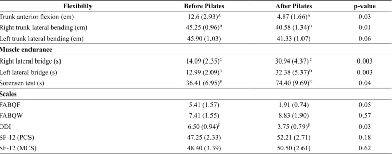
In relation to the tests of lexibility and endurance, a sig-niicant difference was observed in the lexion and right lateral bending movements, Sorensen test, and right and left lateral bridge (Table 1). Among the scales used to measure disability, quality of life and beliefs/ fears in relation to work and physical activity, there was an improvement in the scores of all ques-tionnaires. However, there was statistical signiicance only for disability assessed using the Oswestry Disability Index (Table 1). Except for the SF-12, all of these tests had a large effect size comparing results before and after the intervention.
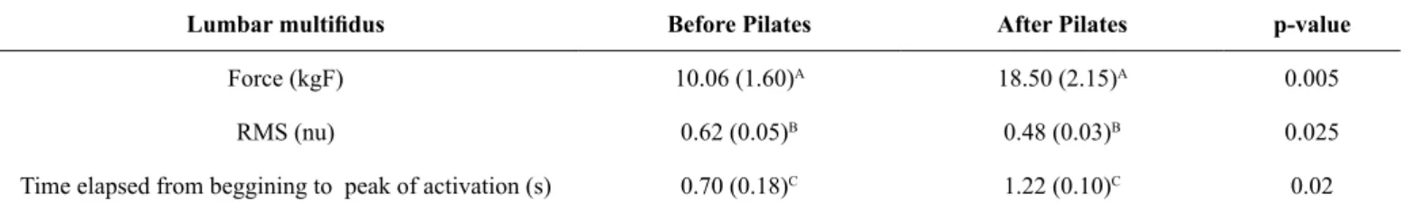
Concerning muscle activation of lumbar multiidus, there was a signiicant decrease in the normalized RMS value (p = 0.025, ES-d = 0.62) in addition to an increase in trunk extension force (p = 0.005, ES-d = 1.33). When analyzing the time elapsed from the beginning to the peak of the activation of the LM, there was a signiicant increase in this period (p = 0.023, ES-d = 0.78) (Table 2).
Table 1. Mean (standard error of measurement) of lexibility, muscle endurance and speciic scales before and after the Pilates.
Flexibilily Before Pilates After Pilates p-value
Trunk anterior lexion (cm) 12.6 (2.93)A 4.87 (1.66)A 0.03
Right trunk lateral bending (cm) 45.25 (0.96)B 40.58 (1.34)B 0.01
Left trunk lateral bending (cm) 45.90 (1.03) 41.33 (1.07) 0.06
Muscle endurance
Right lateral bridge (s) 14.09 (2.35)C 30.94 (4.37) C 0.003
Left lateral bridge (s) 12.99 (2.09)D 32.38 (5.37)D 0.003
Sorensen test (s) 36.41 (6.95)E 74.40 (9.69)E 0.04
Scales
FABQF 5.41 (1.57) 1.91 (0.74) 0.05
FABQW 7.41 (1.55) 8.83 (1.90) 0.57
ODI 6.50 (0.94)F 3.75 (0.79)F 0.03
SF-12 (PCS) 47.25 (2.33) 52.21 (2.71) 0.18
The comparison of the LM and TrA/IO temporal parameters of muscle activity before the Pilates protocol showed a difference in the time from the beginning to the peak of the sEMG signal and in the duration of muscle contraction between the two musculature (p < 0.05): the LM reached the peak of activation earlier than the TrA/IO and with a lower period in contraction with medium to large effect size (Table 3). After the Pilates protocol, both muscles
presented the same behavior: the LM increased the duration of contraction, in addition to increasing the time to reach the peak of activation (p > 0.05), remaining with temporal parameters similar to the TrA/IO (Table 3) but with small effect size (ES-d < 0.40).
The level of pain through VAS was measured daily before and after the Pilates exercises and during the eight weeks, there was a decrease in these parameters (igure 3).
Table 2. Mean (standard error of measurement) of trunk extension force, RMS and time elapsed from beggining to peak of activation of LM.
Lumbar multiidus Before Pilates After Pilates p-value
Force (kgF) 10.06 (1.60)A 18.50 (2.15)A 0.005
RMS (nu) 0.62 (0.05)B 0.48 (0.03)B 0.025
Time elapsed from beggining to peak of activation (s) 0.70 (0.18)C 1.22 (0.10)C 0.02
kgF: kilogram force; RMS: Root Mean Square; nu: normalized unit; A-C: signiicant difference.
Table 3. Mean (standard error of measurement) of the comparison between temporal parameters of EMG activity of LM and TrA/IO before and after the Pilates protocol.
Before Pilates Protocol
EMG data LM TrA/IO p-value
Time elapsed from beggining to peak of activation (s) 0.70 (0.18)A 2.15 (0.53)A 0.01
Duration of contraction (s) 4.99 (0.31)B 6.66 (0.69)B 0.03
After Pilates Protocol
EMG data LM TrA/IO p-value
Time elapsed from beggining to peak of activation (s) 1.22 (0.10) 1.79 (0.59) 0.34
Duration of contraction (s) 5.69 (0.38) 6.72 (0.71) 0.21
s: seconds; A, B: signiicant difference.
Figure 3. Evolution of pain at visual analogue scale (VAS) throughout the 16 days of Pilates.
1 2 3 4 5 6 7 8 9 10 11 12 13 14 15 16
0 1 2 3 4 5 6 7 8 9 10
Discussion
The aim of this study was to evaluate the inluence of Pilates on clinical aspects and muscle activation of LM and TrA/IO of individuals with nonspeciic low back pain. After eight weeks of a Pilates protocol, there was an improvement in pain, dis-ability, lexibility, motor control, the force of trunk extension, and muscle endurance of the trunk stabilizers.
The results showed that Pilates was effective for the reduc -tion of pain and disability in individuals with non-speciic LBP corroborating with similar results found in the literature7,30,31. Kofotolis, Kellis, Vlachopoulos, Gouitas, Theodorakis32 reported that Pilates was more effective than trunk strengthening in increasing the quality of life and decreasing the functional dis-abilities after an 8-week protocol and the effects were retained for a three-month follow-up32. Studies that evaluated the ef-fectiveness of a Pilates protocol compared to general exercises or self-management for eight weeks concluded that the method was more effective in reducing pain, disability, lexibility and balance in individuals with chronic LBP21,22. These authors also emphasized the importance of subgrouping these patients in order to create effective programs aimed at the speciic mechanisms of pain, acting even more in the reduction of disability22.
A preliminary study evaluating muscle endurance using the Sorensen test in patients with pain during movement or lexion posture suggested that decreased muscle endurance, increased sitting time, bad posture, decreased lexibility, and that inactiv -ity may contribute to the onset of non-speciic low back pain33. Similar results were found in our study, in which 58.33% of the individuals had pain in the anterior lexion movement of the trunk, decreased lexibility, and long periods of sitting. Phrompaet, Paungmali, and Pirusan34 evaluated the lexibility and pelvic stability of healthy individuals before and after a Pilates program of eight weeks and found that there was an increase in these parameters after the intervention, suggesting that the improvement of stability may be due to the improvement of lo-cal muscle control, motor learning and physiologilo-cal response to exercise. Our indings corroborate to these authors because there was an improvement of muscle force and lexibility after the intervention protocol.
Regarding the EMG, the results showed lower muscle activa-tion of multiidus and greater force of trunk extension after the Pilates protocol. This suggests that there was an improvement in motor control and a lower propensity to fatigue because there was a need to recruit fewer motor units to achieve greater force. The literature has demonstrated that people with low back pain show a temporal contraction pattern different from healthy people and similar to healthy but fatigued individuals35. It is known that lumbar pain causes inhibition and atrophy of the deep ibers of multiidus, the pathological mechanism of which can be identiied in electromyography as an increase in muscle activation and a decrease in iber conduction velocity36,37. For the evaluation of the time elapsed from the beginning to the peak of the EMG signal of the LM, an increase of this period occurred, showing that the activation happened in a smoother
iber predominant. Motor units composed of type I ibers have more ordered iring characteristics and larger inter-peak periods, and therefore a more synchronous activation. While motor units composed of type II ibers present shorter intervals, frequently present in individuals with low back pain, it can be visualized as an increase in the amplitude of the EMG signal38.
When the temporal EMG parameters were compared to LM and TrA/IO before the Pilates protocol, the time from onset to peak and the duration of contraction were different. The multiidus had a shorter duration of contraction and reached the peak of activation earlier than the TrA/IO. After the Pilates protocol, both muscles had the same behavior, increasing the duration of contraction of the multiidus and also the time to reach the peak of activation, remaining with the temporal parameters similar to the TrA/IO. Thus, after the intervention, there was a balance of muscular action between LM and TrA/IO suggesting a co-activation of the trunk stabilizing muscles. The postural preparation physiologically occurs before the begin-ning of the movement and the LM and TrA/IO are responsible for this dynamic stability26. The stability of the trunk may be compromised by delayed activation of the musculature of this region39. Furthermore, it is not yet known if the lack of pre-activation is a cause or a manifestation of low back pain8. By our indings, it can be observed that there was pre-activation of the lumbar musculature in relation to the abdominal musculature determined by the time elapsed from the beginning to the peak of the activation before the treatment protocol; and after treat-ment, the multiidus started to have the activation together with the abdomen, suggesting that the lack of a previous activation or co- activation of the abdomen may be the cause of LBP. However, the effect-size of the comparison between LM and TrA /IO after Pilates was small, so caution should be taken when extrapolating these data to the population. It is recommended, therefore, that future studies may recruit more individuals so that this comparison can be performed.
Our results allow us to infer that Pilates promoted an im-provement in clinical parameters of LBP patients, in addition to a decrease in muscle fatigue of LM. Furthermore, the lack of co-activation of lumbar and abdominal muscles could be reversed with the Pilates protocol, since individuals started to present similar activation between the multiidus and abdomen, regarding the temporal parameters. With the results obtained by this study and with data already reported in the literature, it is suggested that the Pilates treatment is effective for improv-ing motor behavior and clinical parameters in individuals with low back pain who present instability according to the System of Treatment-based Classiication of low back pain. Thus, conservative treatment is effective and may decrease costs on pharmacological or the risks of surgical treatment.
Conclusions
The results allow us to conclude that an exercise protocol using the Pilates method for eight weeks is effective for improving the motor behavior of the trunk stabilizing muscles and for the clinical signs of pain, disability, lexibility, force and muscular endurance in patients with non-speciic low back pain. This sug-gests that the Pilates Method may be indicated for the treatment of these individuals.
References
1. Natour J, Cazotti LA, Ribeiro LHR, Baptista AS, Jones A. Pilates improves pain, function and quality of life in patients with chronic low back pain: a randomized controlled trial. Clin Rehabil. 2015; 29(1):59-68. < http://dx.doi.org/10.1177/0269215514538981>. 2. Allegri M, De Gregori M, Minella C, Klersy C, Wang W, Sim M,
et al. ‘Omics’ biomarkers associated with chronic low back pain: protocol of a retrospective longitudinal study. BMJ Open. 2016; 6(10):1-8. <http://dx.doi.org/10.1136/bmjopen-2016-012070>. 3. Maher C, Underwood M, Buchbinder R. Non-specific low
back pain. The Lancet. 2016; 1-12. < http://dx.doi.org/10.1016/ S0140-6736(16)30970-9>.
4. Hoy D, March L, Brooks P, Blyth F, Woolf A, Bain C, et al. The global burden of low back pain: estimates from the Global Burden of Disease 2010 study. Ann Rheum Dis. 2014; 73(6):968-974.< http://dx.doi.org/10.1136/annrheumdis-2013-204428>.
5. Nascimento PRC, Costa LOP. Prevalência da dor lombar no Brasil: uma revisão sistemática. Cad. Saúde Pública. 2015; 31(6):1141-1155. <http://dx.doi.org/10.1590/0102-311X00046114>. 6. Silva MC, Fassa AG, Valle NCJ. Dor lombar crônica em uma
população adulta do Sul do Brasil: prevalência e fatores associa -dos. Cad. Saúde Pública. 2004; 20(2):377-385. <http://dx.doi. org/10.1590/S0102-311X2004000200005>.
7. Conceição JS, Mergener CR. Eicácia do método Pilates no solo em pacientes com lombalgia crônica: relato de casos. Revista Dor. 2012; 13 (4):385-388. <http://dx.doi.org/10.1590/ S1806-00132012000400015>.
8. Marshall P, Murphy B. The validity and reliability of surface EMG to assess the neuromuscular response of the abdominal muscles to rapid limb movement. Journal Of Electromyography And Kinesiology. 2003; 13(5):477-489. <http://dx.doi.org/10.1016/ S1050-6411(03)00027-0>.
9. Alaranta H, Luoto S, Heliövaara M, Hurri H. Static back endurance and the risk of low-back pain. Chid Biomechanics. 1995; 10(6):323-324. <http://dx.doi.org/ 10.1016/0268-0033(95)00002-3>. 10. Ramos LAV. Avaliação da fadiga do músculo multíido lombar e
ativação do transverso do abdome em indivíduos com hérnia discal lombar. São Paulo. Dissertação. [Mestrado Curso de Ciências da Reabilitação] - Faculdade de Medicina da Universidade de São Paulo; 2012.
11. Ekstron RA, Donatelli R A, Carp KC. Electromyographic Analysis of Core Trunk, Hip, and Thigh Muscles During 9 Rehabilitation Exercises. J Orthop Sports Phys Ther. 2007; 37(12):754-762. <http://dx.doi.org/10.2519/jospt.2007.2471>.
12. Hebert JJ, Koppennhaver SL, Walker BF. Subgrouping Patients With Low Back Pain: A Treatment-Based Approach to Classiication. Sports Health: A Multidisciplinary Approach. 2011; 3(6):534-542. <http://dx.doi.org/10.1177/1941738111415044>. 13. Maher CG. Effective physical treatment for chronic low back
pain. Orthop Clin North Am. 2004; 35(1):57-64. <http://dx.doi. org/10.1016/S0030-5898(03)00088-9>.
14. Qaseem A, Wilt TJ, McLean RM, Forciea MA. Noninvasive treat -ments for acute, subacute and chronic low back pain: a clinical practice guideline from American College of Physicians. Ann Intern Med. 2017, 166(7):514-530. <http://dx.doi.org/10.7326/ M16-2367>
15. Yamato TP, Maher CG, Saragiotto BT, Hancock MJ, Ostelo RW, Cabral CM, et al. Pilates for low back pain. Cochrane Database Syst Rev. 2015; 2(7):1-71. <http://dx.doi. org/10.1590/1516-3180.20161344T1>
16. Menacho MO, Obara K, Conceição JS, Chitolina ML, Krantz DR, da Silva RA, et al. Electromyographic Effect of Mat Pilates Exercise on the Back Muscle Activity of Healthy Adult Females. J Manipulative Physiol Ther. 2010; 33(9):672-678. <http://dx.doi. org/ 10.1016/j.jmpt.2010.08.012>.
17. Pereira IL, Queiroz B, Loss J, Amorim C, Sacco IC. Trunk muscle EMG during intermediate Pilates Mat exercises in beginner healthy and chronic low back pain individuals. J Manipulative Physiol Ther. 2017; 40(5): 350-357. http://dx.doi. org/10.1016/j.jmpt.2017.02.010.
18. Yamato TP, Maher CG, Saragiotto BT, Hancock MJ, Ostelo RWJG, Cabral CMN, Costa LCM, Costa LOP. Pilates for low back pain: complete republication of a Cochrane review. Spine. 2016; 41(12): 1013-1021. http://dx.doi.org/10.1097/brs.0000000000001398 19. Lin HT, Hung WC, Hung, JL, Wu PS, Liaw LJ, Chang JH. Effects
of Pilates on patients with chronic non-speciic low back pain: a systematic review. J Phys Ther. 2016; 28(10): 2961-2969. <http:// doi.org/10.1589/jpts.28.2961>
20. Liebenson C. Spinal stabilization – an update. Part 2 – functional assessment. J Bodyw Mov Ther. 2004; 8(3):199-210. <http:// dx.doi.org/10.1016/j.jbmt.2004.03.002>.
21. Valenza MC, Rodríguez-Torres J, Cabrera-Martos I, Díaz-Pelegrina A, Aguilar-Ferrándiz ME, Castellote-Caballero Y, et al. Results of a Pilates exercise program in patients with chronic non-specific low back pain: A randomized controlled trial. Clin Rehabil. 2016; 1-8. <http://dx.doi.org/ 10.1177/0269215516651978>.
22. Brooks C, Kennedy S, Marshall PW. Specific Trunk and General Exercise Elicit Similar Changes in Anticipatory Postural Adjustments in Patients With Chronic Low Back Pain. Spine. 2012; 37(25):1543-1550. <http://dx.doi.org/10.1097/ BRS.0b013e31826feac0>.
23. Ware JE, Kosinski M, Keller SD. A 12-Item Short-Form Health Survey: Construction of Scales and Preliminary Tests of Reability and Validity. Med Car. 1996; 34(3):220-233. <http://dx.doi. org/10.1097/00005650-199603000-00003>.
25. Hermens HJ, Freriks B, Disselhorst-Klug C, Rau G. Development of recommendations for SEMG sensors and sensor placement procedures. J Electromyogr Kinesiol. 2000; 10:361–74. <http:// dx.doi.org/10.1016/S1050-6411(00)00027-4>.
26. Rossi DM, Morcelli MH, Marques NR, Hallal CZ, Gonçalves M, Laroche DP, et al. Antagonist coactivation of trunk stabilizer muscles during Pilates exercises. J Bodyw Mov Ther. 2014; 18(1):34-41. <http://dx.doi.org/10.1016/j.jbmt.2013.04.006>. 27. Isacowitz R, Clippinger K. Anatomia do pilates. São Paulo:
Manole, 2013.
28. Wells C, Kolt GS, Bialocerkowski A. Deining Pilates exercise: a systematic review. Complement Ther Med. 2012; 20(4):253-262. <http://dx.doi.org/10.1016/j.ctim.2012.02.005>.
29. Espirito-Santo H, Daniel F. Calcular e apresentar tamanhos do efeito em trabalhos cientíicos (1): as limitações do p< 0, 05 na análise de diferenças de médias de dois grupos. RPICS. 2015; 1(1):3-16. <http://dx.doi.org/10.7342/ismt.rpics.2015.1.1.14>. 30. Miyamoto GC, Costa LOP, Cabral CMN. Eficacy of the Pilates
method for pain and disability in patients with chronic nonspeciic low back pain: a systematic review with meta-analysis. Braz J Phys Ther. 2013; 17(6):517-532. <http://dx.doi.org/10.1590/ S1413-35552012005000127>.
31. Patti A, Bianco A, Paoli A, Messina G, Montalto M A, Bellaiore M, et al. Effects of Pilates Exercise Programs in People With Chronic Low Back Pain: A Systematic Review. Medicine. 2015; 94(4):383-394. <http://dx.doi.org/10.1097/ MD.0000000000000383>.
32. Kofotolis N, Kellis E, Vlachopoulos SP, Gouitas I, Theodorakis Y. Effects of Pilates and trunk strengthening exercises on health-related quality of life in women with chronic low back pain. J Back Musculoskelet Rehabil. 2016; 29(4): 649-659. <http:// dx.doi.org/10.3233/BMR-160665>.
33. O’Sullivan PB, Mitchell T, Bulich P, Waller R, Holte J. The rela-tionship between posture and back muscle endurance in industrial workers with lexion-related low back pain. Man Ther. 2006; 11(4):264-271. <http://dx.doi.org/10.1016/j.math.2005.04.004>.
34. Phrompaet S, Paungmali A, Pirunsan U. Effects of Pilates Training on Lumbo-Pelvic Stability and Flexibility. Asian J Sports Med. 2011; 2(1):16-22. <http://dx.doi.org/10.5812/asjsm.34822>. 35. Jubany J, Danneels L, Angulo-Barroso R. The influence of
fatigue and chronic low back pain on muscle recruitment pat -terns following an unexpected external perturbation. BMC Musculoskelet Disord. 2017; 18(1): 161. <https://doi.org/10.1186/ s12891-017-1523-3>
36. Mcneill W. Core stability is a subset of motor control. J Bodyw Mov Ther. 2010; 14(1):80-83. <http://dx.doi.org/10.1016/j. jbmt.2009.10.001>.
37. Vollestad NK. Measurement of human muscle fatigue. J Neurosci Methods. 1997; 74(2):219-227. <http://dx.doi.org/10.1016/ S0165-0270(97)02251-6>.
38. Miura T, Sakuraba K. Properties of Force Output and Spectral EMG in Young Patients with Nonspeciic Low Back Pain during Isometric Trunk Extension. J. Phys. Ther. Sci. 2014; 26(3):323-329. <http://dx.doi.org/10.1589/jpts.26.323>.
39. Silies SP, Mehta R, Smith SS, Karduna AR. Differences in feed -forward trunk muscle activity in subgroups of patients with me -chanical low back pain. Arch Phys Med Rehabil. 2009; 90(7):1159-1169. <http://dx.doi.org/10.1016/j.apmr.2008.10.033>.
Corresponding author
*Heloyse Uliam Kuriki
LARAL, Laboratório de Avaliação e Reabilitação do Aparelho Locomotor. Rua
Pe-dro João Pereira, 150, Mato Alto, Araranguá-SC Email: [email protected]
Manuscript received on February 24, 2017
Manuscript accepted on December 6, 2017