Letters to the Editor
Radiol Bras. 2017 Mar/Abr;50(2):135–138
135
0100-3984 © Colégio Brasileiro de Radiologia e Diagnóstico por Imagem
Letters to the Editor
What radiologists need to know about 3D printing and its main applications in musculoskeletal imaging
Dear Editor,
The utility of the various imaging methods in the evaluation and diagnosis of musculoskeletal disorders is well established, and those methods play a fundamental role in the planning of the dif-ferent treatments, be they conservative or surgical, providing im-ages that can be manipulated through specific software to create three-dimensional (3D) reconstructions. To date, however, such 3D reconstructions have been made available only as digital files, as images on radiographic films, or as prints on paper. These tra-ditional forms of image documentation do not always allow sur-geons to have a real in-depth sensory notion and knowledge of the 3D anatomical relationships in the planning of different types of surgical procedures. Recently, 3D printing has come to be used with increasingly frequency to obtain a more realistic and more accurate analysis by creating 3D models(1–3).
What really is 3D printing? The definition of 3D printing, also known as rapid prototyping, is the use of a set of methods to create solid three-dimensional objects (models or prototypes) from the data contained in digital files. There are different forms of 3D printing, one of the most popular being the additive processing technique, in which the object is created layer by layer through successive depositions of a highly resistant plastic polymer.
How does 3D printing work? It all begins with the develop-ment of the 3D digital file. The file is obtained through the acqui-sition of sectional images through the use of magnetic resonance imaging, computed tomography, or even (3D or 4D) ultrasound.
The digital file is then analyzed and processed with computer-aided design (CAD) software, according to what is required in each situ-ation. After developing the 3D digital file, the CAD modeling soft-ware divides the prototype into hundreds or thousands of thin hori-zontal layers, thus preparing the file for printing. The digital file can then be loaded into a 3D printer for printing.
Is 3D printing already a reality in clinical practice or only in experimental research? In several countries, it is already a part of the clinical routine, having been shown to have a great impact on the precision and safety of surgical procedures(2–5). There has been
rapid growth in the number of potential applications of 3D print-ing in medicine, which has already been used in several situations, even in Brazil(6–8). We illustrate, as an example, a case in which
3D printing was employed at our facility for the preoperative plan-ning of the surgical treatment of an osteolytic lesion in the man-dible (Figure 1).
What are the applications of 3D printing in musculoskeletal imaging? Among its many potential applications in clinical prac-tice and in the teaching of medicine, we emphasize in this article the use of the technique in musculoskeletal imaging. We high-light its application in the preoperative planning of complex surgi-cal procedures, which require high precision, such as those em-ployed in the treatment of spinal deformities and complex frac-tures, as well as in the creation of models of orthotics and prosthe-ses tailored to the anatomy and needs of each patient(1–9).
We believe it to be inevitable that, in the coming years, there will be growth in the application of the 3D printing technique in the field of medicine as a whole, especially in the area of muscu-loskeletal imaging. The incorporation of this new technique will
Letters to the Editor
Radiol Bras. 2017 Mar/Abr;50(2):135–138
136
http://dx.doi.org/10.1590/0100-3984.2015.0208
Subcapsular splenic hematoma and spontaneous hemoperitoneum in a cocaine user
Dear Editor,
A 23-year-old male patient presented with a 36-h history of intense, sudden, progressive abdominal pain, predominantly in the left hypochondrium, irradiating to the ipsilateral infrascapular re-gion. He reported no previous trauma, fever, headache, fatigue, myalgia, arthralgia, skin alterations, or comorbidities. During the clinical interview, he reported moderate smoking and the routine use of an illicit drug (cocaine), including hours prior to the onset of pain. On physical examination, he was well-oriented, hemody-namically stable, and afebrile. The serology was negative for hepa-titis B, hepahepa-titis C, and dengue, and the results were normal for antineutrophil cytoplasmic antibody, antinuclear factor, the ve-nereal disease research laboratory test, urea, creatinine, erythro-cyte sedimentation rate, C-reactive protein, and coagulation pro-file. Hemoglobin electrophoresis showed no alterations.
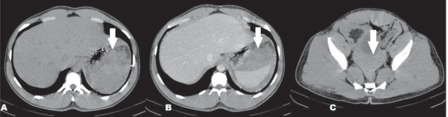
Computed tomography (CT) showed a dense collection, com-patible with hematic material, in close proximity to the spleen, as well as showing hemoperitoneum (Figure 1). Arteriography showed no abnormalities. Exploratory laparotomy revealed sub-capsular splenic hematoma and confirmed the hemoperitoneum, with no evidence of a lesion within the cavity.
Given that there was no perisplenic trauma or adhesions sug-gestive of previous trauma and that the macroscopic aspect of the spleen was normal on the CT scan and in the exploratory laparo-tomy, together with the facts that diseases affecting the splenic parenchyma were ruled out and that the patient had used cocaine immediately prior to the episode, we established the working di-agnosis of nontraumatic splenic hemorrhage secondary to cocaine use. During clinical follow-up, the patient progressed well, with-out complications.
Recent studies in the radiology literature of Brazil have em-phasized the importance of CT and magnetic resonance imag-ing scans to improvimag-ing the diagnosis in nontraumatic abdominal disorders(1–5). Splenic hemorrhages are rarely encountered
with-out prior trauma and can have fatal consequences, which makes their early diagnosis essential. The main nontraumatic conditions include neoplasms, as well as inflammatory/infectious, iatrogenic, and mechanical processes(6).
The clinical signs of nontraumatic splenic hemorrhage are similar to those found in cases resulting from trauma, including pain in the upper left quadrant, with or without irradiation to the left shoulder, caused by diaphragmatic irritation, evolving to he-modynamic instability in the most severe cases. Such manifesta-tions are nonspecific and cannot be characterized solely by physi-cal examination. Therefore, in hemodynamiphysi-cally stable patients,
Francisco Abaeté Chagas-Neto1, Francisco Coracy Carneiro
Monteiro2, Eduardo Lima da Rocha3, Everaldo
Gregio-Junior4, Marcello Henrique Nogueira-Barbosa4
1. Centro Universitário Christus (Unichristus) e Hospital Antônio Prudente, Fortaleza, CE, Brazil. 2. Hospital Albert Sabin, Fortaleza, CE, Brazil. 3. Hospital Antônio Prudente, Fortaleza, CE, Brazil. 4. Faculdade de Medicina de Ribeirão Preto da Universidade de São Paulo (FMRP-USP), Ribeirão Preto, SP, Brazil. Mailing address: Dr. Francisco Abaeté Chagas-Neto. Rua João Adolfo Gurgel, 133, Coco. Fortaleza, CE, Brazil, 60192-345. E-mail: [email protected].
allow the optimization of protocols promoting good practices, of-fering greater effectiveness to the professionals involved and al-lowing better results, with potentially greater safety for patients.
REFERENCES
1. Jones DB, Sung R, Weinberg C, et al. Three-dimensional modeling may improve surgical education and clinical practice. Surg Innov. 2016; 23:189–95.
2. Wu C, Tan L, Lin X, et al. Clinical application of individualized refer-ence model of sagittal curves by three-dimensional printing technique and computed-aided navigation system for lumbar spondylolystesis. Zhongguo Xiu Fu Chong Jian Wai Ke Za Zhi. 2015;29:734–40. 3. Chen X, Zhang G, Lin H, et al. Digital design of standard parts database
for proximal tibia fractures treated with plating via three-dimensional printing. Zhongguo Xiu Fu Chong Jian Wai Ke Za Zhi. 2015;29:704– 11.
4. Mowry SE, Jammal H, Myer C 4th, et al. A novel temporal bone simulation model using 3D printing techniques. Otol Neurotol. 2015;36:1562–5. 5. Xu N, Wei F, Liu X, et al. Reconstruction of the upper cervical spine using a personalized 3D-printed vertebral body in an adolescent with Ewing sarcoma. Spine (Phila Pa 1976). 2016;41:E50–4.
6. Werner H, Rolo LC, Araujo Júnior E, et al. Manufacturing models of
fetal malformations built from 3-dimensional ultrasound, magnetic reso-nance imaging, and computed tomography scan data. Ultrasound Q. 2014;30:69–75.
7. Werner Jr H, Santos JL, Belmonte S, et al. Applicability of three-dimen-sional imaging techniques in fetal medicine. Radiol Bras. 2016;49:281– 7.
8. Araujo Júnior E. Three-dimensional ultrasound in fetal medicine after 25 years in clinical practice: many advances and some questions. Radiol Bras. 2016;49(5):v–vi.
9. AbouHashem Y, Dayal M, Savanah S, et al. The application of 3D print-ing in anatomy education. Med Educ Online. 2015;20:29847.