Hydrostatic pulmonary edema: high-resolution computed
tomography aspects*
CLÁUDIA MARIA CUNHA RIBEIRO1, EDSON MARCHIORI2, ROSANA RODRIGUES3, EMERSON GASPARETTO4, ARTHUR SOARES SOUZA JÚNIOR5, DANTE ESCUISSATO6, LUIZ FELIPE NOBRE7, GLÁUCIA ZANETTI8, CÉSAR DE ARAUJO NETO9, KLAUS IRION10
* Study carried out in the Radiology Department of the Universidade Federal do Rio de Janeiro (UFRJ, Federal University of Rio de Janeiro) Clementino Fraga Filho University Hospital - Rio de Janeiro, Brazil.
1. Masters student in Radiology at the Universidade Federal do Rio de Janeiro (UFRJ, Federal University of Rio de Janeiro) -Rio de Janeiro, Brazil
2. Assistant Professor of Radiology at the Universidade Federal de Santa Catarina (UFSC, Federal University of Santa Catarina) - Florianópolis, Brazil
3. Professor of Pulmonology at the Faculdade de Medicina de Petrópolis (FMP, Petrópolis School of Medicine) - Petrópolis, Brazil
4. Assistant Professor of Radiology at the Universidade Federal da Bahia (UFBA, Federal University of Bahia) - Salvador, Brazil 5. Professor for the Postgraduate Program in Internal Medicine at the Universidade Federal do Rio Grande do Sul (UFRGS, Federal University of Rio Grande do Sul) - Porto Alegre, Brazil; Physician for the The Pennine Acute Hospitals NHS Trust, Manchester, England
6. Adjunct Professor of Radiology at the Faculdade de Medicina de São José do Rio Preto (FAMERP, Sao Jose do Rio Preto School of Medicine) - São José do Rio Preto, Brazil
7. Physician Researcher in the Radiology Department of the Universidade de São Paulo (USP, University of São Paulo) - São Paulo, Brazil
8. Head Professor at the Radiology Department at the Universidade Federal Fluminense (UFF, Fluminense Federal University) - Niterói, Brazil; Adjunct Coordinator of the Postgraduate Program in Radiology at the Universidade Federal do Rio de Janeiro (UFRJ, Federal University of Rio de Janeiro) - Rio de Janeiro, Brazil
9. Assistant Radiology Professor at the Universidade Federal do Paraná (UFPR, Federal University of Parana) - Londrina, Brazil. 10. Physician in the Radiology Department of the Universidade Federal do Rio de Janeiro (UFRJ, Federal University of Rio de Janeiro) Clementino Fraga Filho University Hospital - Rio de Janeiro, Brazil
Correspondence to: Edson Marchiori. Rua Thomaz Cameron, 438. Valparaíso - CEP: 25685-120, Petrópolis, RJ, Brazil. Submitted: 18 November 2005. Accepted, after review: 13 February 2006.
ABSTRACT
Objective: This study aimed to use high-resolution computed tomography scans of the chest to characterize the principal alterations occurring in cases of hydrostatic pulmonary edema. Methods: A retrospective analysis was made of the tomography scans of 15 patients presenting clinical profiles of hydrostatic pulmonary edema. The cases were divided into five groups by etiology: congestive heart failure (n = 7); acute mitral valve disease (n = 2); acute myocardial infarction (n = 2); myocarditis (n = 2); and fibrosing mediastinitis (n = 2). Results: The principal findings in the cases of hydrostatic pulmonary edema were ground-glass opacities (in 100%), interlobular septal thickening (in 100%), pleural effusion (in 87%) and peribronchovascular interstitial thickening (in 80%). Other, less common, findings were increased blood vessel diameter, consolidations and air-space nodules. Conclusion: The predominant pattern found in the patients studied was that of ground-glass opacities accompanied by interlobular septal thickening (mosaic attenuation pattern) and bilateral (predominantly right-sided) pleural effusion.
INTRODUCTION
Pulmonary edema is a common pathological condition that occurs when the rate of interstitial fluid production exceeds the eliminating capacity of the lungs, thereby resulting in fluid accumulation. Pulmonary edema can result from an increase in the extravascular production of fluid or from an inability to remove the interstitial fluid.(1-4)
Pulmonary edema can be classified as one of four types: hydrostatic edema; permeability edema with diffuse alveolar damage; permeability edema without diffuse alveolar damage; and mixed edema (caused by a combination of increased hydrostatic p r e s s u r e a n d g r e a t e r p u l m o n a r y c a p i l l a r y permeability).(1,3) In the current study, assessments
were based exclusively on tomographic findings related to hydrostatic pulmonary edema.
Hydrostatic edema is characterized by a transudative mechanism, and heart failure is the most common cause.(1,5) There are other causes, although
of lesser prevalence, that lead to pulmonary venous obstruction, which is in turn accompanied by hydrostatic edema.(1,3,5)
Although the radiological aspects of hydrostatic edema have been extensively studied, the tomographic findings, especially those from high-resolution computed tomography scans, have received little attention. Nevertheless, knowledge of such findings is important for making the differential diagnosis with other lung diseases, as well as for, under certain circumstances, avoiding unnecessary lung biopsies.(6) The tomographic
findings described in the current literature are similar to those seen in other pulmonary diseases. Therefore, including pulmonary edema in the differential diagnosis in these cases might aid clinicians in reaching an accurate final diagnosis. The present study aims to describe the pulmonary alterations that occur during hydrostatic edema, as observed on high-resolution computed tomography scans of the chest.
METHODS
A retrospective study was conducted on the basis of a review of radiology files from five medical institutions in the state of Rio de Janeiro: Clementino Fraga Filho University Hospital; Barra D'Or Hospital; Copa D'Or Hospital; Central Army
Hospital; and Mário Lioni Clinical Hospital. High-resolution computed tomography scans were acquired from fifteen patients with pulmonary edema of multi-etiological hydrostatic origin, assessed from 2000 to 2005.
Of those fifteen patients, five were male, and five were female. At the time of diagnosis, the mean age of the patients was 54.7 years (range, 15-90 years; median, 57 years). As for the distribution by age bracket, there were more patients in their fifties than in any other age bracket.
In seven of the fifteen patients studied, the edema resulted from chronic cardiovascular diseases, confirmed by clinical data and by improvement after specific treatment for cardiogenic pulmonary edema. In some patients, the confirmation was made through echocardiography (low left ventricle ejection fraction). There were two cases of secondary pulmonary edema leading to severe myocarditis, the diagnosis being based on clinical data, laboratory test results and echocardiography findings. Two cases of pulmonary edema resulted from acute mitral valve disease (rupture of the mitral valve chordae), confirmed through trans-thoracic echocardiogram and surgery: one due to myxomatous valvular dystrophy; and one due to acute myocardial infarction. The two cases were treated as an isolated group (acute mitral valve disease). There were two cases of acute myocardial infarction diagnosed through clinical findings, laboratory test results and echocardiography data. Regarding the two cases of fibrosing mediastinitis, one was confirmed (through histological analysis of a sample obtained via open lung biopsy) as fibrosing mediastinitis secondary to histoplasmosis. In the second case, there was no histological confirmation. However, a presumptive diagnosis of histoplasmosis was made based on the clinical and epidemiological history, which was highly suggestive of such. All patients underwent high-resolution spiral computed tomography scans of the chest in one of the following devices: a Siemens Somaton Plus 4 (Barra D'Or Hospital); a Siemens Somatom Balance (Clementino Fraga Filho University Hospital and Mário Lioni Clinical Hospital); a Toshiba Asteion (Clementino Fraga Filho University Hospital); a GE Light Speed (Hospital Copa D'Or Hospital; and a Philips Secura, Release 1.3 (Central Army Hospital).
TABLE 1
Tomographic findings in hydrostatic edemas
Tomographic findings Patients % (n = 15) Ground-glass opacities 15 100 Interlobular septal thickening 15 100 Pleural effusion 13 87 Peribronchovascular thickening 12 80 Mosaic pattern of attenuation 10 67 Vascular diameter increase 7 47 Consolidations 5 33 Peribronchial nodules 3 20
Figure 1 - High-resolution computed tomography scans, showing slices acquired at the lung bases, using a pulmonary window, in a patient with congestive heart failure presenting ground-glass opacities and smooth interlobular septa thickening, a mosaic pattern of attenuation and bilateral pleural effusion
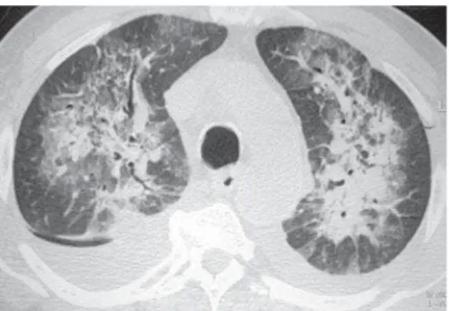
Figure 2 - High-resolution computed tomography scans, showing slices acquired at the aortic arch level, using a pulmonary window, in a patient with acute myocardial infarction presenting consolidations, ground-glass opacities, smooth interlobular septal thickening and bilateral pleural effusion, for patient with. Please observe the medullary distribution of the opacities
the tests shows that appropriate lung windows, as well as, in some cases, mediastinal windows, were used. No venous contrast medium was administered to any of the patients. Complementary ventral decubitus slices were acquired in some of the patients.
Two radiologists assessed the tests independently, and the final decisions were arrived at by consensus. Using a pre-established analysis protocol, tests were analyzed for the presence or absence of parenchymal or nonparenchymal alterations. The following were studied: ground-glass opacities; thickening of the interlobular septa; mosaic pattern of attenuation; thickening of the peribronchial-vascular interstice; consolidation; peribronchial nodules; pleural effusion; and vascular diameter. Findings were identified and classified pursuant to the Brazilian Consensus on Terminology related to Describing Findings on Computed Tomography Scans of the Chest (Brazilian Thoracic Society),(7) as
well as to the Fleischner Society Glossary of Terms.(8)
RESULTS
The most common tomographic findings in the parenchyma were as follows: ground-glass opacities (n = 15; 100%); thickening of the interlobular septa (n = 15; 100%); peribronchovascular thickening (n = 12; 80%); and mosaic pattern of attenuation (n = 10; 66%). Other less common findings were consolidations (n = 5; 33%) and peribronchial nodules (n = 3; 20%). Of the nonparenchymal findings, pleural effusion was the most common (n = 13; 87%), followed by an increase in vascular diameter (n = 7; 46%) (Table 1). Overall, ground-glass opacities and thickening of the interlobular septa were the most common findings, occurring in 100% of the cases (Figures 1 and 2).
Figure 5 - High-resolution computed tomography scans, showing slices acquired at the upper lobe level, using a pulmonary window, revealing asymmetrical consolidations predominantly in the upper right lobe in a patient with ruptured mitral chordae. Note the ground-glass opacities adjacent to the consolidations
Figure 3 - High-resolution computed tomography scans, showing slices acquired at the lung bases, using a pulmonary window, revealing smooth septal thickening in the lower right lobe, increased vascular diameter and bilateral pleural effusion in a patient with congestive heart failure. Please observe the aneurysm on the descending aorta
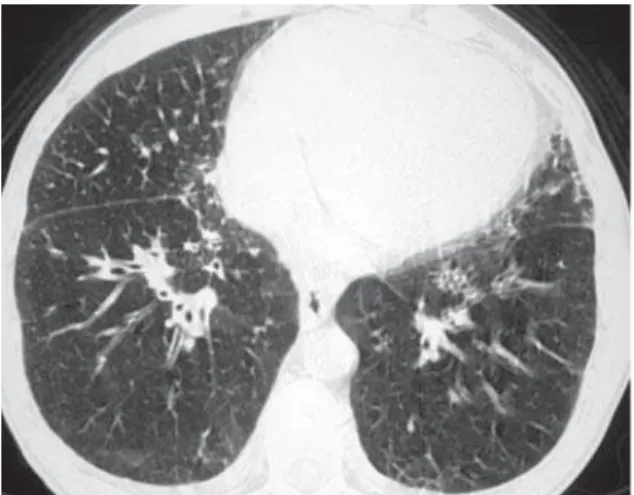
Figure 4 - High-resolution computed tomography scans, showing slices acquired at the level of the lower third of the lungs, using a pulmonary window, revealing peribronchovascular interstitial thickening and discrete thickening of the interlobular septa, more evident in the middle lobe
Nevertheless, the mosaic pattern of attenuation was observed in only ten cases (66%) (Figure 1).
Peribronchovascular interstitial thickening was observed in twelve cases (80%), being bilateral in ten (Figure 4) and unilateral in two (the cases of fibrosing mediastinitis).
Consolidation was detected in only five (33%) of the patients (Figures 2 and 5). All of the patients presented bilateral consolidations, In two cases, the distribution was asymmetric, with a 'bat-wing'
aspect, and predominant in the upper right lobe (cases of mitral valve chordae rupture) (Figure 5). Peribronchial nodules were found in three patients (20%), all with ground-glass attenuation and measuring less than 1 cm in diameter.
Pleural effusion was the third most common tomographic finding overall and was the most common nonparenchymal finding, being found in 13 patients (87%), predominantly in the bilateral form (n = 11) (Figures 1-3). Among the eleven patients with bilateral pleural effusion, nine presented asymmetric pleural effusion, predominantly on the right side. Two cases of exclusively right-sided pleural effusion were identified.
Vascular diameter was increased in seven cases (44%) (Figure 3).
For the two fibrosing mediastinitis patients, we observed calcified mediastinitis masses, which caused a reduction in the diameter of the mediastinal structures, including the right pulmonary veins.
DISCUSSION
tomography scans of the chest, and high-resolution scans in particular.
There is currently a more comprehensive system of classifying pulmonary edema, which defines four types: hydrostatic edema; permeability edema with diffuse alveolar damage; permeability edema without diffuse alveolar damage; and mixed edema (caused by a combination of increased hydrostatic p r e s s u r e a n d g r e a t e r p u l m o n a r y c a p i l l a r y permeability).(1,3)
Hydrostatic edema is secondary to elevated capillary hydrostatic pressure, with the preservation of normal selective permeability of the capillary endothelium and alveolar epithelial barriers. Elevated hydrostatic pressure can result from obstructive lesions of the pulmonary veins, left atrium or mitral valve, or even from dysfunction of the left ventricle, the most common cause being a reduction in the compliance of the left ventricle and increased end-diastolic pressure, which is retrogradely transmitted to the pulmonary venous microcirculation.(3,5)
Classically, two physiopathological and radiological phases of hydrostatic pulmonary edema are recognized: interstitial edema and alveolar edema.(1,3,6,9) The first phase is characterized
by interstitial edema, which is secondary to the transudation of fluid from the vascular space to the interstitium, due to increased capillary pressure. With progressive increases in pressure, the fluid is extravasated to the alveolar spaces. Assuming that the capillary endothelial barrier remains intact, the protein content of the alveolar fluid is lower than that of plasma.
Radiological alterations derived from increased hydrostatic capillary pressure are divided into three stages: vascular pulmonary congestion; interstitial edema; and alveolar edema.(3) However, there are
factors that make it difficult to establish a correlation between radiological findings and pulmonary capillary pressure. It is known that the movement of fluid within the pulmonary spaces occurs slowly. Therefore, even when hydrostatic pressure is high, radiological findings might not be evident. The opposite is also true: in patients presenting recent improvement in pulmonary capillary pressure, the pulmonary fluid might not have yet mobilized, and such patients can present images of edema and normal pressure levels.(1,10)
Despite the fact that vascular pulmonary
congestion can be the earliest finding, its finding on chest X-rays is limited, since it is a subjective assessment requiring appropriate (orthostatic) positioning of the patient. It is identified by determining the number and diameter of vessels in the upper and lower zones of the lung, equidistant from the pulmonary hilum. The use of prior tests for comparison increases the detection of this signal.(3)
On high-resolution computed tomography scans, increased diameter of arteries and veins can be seen, facilitating their recognition in the perihilar regions, in which arterial diameter is usually similar to that of the respective bronchi.(3,9)
The second phase in the evolution of hydrostatic edema is interstitial edema, which provokes thickening of the peribronchovascular interstice (central fluid-draining form). This in turn, on chest X-rays, manifests as poor definition of the pulmonary vessels and apparent thickening of the bronchial walls (peribronchial cuff).(3) On
high-resolution computed tomography scans, thicker bronchial walls are seen, together with an apparent increase in the diameter of the central and peripheral pulmonary vessels.(9) When interstitial
pressure remains high, the interlobular septa appear due to having been thickened by the infiltration of fluid. Another aspect of interstitial edema is the presence of ground-glass opacities.(1,3,9,11)
When the fluid reaches the alveolar space, characterizing the last phase of hydrostatic edema (alveolar edema), small nodules form. These are accompanied by acinar areas, which are prone to confluence, forming pronounced, well-defined bilateral consolidations. Probably due to the gravitational effect, these consolidations are predominantly found in the central and lower potions of the lungs.(1,3)
Hydrostatic pulmonary edema that occurs suddenly can present as a distinct pattern known as a 'bat-wing' or 'butterfly-wing' pattern. The increase in pressure occurs abruptly, with a rapid passage from the first phase (interstitial) to the second (alveolar), and the first phase is therefore not seen radiologically. This aspect occurs mainly in cases of acute myocardial infarction, chordal rupture, infarction of the papillary muscles or renal failure.(3,12)
principal ones are heart enlargement and pleural effusion. Heart enlargement is a common radiological finding, since the most frequent cause of hydrostatic pulmonary edema is left ventricle dysfunction. This finding is generally absent in hydrostatic edema resulting from pulmonary venous abnormalities, pure mitral stenosis, acute myocardial infarction or acute mitral regurgitation.(3) In high-resolution computed
tomography scans, the assessment of the cardiac dimensions is defined on the axial planes, at the largest diameter perpendicular to the long axis of the cavity. The principal cardiac measurement in pulmonary edema is that of the left atrium, defined as the longest transversal axis at the level of the pulmonary veins and aortic emergence, a diameter of 6.6 cm, with a mean deviation of ±1 cm, being considered normal.(13)
Pleural effusion is a common finding, especially in severe cases of hydrostatic pulmonary edema. It is believed that greater, right-sided pleural effusion is the most common. However, such effusion can also be left-sided or bilateral.(3)
In this study, fifteen patients with hydrostatic pulmonary edema were assessed. Of those fifteen, seven (47%) presented chronically acquired heart disease as the underlying disease. The remaining eight patients were classified as fitting one of four profiles: acute myocardial infarction (n = 2); acute mitral valve disease with rupture of the mitral chordae (n = 2), acute cardiomyopathy (n = 2); and fibrosing mediastinitis due to histoplasmosis, accompanied by a reduction in pulmonary vein diameter and a unilateral increase in hydrostatic pressure. Fibrosing mediastinitis can be caused by a variety of agents or conditions. However, the most common cause is infection with Histoplasma capsulatum.(14-15)
Ground-glass opacities were detected in all fifteen patients (100%) and were bilateral in thirteen. Ground-glass opacities were unilateral only in the two fibrosing mediastinitis patients. In another study,(9) ground-glass opacities were
observed in six of seven patients with hydrostatic edema.
Thickening of the interlobular septa was a common finding accompanying hydrostatic edema, o c c u r r i n g i n 1 0 0 % o f t h e p a t i e n t s a n d corresponding to the interstitial pathway for the drainage of excess pulmonary fluid. In thirteen patients (87%), the thickening was bilateral, whereas it was unilateral in two (13%). The
interlobular septal thickening was smooth in all of the cases (100%). In another study(9) involving four
patients, only one presented a nodular aspect, which was likely attributable to an increase in septal vein diameter. Nodular thickening of the interlobular septa is a common finding in a number of diseases, particularly in sarcoidosis and lymphangitic carcinomatosis.(16)
Septal thickening was not found in isolation. In all patients presenting septal thickening, there were also ground-glass opacities, and the mosaic pattern of attenuation was observed in ten patients (67%). In another study,(9) interlobular septal thickening was
also not observed as an isolated tomographic finding, although its presence was closely correlated with peribronchovascular interstitial thickening.
Among the image findings, pleural effusion was one of the findings most commonly associated with hydrostatic edema, occurring in thirteen cases. The pleural effusion was bilateral in twelve patients, presenting an asymmetrical, predominantly right-sided distribution in ten of those twelve. The association between pleural effusion and hydrostatic edema has been well established in literature. Some authors(9) reported pleural effusion in four of seven
patients with hydrostatic edema, the majority of the effusion being unilateral and right-sided, only one patient presenting bilateral effusion. In our study, only the patients with fibrosing mediastinitis presented unilateral pleural effusion.
Peribronchovascular interstitial thickening was seen in twelve patients (80%). In ten patients, the thickening was bilateral and smooth. In the two cases of fibrosing mediastinitis, the peribronchovascular interstitial thickening was unilateral. Such thickening is identified by determining the thickness of the bronchial walls. The presence of peribronchovascular interstitial thickening reflects, for hydrostatic edema patients, drainage of the fluid via central interstitial pathways.(3) Em outro estudo,(9) o espessamento foi
achado na tomografia computadorizada de alta resolução em quatro de sete pacientes.
In another study,(9) such thickening was
detected in the high-resolution computed tomography scans in four of seven patients.
In the present study, an increase in vascular diameter was identified in seven patients (47%). Some authors(3) consider increased vascular
is nevertheless difficult. Tomography allows vascular diameter to be measured in a more straightforward manner, comparing the diameter of the vessels with that of the corresponding bronchi. Other authors(9) have also observed an
increase in vascular diameter in four of seven hydrostatic edema patients.
In the present study, consolidations were seen in only five patients. This could be explained by the fact that the great majority of cases occur in patients with diseases of lesser severity, probably during the early interstitial and alveolar phases of the pulmonary edema. Consolidations appear during the later phases of alveolar edema, when hydrostatic pressure levels rise sufficiently to promote the infusion of the air spaces.(3,19) In
another study,(9) no consolidations were identified
in hydrostatic edema patients.
We observed the 'bat-wing' distribution of consolidations in two patients with acute rupture of the mitral chordae, one due to myxomatous degeneration and the other due to acute myocardial infarction. According to some authors,(1)
this 'bat-wing' aspect is only seen in cases of acute hydrostatic pulmonary edema. The imaging aspect is that of a consolidation that spares the lung periphery. In the cases of rupture of the mitral chordae, the consolidation was asymmetric and predominantly in the upper right lobe. Other a u t h o r s(18) r e p o r t e d f o u r c a s e s o f m i t r a l
regurgitation with asymmetric edema in the upper right lobe. Still other authors(19) determined that
the frequency of upper right lobe involvement in patients with mitral reflux was 9%. The regurgitated blood moves to the posterior wall of the left atrium, where the entrance of the upper right pulmonary vein is located, resulting in more pronounced edema within the region drained by this vein.(18,20)
The differential diagnosis of asymmetric pulmonary edema with other diseases that present as pulmonary consolidations is fundamental in therapeutic practice. Tomographic findings consistent with hydrostatic edema (ground-glass opacities, thickening of the interlobular septa and peribronchovascular interstitial thickening), without consolidations, in the pulmonary parenchyma suggests a diagnosis of pulmonary edema.(18) Patients in the present study, the patients
p r e s e n t i n g a s y m m e t r i c a l c o n s o l i d a t i o n s predominantly in the upper right lobe also
displayed thickening of the interlobular septa and peribronchovascular interstitial thickening, as well as ground-glass opacities, increased pulmonary vascular diameter and a mosaic pattern of attenuation.(3)
A s w a s f o u n d f o r t h e c o n s o l i d a t i o n s , peribronchial nodules were not common, occurring only in three cases. This was due to the fact that such nodules reflect the later (alveolar) phase of the hydrostatic edema. According to some authors,(1) nodular opacities form early in the
alveolar edema phase and converge to create pulmonary consolidations.
One of the challenges in this study was the comparison made between the findings observed in the tests we studied and reports in literature, mainly when the alterations were assessed in terms of their extent and characteristics. Studies in which images of pulmonary edema are evaluated have not fully explored their aspects, focusing the discussion rather on the clinical and physiopathological factors. In addition, due to the characteristics of the disease itself, none of the patients were submitted to lung biopsy, therefore rendering it impossible to establish any anatomopathological correlations with the image findings.
An additional shortcoming of this study was the small number of studied cases. Since these patients were often in a critical condition and on ventilatory support, it was not always possible to transport them to the radiology department. Furthermore, some patients, although lucid, were unable to remain in dorsal decubitus during the acquisition of the images. Since this was a retrospective study, it was also impossible to adequately assess heart volume. In order to do so, a mediastinal window would have to have been used in all of the patients, which did not occur.
REFERENCES
1. Gluecker T, Capasso P, Schnyder P, Gudinchet F, Schaller MD, Revelly JP, et al. Clinical and radiologic features of pulmonary edema. Radiographics. 1999;19(6):1507-31; discussion 1532-3.
2. Guyton AC, Hall JE. Circulação pulmonar, edema pulmonar, derrame pleural. In: Guyton AC, Hall JE. Tratado de fisiologia médica. 9a edição. Rio de Janeiro: Guanabara Koogan; 1997. p.445-52.
3. Morgan PW, Goodman LR. Pulmonary edema and adult respiratory distress syndrome. Radiol Clin North Am. 1991;29(5):943-63. Erratum in: Radiol Clin North Am. 1991;29(6):ix.
4. Smith WS, Matthay MA. Evidence for a hydrostatic mechanism in human neurogenic pulmonary edema. Chest. 1997;111(5):1326-33.
5. Calenoff L, Kruglik GD, Woodruff A. Unilateral pulmonary edema. Radiology. 1978;126(1):19-24.
6. Grover M, Slutsky RA, Higgins CB, Shabetai R. Extravascular lung water in patients with congestive heart failure. Difference between patients with acute and chronic myocardial disease. Radiology. 1983;147(3):659-62. 7. Pereira-Silva JL, Kavakama J, Terra Filho M, Porto NS, Souza Jr
AS, Marchiori E, et al. Consenso brasileiro sobre a terminologia dos descritores de tomografia computadorizada do tórax. J Bras Pneumol. 2005;31(2):149-56.
8. Austin JH, Muller NL, Friedman PJ, Hansell DM, Naidich DP, Remy-Jardin M, et al. Glossary of terms for CT of the lungs: recommendations of the Nomenclature Committee of the Fleischner Society. Radiology. 1996;200(2):327-31. 9. Storto ML, Kee ST, Golden JA, Webb WR. Hydrostatic pulmonary edema: high-resolution CT findings. AJR Am J Roentgenol. 1995;165(4):817-20.
10. Milne EN. Hydrostatic versus increased permeability pulmonary edema. Radiology. 1989 Mar;170(3 Pt 1):891-4.
11. Scillia P, Delcroix M, Lejeune P, Melot C, Struyven J, Naeije R, et al. Hydrostatic pulmonary edema: evaluation with thin-section CT in dogs. Radiology. 1999;211(1):161-8. 12. Aberle DR, Wiener-Kronish JP, Webb WR, Matthay MA.
Hydrostatic versus increased permeability pulmonary edema: diagnosis based on radiographic criteria in critically ill patients. Radiology. 1988;168(1):73-9. 13. Keats TE, Sistrom C. Heart and great vessels. In: Keats TE,
Sistrom C. Atlas of radiologic measurement. 7th ed. St Louis: Mosby 2001. p.335-59.
14. Sherrick AD, Brown LR, Harms GF, Myers JL. The radiographic findings of fibrosing mediastinitis. Chest. 1994;106(2):484-9. Comment in: Chest. 2001;120(5):1750-1.
15. Landay MJ, Rollins NK. Mediastinal histoplasmosis granuloma: evaluation with CT. Radiology. 1989;172(3):657-9.
16. Webb WR, Muller NL, Naidich DP. High-resolution CT of the lung. 3th. ed. Philadelphia: Williams and Wilkins; 2001. 17. Golden FS, Tipton MJ, Scott RC. Immersion, near-drowning
and drowning. Br J Anaesth. 1997;79(2):214-25. 18. Gurney JW, Goodman LR. Pulmonary edema localized in
the right upper lobe accompanying mitral regurgitation. Radiology. 1989;171(2):397-9.
1 9 . Schnyder PA, Sarraj AM, Duvoisin BE, Kapenberger L, Landry MJ. Pulmonary edema associated with mitral regurgitation: prevalence of predominant involvement o f t h e r i g h t u p p e r l o b e . A J R A m J R o e n t g e n o l . 1993;161(1):33-6.