Anastomosing hemangioma simulating renal cell
carcinoma
_______________________________________________
Mariana Athaniel Silva Rodrigues
1, Eduardo Kaiser Ururahy Nunes Fonseca
1, Fernando Ide Yamauchi
1,
Ronaldo Hueb Baroni
11 Departamento de Imagem, Hospital Israelita Albert Einstein, São Paulo, SP, Brasil
ABSTRACT
ARTICLE
INFO
______________________________________________________________ ______________________
987
RADIOLOGY PAGE
CASE PRESENTATION
A 53-year-old man underwent computed tomography (CT) for renal stone evaluation. His physical examination was otherwise unremarkable. His creatinine level was 1.0mg/dL and his fasting glucose was 91mg/dL. An incidental left renal mass was identified (Figure-1), that was further evalua-ted with magnetic resonance imaging (MRI).
MRI showed a renal mass with thick sep-ta and progressive enhancement after gadoli-nium injection. The lesion was interpreted as a complex renal cystic lesion, classified as Bosniak IV (Figures 2-5).
After MRI results, patient underwent video-laparoscopic resection of the lesion, later confirmed to be a renal anastomosing hemangioma by histo-pathological analysis (Figure-6).
Vol. 43 (5): 987-989, September - October, 2017
doi: 10.1590/S1677-5538.IBJU.2016.0653
The anastomosing hemangioma is a recent described rare variant, which histologically simulates an angiosarcoma and occurs primarily in the genitourinary tract. We present a case of renal anastomosing hemangioma from a radiologic perspective, describing its imaging features and reviewing its presentation and management.
Keywords: Radiology; Kidney; Magnetic Resonance Imaging
Int Braz J Urol. 2017; 43: 987-9
_____________________ Submitted for publication: December 06, 2016 _____________________ Accepted after revision: January 24, 2017 _____________________ Published as Ahead of Print: June 01, 2017
DISCUSSION
Renal vascular tumors are extremely rare, with hemangiomas being the most frequent lesion in this subgroup (1).
The vast majority of renal hemangiomas are smaller than 2cm, asymptomatic and inciden-tally found on imaging exams. Symptomatic pa-tients may have recurrent episodes of hematuria and abdominal pain (1, 2).
IBJU| RADIOLOGY PAGE
988
Figure 1 – Corticomedullary phase from the urotomography demonstrates the lesion (arrow) with wall and septa enhancement.
Figure 6 - Histologic sample of the ressected lesion shows anastomosing proliferation of capillary sized vessels, reminiscent of splenic sinusoids and scattered hobnailed endothelial cells, confirming the diagnosis of an anastomosing hemangioma
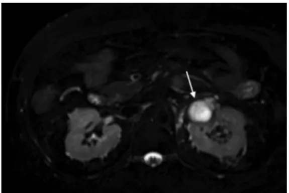
Figure 2 - Axial T2 imaging with fat saturation shows an expansive, exophytic lobulated mass with high signal, (arrow) in the upper pole left kidney.
Figure 3 - Coronal T1 pre-contrast imaging – the lesion is hypointense to adjacent renal parenchyma (arrow).
Figure 4 - Coronal T1 post-contrast arterial phase imaging showing peripheral enhancement (arrow).
IBJU| RADIOLOGY PAGE
989
solid heterogeneous lesions, with intense and progressive enhancement (3).
On MRI, hemangiomas show hyperinten-sity on T2 and variable degrees of enhancement after contrast administration. Presentations may resemble cystic lesions with solid component, mimicking cystic renal cell carcinoma as the present case (1, 2, 4). When large, these lesions are indistinguishable from malignant lesions
such as angiosarcomas and renal cell carcino-mas with central necrosis.
Treatment is controversial since preope-rative diagnosis is not possible based on ima-ging exams. When biopsy results are available, it may vary from expectation to partial ne-phrectomy, embolization and radical nephrec-tomy, depending on the lesion size, location and presence of symptoms.
REFERENCES
1. Katabathina VS, Vikram R, Nagar AM, Tamboli P, Menias CO, Prasad SR. Mesenchymal neoplasms of the kidney in adults: imaging spectrum with radiologic-pathologic correlation. Radiographics. 2010;30:1525-40.
2. Prasad SR, Surabhi VR, Menias CO, Raut AA, Chintapalli KN. Benign renal neoplasms in adults: cross-sectional imaging findings. AJR Am J Roentgenol. 2008;190:158-64.
_______________________ Correspondence address: Eduardo Kaiser Ururahy Nunes Fonseca, MD Departamento de Imagem, Hospital Israelita Albert Einstein Av. Albert Einstein, 627 São Paulo, SP, 05652-901, Brasil Fax: + 55 11 2151-0195 E-mail: [email protected]
3. Omiyale AO. Anastomosing hemangioma of the kidney: a literature review of a rare morphological variant of hemangioma. Ann Transl Med. 2015;3:151.