outcomes of ten patients (eight men and two women) in the age range between 28 and 72 years (mean, 55 years) submitted to at least two procedures of percutaneous sclerotherapy for symptomatic simple renal cysts. Main presentation was refractory flank pain in all patients, seven of them with some degree of hydronephrosis because of the parapyelic localization of the cyst. The aspirated volume ranged from 20 to 1300 ml (mean, 200 ml). The mean follow-up period following the procedure was of seven months. Two patients had ultrasoud-guided sclerotherapy, and eight, computed tomography-guided sclerotherapy. The hospital stay period ranged between 24 and 72 hours. Complete success corresponded to total cyst regression, and partial success corresponded to recurrence of less than half the original cyst volume. RESULTS: Complete cyst ablation was achieved in seven patients and partial resolution in three. No complication was observed and the therapy was well tolerated by all of the patients. CONCLUSION: Percutaneous ethanol sclerotherapy of symptomatic simple renal cysts is a safe, effective and minimally invasive procedure, with results similar to the ones reported by other studies.
Keywords: Kidney diseases – therapy; Sclerotherapy – methods; Ethanol – administration and dosage.
OBJETIVO: Demonstrar a eficácia e os resultados práticos da alcoolização percutânea de pacientes com cistos renais simples sintomáticos. MATERIAIS E MÉTODOS: Foram revistos os resultados obtidos em dez pacien-tes com cistos renais simples sintomáticos (oito homens e duas mulheres), com idade entre 28 e 72 anos (média de 55 anos), submetidos a pelo menos duas alcoolizações percutâneas. A indicação do procedimento foi dor no flanco refratária nos dez pacientes, sete deles com algum grau de hidronefrose pela localização parapiélica do cisto. O volume aspirado variou entre 20 e 1.300 ml (média de 200 ml). O tempo médio de seguimento após o procedimento foi de sete meses. O procedimento foi dirigido por ultrassonografia em dois casos e por tomografia computadorizada em oito. O tempo de internação variou entre 24 e 72 horas. Foi considerado sucesso completo do tratamento o desaparecimento do cisto, e parcial a redução maior que 50%. RESULTADOS: Durante o seguimento houve redução completa do cisto em sete pacientes e redução parcial em três. Em nenhum caso foram registradas complicações e os pacientes toleraram bem o procedi-mento. CONCLUSÃO: A alcoolização percutânea de cistos renais sintomáticos é um procedimento seguro, efetivo e pouco invasivo e com resultados semelhantes aos obtidos por outros autores.
Unitermos: Doenças renais – terapia; Escleroterapia – métodos; Etanol – administração e dosagem.
Resumo
* Study developed at Hospital São Luiz, and Department of Diagnostic Imaging – Universidade Federal de São Paulo/Escola Paulista de Medicina (Unifesp/EPM), São Paulo, SP, Brazil.
1. Professor at Department of Diagnostic Imaging – Universi-dade Federal de São Paulo/Escola Paulista de Medicina (Unifesp/ EPM).
2. Fellow at Department of Diagnostic Imaging – Universidade Federal de São Paulo/Escola Paulista de Medicina (Unifesp/EPM), MD, Radiologist, Unit of Diagnostic Imaging – Hospital São Luiz, São Paulo, SP, Brazil.
3. Radiologist, Hospital São Luiz, São Paulo, SP, Brazil. 4. Professor at Urology Department – Universidade Federal de São Paulo/Escola Paulista de Medicina (Unifesp/EPM), São Paulo, SP, Brazil.
5. Professor at Urology Department – Universidade Federal de São Paulo/Escola Paulista de Medicina (Unifesp/EPM), São Paulo, SP, Brazil.
incidentally found in elderly patients. Treatment starts being considered as these lesions are associated with flank pain, hy-pertension, hematuria, infection and col-lecting system obstruction(1,2).
Percutaneous ethanol sclerotherapy is a minimally invasive procedure, and is cur-rently considered as one of the main op-tions for the management of symptomatic renal cysts(1,2).
Along the last two decades, a great num-ber of authors have published reports on
D’Ippolito G, Torres LR, Ribeiro ACR, Roque AJ, Ortiz V, Ajzen S. Percutaneous ethanol sclerotherapy of renal cysts: treat-ment outcomes and literature review. Radiol Bras. 2009;42(4):225–230.
6. Professor at Department of Diagnostic Imaging – Universi-dade Federal de São Paulo/Escola Paulista de Medicina (Unifesp/ EPM), São Paulo, SP, Brazil.
Mailing address: Dr. Giuseppe D’Ippolito. Rua Doutor Alceu de Campos Rodrigues, 95, subsolo, Vila Nova Conceição. São Paulo, SP, Brazil, 04544-000. E-mail: [email protected]
Received February 26, 2009. Accepted after revision May 8, 2009.
INTRODUCTION
their experiments with percutaneous treat-ment of symptomatic renal cysts with dif-ferent strategies and not always coinciden-tal results(1–21).
The objective of this study is to present the practical results from percutaneous etha-nol sclerotherapy of renal cysts in a group of ten symptomatic patients submitted to this treatment in the authors’ Institution.
MATERIALS AND METHODS
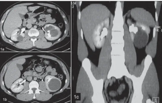
In the period between 2005 and 2007, ten patients (eight men and two women), with a total of ten renal cysts, were submit-ted to percutaneous sclerotherapy with 95% ethanol. The patients’ ages ranged from 22 to 78 years (mean, 55 years). In all of the cases, the indication for the proce-dure was ipsilateral lumbar pain refractory to other treatments. At least one sono-graphic examination was performed for each patient to evaluate the number, inter-nal content and walls of cysts. Also, the patients were submitted to contrast-en-hanced computed tomography (CT) for evaluation of the relationship between cyst and collecting system (Figure 1). All the lesions were classified as simple cysts (Bosniak category I)(22). The cysts volume ranged between 20 and 1300 ml (mean, 200 ml). Seven patients presented parapyelic cysts, and three, cortical cysts. All the
Figure 1. Contrast-enhanced CT image (pyelographic phase) in the axial plane (a,b) and coronal recon-struction (c) demonstrating para-pyelic and cortical cyst compressing and deforming the pericalyceal sys-tem. No communication between the cyst and the collecting system is observed.
Figure 2. CT-guided cyst puncture (a) with the aspirate being sent for oncotic cytology and culture (b).
parapyelic cysts caused some degree of dilatation of the pyelocalyceal system by extrinsic compression (Figure 1).
Ultrasonography-guided and CT-guided procedures were performed, respectively in two and eight cases. The percutaneous pro-cedure comprised six steps, as follows:
1. Determination of the presence of con-nection between the cyst and the collecting system by intravenous contrast injection (through CT) and calculation of the cyst volume either by US or CT (Figure 1).
2. Cyst puncture, with the aspirate be-ing sent for oncotic cytology and culture (Figure 2).
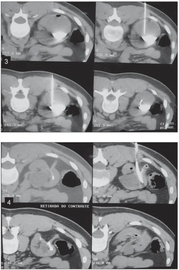
3. Contrast agent injection into the cyst to confirm the connection between the le-sion and the colleting system (Figure 3).
4. Percutaneous drain insertion for com-plete drainage of the cyst content, with measurement of the total aspirate volume (Figure 4).
Figure 4. After percutaneous drain insertion, the cyst is completely drained with measurement of the aspirate volume.
Figure 3. Percutaneous drain inser-tion and 5–10 ml iodinated contrast injection, confirming that here is no communication between the cyst and the collecting system.
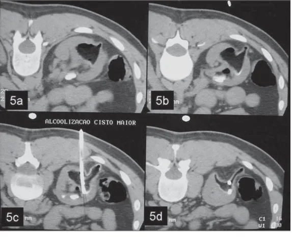
the aspirate volume, up to a maximum vol-ume of 250 ml (Figure 5).
6. Holding definitely the ethanol within the cyst or aspirating it, repeating the pro-cedure within 24 and 48 hours, with a
per-cutaneous 6F or 8.5F drainage catheter (Figures 5 and 6).
Steps 2, 3, 4 and 5 were performed un-der general anesthesia. The procedures rep-etition was performed under radioscopic
etha-nol was aspirated. More than one procedure was performed for all the patients (on av-erage, two procedures per patient), with a percutaneous 6F or 8.5F drainage catheter that remained in position after the first as-piration.
Complete success corresponded to to-tal cyst regression observed at the follow-up assessments (minimum six months)(1–3). Statistical data analysis was not per-formed because of the small number of cases, not allowing the calculation of sta-tistical significance.
RESULTS
All the procedures were performed with no technical difficulty. The length of the patients’ hospital stay ranged between 24 and 72 hours, depending on the number of repetitions (one procedure performed every 24 hours).
The procedure repetitions were per-formed under fluoroscopic guidance and their frequency was established as a func-tion of the cysts size and radiographic as-pect; cysts whose size remained unchanged after the first sclerotherapy procedure were immediately submitted to another proce-dure until a clear decrease in the remain-der cyst volume was observed at radios-copy.
A patient with a 1300 ml cyst, initially received a 250 ml 95% ethanol injection
(about 20% of the cyst volume), with three repetitions along three consecutive days.
No complication was observed. The procedure was well tolerated by all the pa-tients.
As far the reduction in the cysts dimen-sions is concerned, a complete success was achieved in seven patients, and partial suc-cess was achieved in three. The ten patients submitted to percutaneous sclerotherapy presented improvement in clinical symp-toms.
DISCUSSION
The main indications for treatment of simple renal cysts are the following: a) voluminous cysts causing hydronephrosis; b) refractory pain; c) hematuria; d) hyper-tension(1).
Studies have demonstrated that a the simple drainage of a cyst leads to recur-rence of symptoms in up to 80% of cases, therefore this is not considered as an effec-tive therapeutic approach(3). On the other hand, US, CT, or fluoroscopy-guided per-cutaneous sclerotherapy have been suc-cessfully applied with a variety of
sub-Figure 5. Ethanol is injected in an amount corresponding to about 25%–30% of the aspirate volume. The alcohol remains within the cyst for about 20 minutes (a,b) and subsequently is completely drained. The drainage catheter remains in place (c,d).
proposed basically varying in relation to the number of sessions (single × multiple)(2,3, 10–12) and prolonged(13) or persistent
reten-tion(14) of the sclerosing agent with some-what conflicting outcomes.
The described six-step technique is pro-posed by several authors and was adopted in the present study(1,12–15). The reason for its choice is related to the good results ini-tially achieved.
The procedure may be performed either under US or CT guidance. Advantages of US are: low cost, wide availability and real-time capability. The CT advantages are re-lated to the higher anatomic resolution of the method, allowing the evaluation of the communication between the cyst and the renal collecting system simultaneously with the sclerotherapy.
Some authors perform the procedure under local(14) and other under general an-esthesia arguing for the fact that this mea-sure is better tolerated by the patient, con-sidering that the injection of the alcohol into the cyst may be quite painful(11).
Several authors have studied different technical approaches. Hanna & Dahniya
is kept within the cyst for 24–48 hours, which supposedly would reduce the recur-rence rate(11); in the authors’ experience, this strategy can make the alcohol reinjec-tion through the percutaneous drain more difficult, since the catheter may be ob-structed by the aspirated debris. Most re-cently, a sclerosing treatment without etha-nol aspiration was proposed, that is to say, that the sclerosing agent would be left within the cyst for an indeterminate period with results similar to the ones achieved with other more complex techniques, sug-gesting that this approach is quite practical and effective(14). In a more recent study, results from multiple ethanol sclerotherapy sessions were compared with those of single session OK-432 sclerotherapy. This substance is a blend of low virulence strains of group A streptococcus (Streptococcus pyogenes). According to this study, after one-year treatment, higher efficacy was observed with the utilization of OK-432(21). However, it is important to note that OK-432 is not available in Brazil yet.
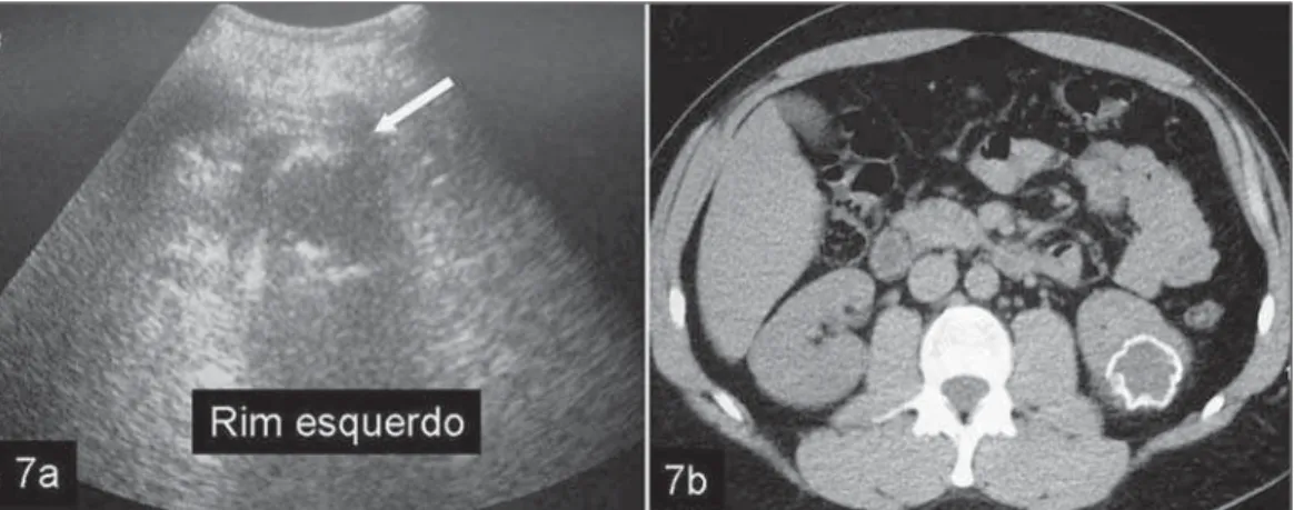
The post-treatment follow-up is gener-ally performed by means of serial US at the
7). In the literature, the complete success rate for sclerotherapy ranges between 70% and 90%(1,3,10–14,16,17), independently from the technique adopted, but with better re-sults for smaller cysts(18,19).
Complications resulting from the pro-cedure rarely occur and generally are self-limited. Most frequent complications are fever, pain, hematuria and small peri-nephric hematomas(1,12,13,17,18).
Contraindications for renal cysts sclero-therapy are those related to percutaneous procedures, such as severe coagulation dis-orders, communication between the cyst and the renal collection system, and lesions that do not meet the simple cysts criteria, with suspicion of infection or with pres-ence of blood in the aspirate(4,17,20).
CONCLUSION
Renal cysts management by means of percutaneous ethanol sclerotherapy is a safe, effective and minimally invasive method that is well tolerated by the pa-tients, with satisfactory mid- and long-term results, and thus it should be considered as
a feasible therapeutic approach for the management of simple renal cysts in symp-tomatic patients.
REFERENCES
1. Mohsen T, Gomha MA. Treatment of symptomatic simple renal cysts by percutaneous aspiration and ethanol sclerotherapy. BJU Int. 2005;96:1369–72. 2. Gasparini D, Sponza M, Valotto C, et al. Renal cysts: can percutaneous ethanol injections be considered an alternative to surgery? Urol Int. 2003;71:197–200.
3. Hanna RM, Dahniya MH. Aspiration and sclero-therapy of symptomatic simple renal cysts: value of two injections of a sclerosing agent. AJR Am J Roentgenol. 1996;167:781–3.
4. Yamamoto K, Sakaguchi H, Anai H, et al. Scle-rotherapy for simple cysts with use of ethanola-mine oleate: preliminary experience. Cardiovasc Intervent Radiol. 2005;28:751–5.
5. Kim SH, Moon MW, Lee HJ, et al. Renal cyst ablation with n-butyl cyanoacrylate and iodized oil in symptomatic patients with autosomal domi-nant polycystic kidney disease: preliminary re-port. Radiology. 2003;226:573–6.
6. Peyromaure M, Debré B, Flam TA. Sclerotherapy of a giant renal cyst with povidone-iodine. J Urol. 2002;168:2525.
7. Seo TS, Oh JH, Yoon Y, et al. Acetic acid as a
sclerosing agent for renal cysts: comparison with ethanol in follow-up results. Cardiovasc Intervent Radiol. 2000;23:177–81.
8. Uemasu J, Fujihara M, Munemura C, et al. Cyst sclerotherapy with minocycline hydrochloride in patients with autosomal dominant polycystic kid-ney disease. Nephrol Dial Transplant. 1996;11: 843–6.
9. van der Ent CK, van Dalen A, Enterman JH. An-tibiotic sclerotherapy for renal cysts. Rofo. 1989; 150:339–41.
10. Delakas D, Karyotis I, Loumbakis P, et al. Long-term results after percutaneous minimally inva-sive procedure treatment of symptomatic simple renal cysts. Int Urol Nephrol. 2001;32:321–6. 11. De Dominicis C, Ciccariello M, Peris F, et al.
Percutaneous sclerotization of simple renal cysts with 95% ethanol followed by 24-48 h drainage with nephrostomy tube. Urol Int. 2001;66:18–21. 12. Akinci D, Akhan O, Ozmen M, et al. Long-term results of single-session percutaneous drainage and ethanol sclerotherapy in simple renal cysts. Eur J Radiol. 2005;54:298–302.
13. Lin YH, Pan HB, Liang HL, et al. Single-session alcohol-retention sclerotherapy for simple renal cysts: comparison of 2- and 4-hr retention tech-niques. AJR Am J Roentgenol. 2005;185:860–6. 14. Falci-Júnior R, Lucon AM, Cerri LM, et al. Treat-ment of simple renal cysts with single-session percutaneous ethanol sclerotherapy without
drainage of the sclerosing agent. J Endourol. 2005;19:834–8.
15. Akinci D, Gumus B, Ozkan OS, et al. Single-ses-sion percutaneous ethanol sclerotherapy in simple renal cysts in children: long-term follow-up. Pediatr Radiol. 2005;35:155–8.
16. Chung BH, Kim JH, Hong CH, et al. Compari-son of single and multiple sessions of percutane-ous sclerotherapy for simple renal cyst. BJU Int. 2000;85:626–7.
17. Paananen I, Hellström P, Leinonen S, et al. Treat-ment of renal cysts with single-session percuta-neous drainage and ethanol sclerotherapy: long-term outcome. Urology. 2001;57:30–3. 18. el-Diasty TA, Shokeir AA, Tawfeek HA, et al.
Ethanol sclerotherapy for symptomatic simple renal cysts. J Endourol. 1995;9:273–6. 19. Agostini S, Dedola GL, Gabbrielli S, et al.
Per-cutaneous treatment of simple renal cysts with sclerotherapy and extended drainage. Radiol Med. 2004;108:522–9.
20. Gelet A, Sanseverino R, Martin X, et al. Percuta-neous treatment of benign renal cysts. Eur Urol. 1990;18:248–52.
21. Ham WS, Lee JH, Kim WT, et al. Comparison of multiple session 99% ethanol and single session OK-432 sclerotherapy for the treatment of simple renal cysts. J Urol. 2008;180:2552–6. 22. Bosniak MA. The current radiological approach