ABSTRACT
www.scielo.br/jaos
Analysis by confocal laser scanning microscopy
of the MDPB bactericidal effect on
S. mutans
bioilm CLSM analysis of MDPB bactericidal effect
on bioilm
Fabíola Galbiatti de CARVALHO1, Regina Maria PUPPIN-RONTANI2, Suzana Beatriz Portugal de FÚCIO3, Thais de Cássia NEGRINI4, Hugo Lemes CARLO5, Franklin GARCIA-GODOY6
1- DDS, MSc, PhD Professor, Health and Technology Rural Center, Federal University of Campina Grande, Patos, PB, Brazil.
2- DDS, MSc, PhD Professor, Department of Pediatric Dentistry, School of Dentistry of Piracicaba, State University of Campinas, Piracicaba, SP, Brazil. 3- DDS, MSc, PhD student, Department of Restorative Dentistry, Dental Materials Area, School of Dentistry of Piracicaba, State University of Campinas, Piracicaba, SP, Brazil.
4- DDS, MSc, PhD student, Department of Microbiology, School of Dentistry of Piracicaba, State University of Campinas, Piracicaba, SP, Brazil. 5- DDS, MSc, PhD Professor, Department of Operative Dentistry, Health Science Center, Federal University of Paraíba, João Pessoa, PB, Brazil.
6- DDS, MSc, Professor and Senior Executive Associate Dean for Research and Director, Bioscience Research Center, College of Dentistry, University of Tennessee, Memphis, TN, USA.
Corresponding address: Profa. Dra. Fabíola Galbiatti de Carvalho - Curso de Odontologia - Centro de Saúde e Tecnologia Rural - Universidade Federal de Campina Grande - Av. Universitária s/n - Santa Cecília - Patos - PB - 58708-110 - Phone +55-83-3511-3064 - Fax: +55-83-3511-3009 - e-mail: fabigalbi@ yahoo.com.br
Received: April 11, 2012 - Modiication: August 21, 2012 - Accepted: September 14, 2012
S
ince bacteria remain in the dentin following caries removal, restorative materials with antibacterial properties are desirable to help maintaining the residual microorganismsinactive. The adhesive system Clearil Protect Bond (PB) contains the antibacterial monomer
12-methacryloyloxydodecylpyridinium bromide (MDPB) in its primer, which has shown
antimicrobial activity. However, its bactericidal effect against bioilm on the dentin has
been little investigated. Objective: The aim of this study was to analyze by confocal laser scanning microscopy (CLSM) and viable bacteria counting (CFU) the MDPB bactericidal effect against S. mutans bioilm on the dentin surface. Material and Methods: Bovine
dentin surfaces were obtained and subjected to S. mutans bioilm formation in BHI broth
supplemented with 1% (w/v) sucrose for 18 h. Samples were divided into three groups,
according to the primer application (n=3): Clearil Protect Bond (PB), Clearil SE Bond, which does not contain MDPB, (SE) and saline (control group). After the bioilm formation, Live/
Dead stain was applied directly to the surface of each sample. Next, 10 µL of each primer were applied on the samples during 590 s for the real-time CLSM analysis. The experiment was conducted in triplicate. The primers and saline were also applied on the other dentin samples during 20, 90, 300 and 590 s (n=9 for each group and period evaluated) and the CFU were assessed by colonies counting. Results: The results of the CLSM showed that with the Se application, although non-viable bacteria were detected at 20 s, there was no increase in their count during 590 s. In contrast, after the PB application there was a gradual increase of non-viable bacteria over 590 s. Conclusions: The quantitative analysis
demonstrated a signiicant decrease of S. mutans CFU at 90 s PB exposure and only after 300 s of Se application. Protect Bond showed an earlier antibacterial effect than Se Bond.
INTRODUCTION
The use of cavity disinfectants before restoration is an important clinical step because bacteria can remain in the smear layer or in the dentinal tubules, and can potentially multiply16. The
ability of restorative materials to control bacteria by antimicrobial components is also desirable to help maintain residual microorganisms inactive14. The self-etching adhesive system, Clearil Protect Bond, contains the antibacterial
monomer 12-methacryloyloxydodecylpyridinium bromide (MDPB) in its primer. This monomer was incorporated to inactivate residual microorganisms on the dentin surface and to prevent bacterial invasion through the gap tooth/restoration7.MDPB
monomer is a compound of the antibacterial agent quaternary ammonium and a methacryloyl group, which is covalently bound to the polymer matrix by its copolymerization with other monomers7.
MDPB has shown antimicrobial activity against oral streptococci, lactobacilli and microorganisms clinically isolated from root caries23.However, studies
that evaluated the MDPB antibacterial effect were usually conducted in bacterial suspension and by agar diffusion testing7,9,11.
Since in the clinical practice adhesive systems are directly applied to the dentin with some level of contamination12,14,21, it would be crucial to
evaluate the MDPB antibacterial effect against bacteria colonized on the dentin surface. This fact is also relevant because the bacteria adhered on
the surfaces, forming bioilms, are less susceptible
to antimicrobials than planktonic microorganisms (bacteria in suspension)4. However, few studies
evaluated the antibacterial effect of adhesive
systems containing MDPB against the bioilm10,15.
Roland, et al.15 (2006) investigated the antibacterial effect of MDPB and luoride on bioilm formation of
dental adhesive surfacesto establish whether these
components had the potential to reduce bioilm
formation on adhesive system surfaces. It was found that the antibacterial agents within the resins have
a minimal effect on bioilm formation15.Izutani, et
al.10 (2011) evaluated the antibacterial effect of
MDPB on S. mutans bioilm and showed that high
concentrations of MDPB in solution are necessary
to effectively kill bioilm S. mutans cells within an acceptable clinical time. Based on these results, it becomes necessary to evaluate if the application time of MDPB-containing a primer recommended by
the manufacturer (20 s) is suficient to decrease or
eliminate microorganisms colonized on the dentin surface or for how long the bactericidal effect of MDPB-containing primer acts against these microorganisms.
Confocal laser scanning microscopy (CLSM) has been used to evaluate bacterial viability on
dentin bioilms15.Fluorescence dyes are applied on the bioilm to differentiate live and dead bacteria,
allowing bacteria to be distinguished according to cytoplasmic membrane permeability9. This holds
particular relevance for studying the antimicrobial action of adhesive systems containing MDPB, because MDPB causes cell death through disruption of the cytoplasmic membrane7-9,15. Furthermore,
CLSM can capture a series of image-scans showing changes in the viability of the bacterial cell over time, making the visualization of real-time death of microorganisms possible.
The aim of this study was to evaluate the bactericidal effect against S. mutans bioilm between
self-etching priming solutions with and without
MDPB (Clearil Protect Bond and Clearil SE Bond,
respectively) over 590 s by real time CLSM analysis and count of viable bacteria test. The hypothesis tested in this study was that there is a difference in the bactericidal effect against S. mutans bioilm
between primers with or without MDPB during 590 s by CLSM analysis and count of viable bacteria test.
MATERIAL AND METhODS
Adhesive systems
Two self-etching/priming solutions were tested, an antibacterial primer containing a M D P B m o n o m e r - C l e a r f i l P r o t e c t B o n d (Kuraray, Tokyo, Japan) - containing MDPB, MDP (10-methacryloxydecyldihydrogen phosphate), HeMA (2-hydroxyethylmethacrylate) and water, and a primer without an antibacterial monomer -
Clearil SE Bond (Kuraray, Tokyo, Japan) - containing
MDP, HeMA, dl-camphorquinone, N,N-diethanol-p-toluidine and water.
Samples preparation
One hundred fourteen bovine incisors were obtained and stored in 0.1% thymol solution. The buccal portion of the enamel was wet ground (Arotec, São Paulo, SP, Brazil) using 400 and 600 grit silicon carbide paper (Saint-Gobain, São Paulo, SP, Brazil). Dentin samples with 4x4 mm were obtained of each tooth using a low-speed diamond saw (Isomet, Buehler, Lake Bluff, IL, USA) and ground with 240 grit silicon carbide paper to reach 1.5 mm thickness. All samples were sterilized by steam autoclave (Phoenix, Araraquara, SP, Brazil) at 121°C for 15 min.
Bacterial colonization on the dentin
hrs. An overnight culture was adjusted to 1x10⁶
[colony forming units (CFU)/mL] with an optical density of 0.6 at 600 nm. The dentin surface of each sample was exposed to 20 µL aliquot of S. mutans suspension in brain heart infusion broth (BHI) (Becton Dickinson, Sparks, MD, USA) without any disturbance, to allow for bacterial adherence. After 2 hrs at room temperature, the non-adhering cells were removed by washing three times with saline solution5.A sample was placed in each well
of a 24-well multi-dish polystyrene plate (Nunclon,
Thomas Scientiic, Swedesboro, NJ, USA). Two mL
of BHI broth supplemented with 1% (w/v) sucrose (LabSynth, Diadema, SP, Brazil) were then added to the wells to simulate bacterial colonization5. The
plates were incubated for 18 h at 37ºC in a 5% CO2 environment. The lack of contamination in the media
of each well was veriied using Gram staining and by
plating the samples. The 18 h incubation period was
established by a previous study, which veriied that it was suficient to develop a thin S. mutans bioilm
(containing 108 colony unit formation) without
producing an intense demineralization of the dentin specimens, since the demineralization process could
increase the dentin autoluorescence3 and harm the bioilm visualization by CLSM.
Assessment of bactericidal effects by CLSM
Live/Dead Baclight bacterial viability stain (L13152) (Molecular Probes, eugene, OR, USA) was used in this study. It consists of a two nucleic acid-binding stains mixture: Syto 9 and propidium iodide. Syto 9 stains all viable bacteria in green, while propidium iodide stains in red the bacteria whose membranes were damaged (non-viable bacteria). Three samples were used for each solution
application: Clearil Protect Bond primer, Clearil
Se Bond primer and saline (control group). After 18 hrs of bacterial colonization, the non-adhering cells were removed by washing three times with saline solution5. Live/Dead was mixed according to
the manufacturer’s instructions and one drop was applied directly to the surface of each sample15.
After 15 min in dark incubation5, the stain surplus
was removed by absorbent paper and 10 µL of each solution was applied on the samples’ surface, according to each group. Immediately after, samples were analyzed by CLSM for 590 s (LSM510 MeTA,
Zeiss, Jena, Germany). This period was used based
on the Imazato, et al.9 (2008) study, which found
rapid killing of planktonic bacteria after contact with MDPB for 590 s. An excitation wavelength of 488 nm was used, and all light emitted between 500 – 550 nm and over 560 nm was collected by different
ilters. The scan mode time series was used to take a series of time-lapse scans (xyt) at intervals of 10 s during 590 s using continuous scanning with 10x objective lens. Scans were taken in 8 bits at
a resolution of 512 by 512 pixels. To standardize the gain and offset values to take a series scan, a preliminary sample prepared as described before was used. The experiment was conducted in triplicate.
Assessment of bactericidal effects by viable bacteria count
The primer application time was tested based on the CLSM results (20 s, 90 s, 300 s, 590 s). One-hundred eight dentin samples were used and twelve groups were obtained according to the time application of each primer or saline (n=3): saline application for 20 s, 90 s, 300 s and 590 s; Protect Bond application for 20 s, 90 s, 300 s and 590 s and Se Bond application for 20 s, 90 s, 300 s and 590 s. The groups with the saline application were considered the negative control groups (no primer application). The experiment was repeated three times (triplicate). After 18 h of bacterial colonization as described before, 10 µL of each primer or 0.9% NaCl solution was applied on the sample surface. The solutions were kept in contact with the S. mutans bioilm, without any disturbance for each
group. The time was recorded by a chronometer. After each experimental time, the excess solutions were removed by sterile absorbent paper. Samples were immersed in individual eppendorfs with 1 mL 0.9% NaCl solution and sonicated for 30 s at 30% amplitude. The suspension was diluted in decimal series from 10−1 to 10−6 in 0.9% NaCl solution and
inoculated on BHI agar plates containing bacitracin (2 U/mL)13. The plates were incubated for 48 h at
37ºC in a 5% supplemented CO2 environment and the number of viable bacteria (CFU) were assessed by counting the colonies formed. The data of CFU were submitted to the Kruskal-Wallis and Dunn’s
Multiple Comparisons tests at a signiicance level
of p<0.05.
RESULTS
CLSM images showed S. mutans biofilm on the dentin surface with viable and non-viable colonies (green and yellow/red, respectively) and non-stained (black) bubble-like structures within
the bioilm architecture (Figures 1, 2 and 3). The
control group demonstrated viable bacteria during 590 s, all colonies remained green, indicating that
S. mutans were viable during the period evaluated (Figure 1). Non-viable bacteria (red colonies) were
seen after the Clearil SE Bond primer application
(Figures 2A and 2B), nonetheless, there was no increase of these bacteria during 590 s and viable bacteria were observed at this time (Figure 2C).
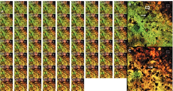
After the Clearil Protect Bond primer application
time recommended by the manufacturer) viable bacteria was still visualized, and no difference was found between the viable and non-viable bacteria in 0 s and 20 s time-lapse scans (Figure 3A). The color change of S. mutans colonies to a yellowish
or orange indicated the death of bacteria after the
Clearil Protect Bond primer application, (Figure 3A)
showing a gradual increase of non-viable bacteria over 590 s. At 590 s, yellowish and red colonies were predominant (Figure 3C).
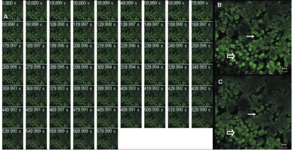
Figure 1- S. mutans bioilm on dentin surface after saline application (control group) visualized by confocal laser scanning
microscopy (CLSM). Images were recorded in real-time over 590 s. Viable bacteria are stained green and non-viable bacteria are stained yellowish/red color. Figure A shows image series of time-lapse scans according to the time (upper-left side of each scan). No bactericidal effect was observed because all colonies were stained green over 590 s. Figures B and C are higher magniication of image at zero time (Figure B) and at 590 s (Figure C), showing the presence of dead bacteria (open arrow) and non-stained (black) bubble-like structures within the bioilm architecture (arrow)
Figure 2- S. mutans bioilm on dentin surface after the Clearil SE Bond primer application visualized by confocal laser
Table 1 shows a quantitative analysis of the number of viable bacteria (CFU) of S. mutans
bioilm on the dentin after the Clearil Protect Bond, Clearil SE Bond and saline application. There was
no statistical difference among CFU after 20, 90,
300 and 590 s for the control and Clearil SE Bond groups. In the contrary, the Clearil Protect Bond
group at 300 s and 590 s primer exposures showed
a signiicant decreasing CFU compared to 20 s.
When the comparisons were made among the primers and saline in each experimental time, at the initial time (20 s), no difference was found among the two primers and saline (Table 1). At 90 s, the
Clearil Protect Bond group showed a signiicant
decreasing CFU compared to the control group and
Clearil SE Bond showed intermediate CFU between the control and Clearil Protect Bond groups (Table 1). For the Clearil SE Bond group the CFU decreased signiicantly after 300 s and 590 s of application, consequently the Clearil Protect Bond showed an
earlier antibacterial effect than the Se Bond (Table 1). At 300 s and 590 s, both primers showed decreasing CFU compared to the control group, but with no difference between them (Table 1).
Groups 20s * 90s 300s 590s
Control 2.5x10⁹A,a 1.0x10⁹A,a 5.3x10⁹A,a 2.5x10⁹A,a
(104/109) (108/109) (108/109) (108/109)
SE Bond 9.0x10⁴A,a 3.6x10³A,ab 8.8x10²A,b 9.6x10²A,b
(104/106) (103/104) (102/103) (102/103)
Protect Bond 1.0x10⁵A,a 1.0x10³AB,b 4.8x10²B,b 5.6x10²B,b
(104/105) (102/103) (10/102) (102/103)
* For each horizontal row: values with identical uppercase letters indicate no statistically signiicant difference (P>0.05). For each vertical column: values with identical lowercase letters indicate no statistically signiicant difference (P>0.05) according to Dunn’s Multiple Comparisons test; n=9 for each time of each primer/saline application
Table 1- Number of viable S. mutans (CFU) recovered after Clearil Protect Bond and Clearil SE Bond primer application
at different times. Values are expressed in median (minimum/maximum)
Figure 3- S. mutans bioilm on dentin surface after the Clearil Protect Bond primer application visualized by confocal
DISCUSSION
The null hypothesis was rejected because there was a difference in the bactericidal effect against
S. mutans bioilm between self-etching priming
solutions with and without MDPB by CLSM analysis and count of viable bacteria test. MDPB monomer can be considered a bactericidal agent against
S. mutans bioilm, since it affected the bacteria
membrane that could be detectable by luorescent
indicators.
Antimicrobial compounds that do not directly affect the cell membrane, such as those that interfere with protein or nucleic acid synthesis, are normally considered bacteriostatic and are not suitable agents to be studied by a viability staining method1. Live/Dead Baclight has been shown to
be an effective stain to distinguish viable and non-viable bacteria according to cytoplasmic membrane permeability2. As MDPB is a derivate of quaternary ammonium, it has a high afinity for negatively
charged bacterial cells by a nitrogen atom on the pyridinium ring, which binds to the cell surface. The cell membrane loses its electrical balance, creating a disruption in the cytoplasmic membrane and the cell dies7.
Few studies evaluated the antibacterial effect of adhesive systems containing MDPB against the
bioilm by CLSM analysis10,15. However, these earlier
studies evaluated the effect of the MDPB monomer
to reduce bioilm formation on the Clearil Protect
Bond adhesive system surface15 and the effect
of high concentrations of unpolymerized MDPB solutions to kill S. mutans in planktonic or bioilm
forms after 20, 40 or 60 s of contact10. The primer
containing-MDPB real-time effect against S. mutans
bioilm by CLSM evaluation over 590 s had never
been tested.
There was a good relation among the CFU results and the elapsed times of the CLSM analysis. The CLSM time-lapse scans and CFU results of the control group showed viable bacteria during 590 s
(Figure 1 and Table 1). After the Clearil SE primer
application some non-viable bacteria were seen, however, the green staining was maintained in all periods tested (Figure 2). No statistical difference was found among the CFU during 590 s for the
Clearil SE Bond group (Table 1). The acidic nature
of the primer of self-etching adhesive systems has been considered as one of the key factors related to bacterial inhibition6. The MDP monomer of Clearil SE and Clearil Protect Bond can have antibacterial
activity because of its low pH level (1.9)6,18.
However, the dentin can act as a buffer, because the acidic dissolution of the dentin apatite may be neutralized by the bonding between the dentin ions and H+ ions of the monomer6. Consequently, the
pH level of the MDP may have been neutralized by
the buffering action of the dentin, leading to the visualization of viable bacteria by CLSM after the
Clearil SE primer application. Likewise, the MDP of the Clearil SE Protect primer may also have been
neutralized; however, the MDPB monomer showed a gradual bactericidal effect over 590 s and non-viable bacteria were predominant at 590 s. The CFU results also showed a decrease after the 90, 300
and 590 s Clearil Protect Bond application. These results conirmed previous studies that a
MDPB-containing primer has an antibacterial effect against
S. mutans colonized in the dentin8,20.
Bacterial bioilms associated with surfaces are
complex three-dimensional structures in which bacteria are embedded in a matrix mainly made of exopolysaccharides (ePS)19. ePS functions as an
ion-exchange matrix and hinders the penetration of positively-charged antimicrobial agents17. The
activation of an adaptive stress response and changes in gene and protein expression of bacteria
are also factors that make bioilms 10 to 1000
times less sensitive to antimicrobial agents than planktonic bacteria1. Three main factors can be
considered to the difference in susceptibility of
planktonic and bioilm bacteria to antimicrobials10.
One is the hindrance of penetration of antimicrobials
into the bioilm, owing to the presence of a copious
amount of an extracellular matrix, which acts as a molecular sieve. A second factor is that the chemical micro-environment, such as
nutrient-depletion or waste products within the bioilm,
may act as antagonists of antimicrobials. A third, and still speculative hypothesis is that some of the bacteria may differentiate into a protected phenotypic state10.
Some studies reported declining S. mutans
counts in dentin chips and bacterial suspension after 20 s of the Clearfil Protect Bond primer application (application time recommended by the manufacturer)7,8,11,20. However, the present
study showed that there were viable bacteria at 20 s similar to 0 s time-lapse scan (Figure 1). Furthermore, the CFU results showed a decreasing
number of bacteria after the 90 s Clearil Protect
Bond primer application (Table 1). Probably, the
EPS among the bioilm channels may have impaired
the penetration of MDPB and consequently its action, needing more time to kill bacteria since the mechanism of action for MDPB is based on the contact of immobilized antibacterial molecules with bacteria7.Thus, a gradual bactericidal effect
was evidenced during the period evaluated. The results of the present study were contradictory to the Imazato, et al.9 (2008)results, which showed
rapid killing of all bacteria (S. mutans) after 590 s contact with MDPB solution using Live/Dead
Baclight bacterial viability stain and luorescence
against planktonic bacteria. Another study10 showed
that high concentrations of MDPB are necessary to
effectively kill planktonic and bioilm S. mutans
cells within 60 s of contact, however, the bioilm
produced under the experimental conditions of the Izutani, et al.10 (2011) study had a sparse structure
(i.e. without a dense extracellular matrix) and this fact may have helped the penetration of MDPB and its antimicrobial effect10. The CLSM analysis showed a dense bioilm with some characteristics
of in vivo bioilm (Figures 1, 2 and 3), such as the
bubble-like structures and dead bacteria that were
initially present. This dense structure of bioilm
may also have impaired the antimicrobial effects
of the Clearil Protect Bond primer. The
bubble-like structures were voids that were possibly
illed with biological substances, such as EPS and
glycoproteins, which are not stainable by the stain used22. Besides, the dead bacteria might have been located in deeper regions of the bioilm and
had reduced access to nutrients from the exposed medium15.
The viability staining method provided a rapid and sensitive way to test the MDPB bactericidal effect against S. mutans bioilm. As the evidence
of in vitro study must not be extrapolated to in vivo situations, clinical studies must be conducted
to evaluate the application time of the Clearil
Protect Bond to disinfect the enamel/dentin cavity to obtain the antimicrobial activity. The collection of softened carious dentin or the use of swabs rubbed on to the base of the cavity before and after the primer application could be performed to clinically measure the antimicrobial activity14. Further
investigations also need to be conducted against
other cariogenic microorganisms in the bioilm or multispecies bioilm. Some attempts were made
in this present study to test the MDPB effect after the bond application and cure, but the polymerized
adhesive (bond) autoluorescence interfered with
the bacteria visualization using CLSM.
CONCLUSIONS
Based on the indings of this study, and within
the limitation of the in vitro investigation, it can
be concluded that the Clearil SE Bond primer
presented a bactericidal effect only after 300 s against S. mutans bioilm and the Clearil Protect
Bond primer had a bactericidal effect against S. mutans bioilm after 90 s and showed a gradual
bactericidal effect over 590 s by real-time CLSM evaluation and viable bacteria counting.
ACKNOwLEDgEMENTS
The authors are grateful to Dr. Toshie Kawano and Alexsander Souza of the Butantan Institute for their technical support for the CLSM analysis and the Kuraray Medical Company for supplying the materials. This study was supported by protocol #05/57268-9 from FAPeSP.
REFERENCES
1- Beckloff N, Laube D, Castro T, Furgang D, Park S, Perlin D, et al. Activity of an antimicrobial peptide mimetic against planktonic and
bioilm cultures of oral pathogens. Antimicrob Agents Chemother.
2007;51:4125-32.
2- Berney M, Hammes F, Bosshard F, Weilenmann H, egli T. Assessment and interpretation of bacterial viability by using the
LIVE/DEAD BacLight kit in combination with low cytometry. Appl
environ Microbiol. 2007;73:3283-90.
3- Carvalho FG, Fucio SB, Sinhoreti MA, Correr-Sobrinho L, Puppin-Rontani RM. Confocal laser scanning microscopic analysis of the depth of dentin caries-like lesions in primary and permanent teeth. Braz Dent J. 2008;19:139-44.
4- Chambless JD, Hunt SM, Stewart PS. A three-dimensional computer model of four hypothetical mechanisms protecting
bioilms from antimicrobials. Appl Environ Microbiol.
2006;72:2005-13.
5- Fúcio SBP, Puppin-Rontani RM, Carvalho FG, Mattos-Graner
RO, Correr-Sobrinho L, Garcia-Godoy F. Analyses of bioilms
accumulated on dental restorative materials. Am J Dent. 2009;22:131-6.
6- Gondim JO, Duque C, Hebling J, Giro EM. Inluence of human
dentine on the antibacterial activity of self-etching adhesive systems against cariogenic bacteria. J Dent. 2008;36:241-8. 7- Imazato S, Kinomoto Y, Tarumi H, Torii M, Russell RRB, McCabe JF. Incorporation of antibacterial monomer MDPB into dentin primer. J Dent Res. 1997;76:768-72.
8- Imazato S, Kuramoto A, Takahashi Y, ebisu S, Peters MC. In vitro antibacterial effects of the dentin primer of Clearil Protect
Bond. Dent Mater. 2006;22:527-32.
9- Imazato S, Ohmori K, Russell RR, McCabe JF, Momoi Y, Maeda N. Determination of bactericidal activity of antibacterial monomer MDPB by a viability staining method. Dent Mater J. 2008;27:145-8. 10- Izutani N, Imazato S, Nakajo K, Takahashi N, Takahashi Y, ebisu S, et al. effects of the antibacterial monomer 12-methacryloyloxydodecylpyridinium bromide (MDPB) on bacterial viability and metabolism. eur J Oral Sci. 2011;119:175-81.
11- Korkmaz Y, Ozalp M, Attar N. Comparison of the antibacterial activity of different self-etching primers and adhesives. J Contemp Dent Pract. 2008;9:57-64.
12- Lima GQ, Oliveira eG, Souza JI, Monteiro Neto V. Comparison
of the eficiency of chemomechanical and mechanical methods
of caries removal in the reduction of Streptococcus mutans and
Lactobacillus spp in carious dentine of primary teeth. J Appl Oral Sci. 2005;13:399-405.
13- Mikami Y, Suzuki N, Takahashi T, Otsuka K, Tsuda H. Bacitracin upregulates mbrAB transcription via mbrCD to confer bacitracin resistance in Streptococcus mutans. J Pharmacol Sci. 2011;117:204-7.
14- Neelakantan P, Rao CVS, Indramohan J. Bacteriology of deep carious lesions underneath amalgam restorations with different pulp-capping materials: an in vivo analysis. J Appl Oral Sci. 2012;20:139-45.
15- Rolland SL, McCabe JF, Robinson C, Walls AW. In vitro bioilm
16- Silva NR, Calamia CS, Coelho PG, Carrilho MR, Carvalho RM,
Cauield P, et al. Effect of 2% iodine disinfecting solution on bond
strength to dentin. J Appl Oral Sci. 2006;14:399-404.
17- Stewart PS. Diffusion in bioilms. J Bacteriol.
2003;185:1485-91.
18- Susin AH, Alves LS, Melo GP, Lenzi TL. Comparative scanning electron microscopic study of the effect of different dental conditioners on dentin micromorphology. J Appl Oral Sci. 2008;16:100-5.
19- Ten Cate JM. Bioilms, a new approach to the microbiology of
dental plaque. Odontology. 2006;94:1-9.
20- Türkün M, Türkün LS, Ergücü Z, Ateş M. Is an antibacterial
adhesive system more effective than cavity disinfectants? Am J Dent. 2006;19:166-70.
21- Wambier DS, Santos FA, Guedes-Pinto AC, Jaeger RG, Simionato MRL. Ultrastructral and microbiological analysis of dentin layers affected by caries lesions in primary molars treated by minimal intervention. Ped Dent. 2007;29:228-34.
22- Wood SR, Kirkham J, Marsh PD, Shore RC, Nattress B, Robinson
C. Architecture of intact natural human plaque bioilms studied by