Experimental model of tracheal stenosis with submucosal
Experimental model of tracheal stenosis with submucosal
Experimental model of tracheal stenosis with submucosal
Experimental model of tracheal stenosis with submucosal
Experimental model of tracheal stenosis with submucosal
resection of cartilaginous rings combined with sodium hydroxide
resection of cartilaginous rings combined with sodium hydroxide
resection of cartilaginous rings combined with sodium hydroxide
resection of cartilaginous rings combined with sodium hydroxide
resection of cartilaginous rings combined with sodium hydroxide
instillations
instillations
instillations
instillations
instillations
Modelo experimental de estenose traqueal mediante ressecção cirúrgica
Modelo experimental de estenose traqueal mediante ressecção cirúrgica
Modelo experimental de estenose traqueal mediante ressecção cirúrgica
Modelo experimental de estenose traqueal mediante ressecção cirúrgica
Modelo experimental de estenose traqueal mediante ressecção cirúrgica
submucosa de anéis traqueais combinada com instilações de hidróxido de sódio
submucosa de anéis traqueais combinada com instilações de hidróxido de sódio
submucosa de anéis traqueais combinada com instilações de hidróxido de sódio
submucosa de anéis traqueais combinada com instilações de hidróxido de sódio
submucosa de anéis traqueais combinada com instilações de hidróxido de sódio
MAURÍCIO GUIDI SAUERESSIG1; AMARILIO VIEIRADE MACEDO NETO, TCBC-RS2; JULIODE OLIVEIRA ESPINEL3; MARIA ISABEL EDELWEISS4; PAULO
ROBERTO STEFANI SANCHES5; ROGÉRIO GASTAL XAVIER6
A B S T R A C T A B S T R A C T A B S T R A C T A B S T R A C T A B S T R A C T
Objective Objective Objective Objective
Objective: To experimentally develop tracheal stenosis and malacia to test new models of tracheal stents. MethodsMethodsMethodsMethodsMethods: Was resected three cartilaginous rings from the cervical trachea of dogs in group A (n = 5) and six rings in group B (n = 4) to produce malacia. The mucosa of the region with malacia then received applications of a solution of sodium hydroxide (NaOH) at 23%, and the animals were accompanied with bronchoscopic examinations to observe the development of luminal narrowing of the airway. When the stenosis was of more than 50% or there were minimal signs of ventilatory failure, the animals were sacrificed. The segment of narrowed airway was then collected for histological analysis and calculation of the area of residual lumen in the tracheal segment with stenosis and malacia. ResultsResultsResultsResultsResults: In histological analysis, fibrosis was found in the submucosa and adventitia, associated with granulomas in the mucosa. The average residual lumen of the segments with stenosis was 9% and 12% in groups A and B, respectively (p >0.05). ConclusionConclusionConclusionConclusionConclusion: The combination of resection of the cartilaginous rings and the application of 23% NaOH in the respiratory mucosa promoted severe tracheal stenosis, but was associated with loss of animals. Further studies are needed to verify that the isolated use of one of the techniques would be safer and more effective to develop tracheal stenosis.
Key words Key words Key words Key words
Key words: Trachea. Tracheomalacia. Tracheal stenosis. Stents. Implants, experimental.
Work performed at the Laboratory of Airway and Lung Research Center, Hospital de Clinicas de Porto Alegre (HCPA), Universidade Federal do Rio Grande do Sul (UFRGS), Porto Alegre, Rio Grande do Sul – RS, Brazil.
1. Thoracic Surgeon, Thoracic Surgery Department, Hospital de Clinicas de Porto Alegre - RS-BR; 2. Thoracic Surgeon, Assistant Professor, Surgery, Faculty of Medicine, Universidade Federal do Rio Grande do Sul - RS-BR; 3. Resident, Department of Thoracic Surgery, Hospital de Clinicas de Porto Alegre-RS-BR; 4. Associate Professor, Pathology, Faculty of Medicine, Universidade Federal do Rio Grande do Sul - RS-BR; 5. Biomedical Engineer, Hospital de Clinicas de Porto Alegre-RS-BR; 6. Associate Professor, Pulmonology, Faculty of Medicine, Universidade Federal do Rio Grande do Sul - RS-BR.
INTRODUCTION
INTRODUCTION
INTRODUCTION
INTRODUCTION
INTRODUCTION
T
racheal inflammatory stenosis and malacia are a consequence of trauma and ischemia of the respiratory mucosa, produced by the endotracheal tube and / or the excessively inflated cuff of the tube or tracheostomy tube1,2.The result of this aggression is inflammation and fibrosis, which may be severe and compromise the entire tracheal wall, leading not only the decrease in airway lumen, but also the loss of its cartilaginous support and tracheal collapse, resulting in malacia.
Surgical resection with end-to-end reconstruction (tracheoplasty) is the treatment of choice for inflammatory tracheal stenosis3. On the other hand, silicone tracheal stents
have become a therapeutic option for patients with increased surgical risk or too long strictures4.
Currently there is no ideal model of a tracheal stent, thus several have been developed4. In our setting,
the high cost of imported stents prompted the authors to develop new models of silicone tracheal stents, such as the HCPA-1 5. In this sense, we seek an animal model of
tracheal stenosis, based on published experimental studies 6-8, which mimics the pathophysiological conditions found in
humans to evaluate our silicone stent. Thus, the purpose of this study is to develop an experimental model of inflammatory tracheal stenosis.
METHODS
METHODS
METHODS
METHODS
METHODS
Fe-deral do Rio Grande do Sul. Bronchoscopic and surgical experiments were performed at the Hospital Veterinário da Universidade Federal do Rio Grande do Sul.
The experiment was conducted under the supervision of the Ethics Committee in Research of Hospi-tal de Clinicas de Porto Alegre, which approved it under number 00-314 and 02-100. All animals received human care in accordance with the “Guide for Care and Use of Laboratory Animals” published in “National Institutes of Health (NIH 1985:85-23, revised). The study received financial incentive of FIPE / HCPA (Fund for the Encouragement of Research and Events of the Hospital de Clinicas de Porto Alegre – file number 00-314).
Animals with food and water fasting for eight hours underwent bronchoscopic and surgical procedures under general anesthesia. They received atropine sulfate (0.044mg/kg/SC) and xylazine (1mg/Kg/IM). Anesthesia was induced with thiopental sodium (12.5mg/kg/IV) and the animals were intubated with a 7mm endotracheal tube. Anesthesia was maintained with halothane (0.5% to 1%), and the animals spontaneously breathed a mixture of air and oxygen (3L/min).
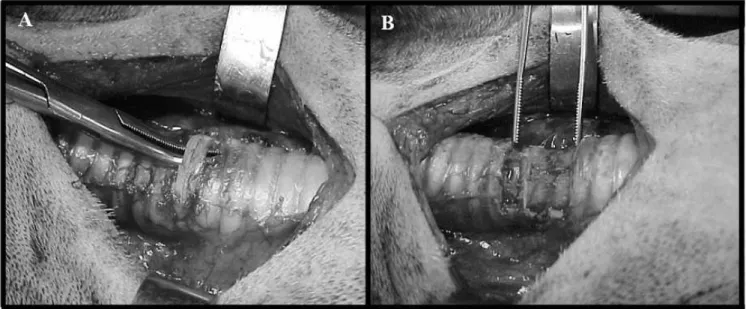
A median cervical incision was performed from the cricoid cartilage close to the upper chest, exposing the third up to the 13th tracheal rings. To develop malacia, we performed an extra-mucosal resection of 70% of the circumference of each tracheal ring (Figure 1), as described by of Korpela et al.7 and Marquette et al.8.
Two experimental groups were created according to the number of resected rings: group A (n = 5), from the sixth to the eighth tracheal rings; group B (n = 4), from the fifth to the tenth tracheal rings. The muscle and skin pla-nes were sutured with 3-0 polyglactin and 4 0 mononylon, respectively. Soon after, rigid bronchoscopy was performed for the first application of 23% NaOH.
Figure 1 Figure 1 Figure 1 Figure 1
Figure 1 - Map showing the cervical incision and the technique of tracheomalacia (dog #2) A) resection of the sixth serosubmucosal cartilaginous ring, B) segment with malacia after resection of three consecutive cartilaginous rings.
In the postoperative period, the animals were daily observed by a veterinarian and by researchers, with special attention to the lack of appetite, vomiting, cough, stridor, tachypnea, and intercostal retractions. We also held weekly bronchoscopies. Analgesia was performed with tramadol (1mg/Kg/SC) postoperatively as required.
The technique of inflammatory stenosis with application of 23% NaOH was previously described and published by two of the authors of this study6: through a
rigid bronchoscope (Karl Storz Endoskope®, Germany) we applied a cotton swab soaked in 23% NaOH solution (pH = 14) on the lining of the trachea with previously surgically induced malacia. We avoided the contact of the NaOH solution to the posterior wall of the trachea. Was subsequently repeated to application of this solution on the region of the previous scar every three (group B) or seven days (group A). The applications were repeated until there were bronchoscopic finding of stenosis greater than 50% or signs of airway obstruction.
Bronchoscopies were performed with rigid tubes, with external diameters from 6.5mm to 8.5mm. The animals were inspected at three or seven days, depending on the group, in order to access the tracheal lumen diameter before instillation of NaOH solution.
The animals were kept under daily observation. Clinical signs of airway obstruction were monitored, as well as other medical or surgical complications. They were sacrificed with a lethal dose of sodium thiopental (100mg/ kg, iv) if there was any significant sign of respiratory failure or ascertained the presence of stenosis above 50% of the tracheal lumen before 50 days of follow-up. After euthanasia, the cervical tracheae were collected and fixed in 10% formalin.
greater stenosis; and level 2: in a location 2cm away from the stenosis, corresponding to the normal diameter of the trachea. The specimens were embedded in paraffin, cut with a 5mm thickness and stained with hematoxylin and eosin. The epithelium, the submucosa and inflammatory reaction were analyzed under an optical microscope with a 10X and 40X objectives.
The latero-lateral and anteroposterior diameters of the tracheal lumen of each specimen were measured using the program Sigmascan Demo-Image Analyser (Sigma®, Chicago, USA). This software allowed us to calculate the area of the tracheal lumen by using the following equation: Area (mm2) = p x [sagittal diameter
(mm) / 2] x [coronal diameter (mm) / 2].
The ratio between the area of greatest stenosis (level 1) and the area of normal trachea (level 2) expressed the degree of narrowing of the tracheal lumen.
Was used the Mann-Whitney test to compare the two groups in the residual lumen at level 1 (area with greater stenosis). The difference was considered statistically significant if p <0.05.
RESULTS
RESULTS
RESULTS
RESULTS
RESULTS
The tracheas of six animals were analyzed. Three animals were excluded from the study: one ani-mal (#1) from group A died from diarrhea two days after the first application of NaOH. Two other dogs (# 8 and #9) in group B were precociously sacrificed 24h after the first application of NaOH due to respiratory failure. Their tracheae had swollen up, but no stenosis greater than 50% had developed. The main results are shown in table 1.
In two animals (#2 and #6), there was a small laceration of the tracheal mucosa during excision of the cartilage rings. These perforations were immediately sutured with 4-0 polyglactin. On the tenth postoperative day, dog #6 developed a limited cervical subcutaneous emphysema, without other consequences. This dog had pneumonia on the eighth postoperative day, which was treated with penicillin.
At the time of the first application of NaOH, we could identify the region of malacia more clearly in group B. In all bronchoscopic examinations following the application of NaOH there was some degree of deposition of fibrinoid material in the tracheal mucosa, as well as scarring and narrowing of the area of application of the caustic solution.
On macroscopic examination, we observed stenosis and fibrosis in all tracheae. In areas of stenosis, there was distortion and blurring of the trachea rings (Figu-re 2). The average longitudinal extent of the stenosis was 1cm and 2cm in groups A and B, respectively.
The residual lumen of the trachea at level 1 (light stenosis) ranged from 1% to 29% (Table 1). The residual lumen level 1 in group A (9.1%) was not statistically different (p = 0.355) from the one of group B (12%). The average lumen area of level 1 in group A (17.92mm2) was not smaller, statistically (p = 0.355) than
in group B (29.8 mm2).
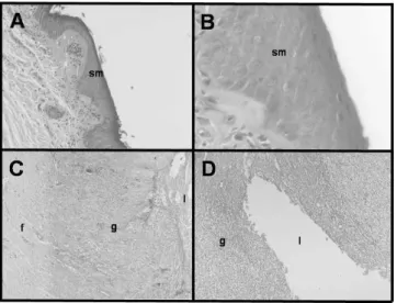
All tracheae displayed wall thickening by fibrosis and inflammatory infiltrate. Fibrosis and necrosis were mainly observed in the submucosa and adventitia, occupying the region of the excised cartilage rings. Granulation tissue predominated in the mucosal surface, leading to partial obstruction of the lumen. The distribution of the sequential phases of the inflammatory process in the tracheal wall was the result of applying the solution to 23% NaOH at different times (Figure 3).
The inflammatory infiltrate, consisting mainly of polymorphonuclear cells, predominated in the mucosa and submucosa. In all specimens, we observed desquamation of respiratory epithelium. Squamous metaplasia was observed in dogs #2 and #6 (Figure 3).
DISCUSSION
DISCUSSION
DISCUSSION
DISCUSSION
DISCUSSION
Although tracheoplasty is the treatment of choice for most cases of inflammatory tracheal stenosis, in some situations, as in patients without clinical conditions, non-surgical procedures are required, such as the placement of silicone tracheal stents.
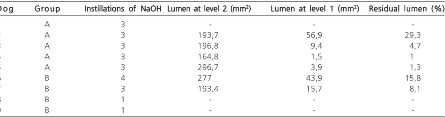
Table 1 Table 1Table 1 Table 1
Table 1 - Number of applications 23% NaOH solution and morphometric analysis.
D o g D o gD o g D o g
D o g G rG r oG rG rG roooo u pu pu pu pu p Instillations of NaOHInstillations of NaOHInstillations of NaOH Lumen at level 2 (mmInstillations of NaOHInstillations of NaOH Lumen at level 2 (mmLumen at level 2 (mmLumen at level 2 (mmLumen at level 2 (mm22222))))) Lumen at level 1 (mmLumen at level 1 (mmLumen at level 1 (mmLumen at level 1 (mmLumen at level 1 (mm22222))))) Residual lumen (%)Residual lumen (%)Residual lumen (%)Residual lumen (%)Residual lumen (%)
1 A 3 - -
-2 A 3 193,7 56,9 29,3
3 A 3 196,8 9,4 4,7
4 A 3 164,8 1,5 1
5 A 3 296,7 3,9 1,3
6 B 4 277 43,9 15,8
7 B 3 193,4 15,7 8,1
8 B 1 - -
-To develop and improve airway orthoses, some experimental models of stenosis and malacia have been described6-13. Once the silicone stents are more satisfactorily
set in stenoses of more than 50% of the lumen and greater than 1 cm in extension4, we seek to adapt and combine
two models of stenosis: resection of cartilage rings in a row, originally described by Mair et al.11, and then modified
by Korpela et al.7 and Marquette et al.8, and the
application of caustic solution by bronchoscopy, published by our group 6.
We thus promote the inflammatory changes found in post-intubation stenosis through an easy to perform and inexpensive method, consisting of application of 23% NaOH in the tracheal mucosa at intervals of seven (Group A)6 or three days (group B) to accelerate the development
of stenosis. Additionally, we adapted, in dogs, the technique of removal of the cartilaginous rings described above for pigs8 and rabbits for7, with the goal of producing a dynamic
narrowing of the airways and a greater reduction of the lumen, since the standalone application NaOH yields only superficial injury, without reaching the perichondrium cartilage6.
The results of our experiments demonstrate the efficacy of combining the two methods described above for the development of an inflammatory stenosis with a reduction of at least 70% of the diameter of the airway. Additionally, there was no statistical difference in relation to the intensity of stenosis produced when comparing the number of rings resected and the interval between applications of NaOH.
Although two surviving animals in group B developed satisfactory 2cm long stenosis, two others of the same group had to be prematurely sacrificed due to airway obstructive symptoms. On the other hand, our study also showed that the resection of only three cartilage rings and the application of NaOH in a span of three rings promotes an apparently less intense stenosis, but shorter (<2 cm), perhaps rendering it inappropriate for the evaluation of orthoses .
In the article by Korpela et al.7, five consecutive
rings were resected and only one animal died, by airway obstruction, between 32 operated. Thus, it appears that the extent of experimental malacia was not the only factor that causeed respiratory dysfunction of the two early euthanized animals in group B. However, an association of six rings malacia and the edema caused by the application of NaOH possibly led to severe obstruction of the airway seen in these dogs.
Perhaps the weekly interval of applications, or even a decrease in concentration of NaOH solution, could result in a slower installation stenosis in group B. In the study by Hanauer et al.6, 23% NaOH was also instilled
once a week (up to three applications), in the larynx and produced a stenosis of at least 50%, with no deaths associated with airway obstruction.
We justify the use of fewer animals in the study due to the early development of severe inflammatory tracheal stenosis. Also, the death of two animals in group B led us to limit the number of animals.
Resection of three or six rings, associated with applications NaOH solution, causes a significant
Figure 2 Figure 2 Figure 2 Figure 2
Figure 2 - Photos of the macroscopic examination of the canine trachea without (n) and with stenosis (s). A) note the change of the external wall at the site of tracheal stenosis and malacia (arrows) produced by topical application of 23% NaOH (dog #6), B, C and D) cross sections of the trachea, showing the differences between the lumen of segment of normal airway (n) and those with stenosis (dogs #7,# 4 and #3, respectively).
Figure 3 Figure 3 Figure 3 Figure 3
narrowing of the trachea. However, future work should show whether the weekly application of NaOH alone for a length of at least 2cm of the airway, or simple resection of six or more tracheal rings, will provide a superior
ex-perimental model, without causing severe acute airway obstruction, greatly reducing the loss of animals, meaning a more safe and effective model to test new tracheal stents.
R E S U M O R E S U M O R E S U M O R E S U M O R E S U M O
Objetivo Objetivo Objetivo Objetivo
Objetivo: Desenvolver, experimentalmente, malácia e estenose traqueal para testar novos modelos de órteses traqueais. Méto-Méto-Méto-Méto- Méto-dos
dos dos dos
dos: Ressecamos três anéis cartilaginosos da traqueia cervical de cães no grupo A (n=5) e seis anéis no grupo B (n=4) para produzir malácia. Logo após, a mucosa da região com malácia recebeu aplicações de uma solução de hidróxido de sódio (NaOH) a 23%, e os animais eram acompanhados com exames broncoscópicos para observar o desenvolvimento de estreitamento da luz da via aérea. Quando a estenose era de mais de 50% da luz, ou havia sinais mínimos de insuficiência ventilatória, os animais eram sacrificados. O segmento de via aérea estreitada foi então coletado para análise histológica e era calculada a área de luz residual do segmento traqueal com estenose e malácia. ResultadosResultadosResultadosResultados: Na análise histológica, foi constatada fibrose na submucosa e adventícia, associadaResultados a granulomas na mucosa. A luz residual média dos segmentos com estenose foi de 9% e 12% nos grupos A e B, respectivamente, (p>0,05). ConclusãoConclusãoConclusãoConclusãoConclusão: A combinação da ressecção de anéis cartilaginosos e da aplicação de NaOH 23% na mucosa respiratória promoveu uma estenose traqueal intensa, porém esteve associada à perda de animais. Novos estudos são necessários para verificar se o emprego isolado de uma das técnicas seria mais seguro e eficaz para desenvolver estenose traqueal.
Descritores: Descritores: Descritores: Descritores:
Descritores: Traqueia. Traqueomalácia. Estenose traqueal. Stents. Implantes experimentais.
REFERENCES
REFERENCES
REFERENCES
REFERENCES
REFERENCES
1. Courey MS. Airway obstruction. The problem and its causes. Otolaryngol Clin North Am 1995; 28(4):673-84.
2. Grillo HC, Donahue DM, Mathisen DJ, Wain JC, Wright CD. Postintubation tracheal stenosis. Treatment and results. J Thorac Cardiovasc Surg 1995; 109(3):486-92; discussion 492-3. 3. Grillo HC. Circumferential resection and reconstruction of the
mediastinal and cervical trachea. Ann Surg 1965; 162(3):374-88. 4. Saueressig M, Macedo Neto AV, Moreschi AH, Xavier RG, Sanches PR. A correção das estenoses traqueobrônquicas mediante o emprego de órteses. J Pneumol 2002; 28(2):84-93.
5. Xavier RG, Sanches PR, Macedo Neto AV, Silva Filho AP, Edelweiss MI, Duarte L, et al. Report of a new silicone stent bronchoscope introducer system (HCPA-1) studied in dogs. Chest 2000; 118(Suppl $):234S.
6. Hanauer AD, Fraga JC, Sousa JK, Sanches PR, Duarte ME, Ulbrich-Kulczynski J, et al. Electrocautery versus 23% NaOH infiltration to induce subglottic stenosis in a canine experimental model. Pediatr Surg Int 2007; 23(12):1227-31.
7. Korpela A, Aarnio P, Sariola H, Törmälä P, Harjula A. Bioabsorbable self-reinforced poly-L-lactide, metallic, and silicone stents in the management of experimental tracheal stenosis. Chest 1999; 115(2):490-5.
8. Marquette CH, Mensier E, Copin MC, Desmidt A, Freitag L, Witt C, et al. Experimental models of tracheobronchial stenoses: a useful tool for evaluating airway stents. Ann Thorac Surg 1995; 60(3):651-6.
9. Fraga JC, Filler RM, Forte V, Bahoric A, Smith C. Experimental trial of balloon-expandable, metallic Palmaz stent in the trachea. Arch Otolaryngol Head Neck Surg 1997; 123(5):522-8.
10. Verkindre C, Brichet A, Maurage CA, Ramon P, Homasson JP, Marquette CH. Morphological changes induced by extensive endobronchial electrocautery. Eur Respir J 1999; 14(4):796-9. 11. Mair EA, Parsons DS, Lally KP, Van Dellen AF. Comparison of
expandable endotracheal stents in the treatment of surgically induced piglet tracheomalacia. Laryngoscope 1991; 101(9):1002-8.
12. Saito Y, Minami K, Kaneda H, Okada T, Maniwa T, Araki Y, et al. New tubular bioabsorbable knitted airway stent: feasibility assessment for delivery and deployment in a dog model. Ann Thorac Surg 2004; 78(4):1438-40.
13. Lee SS, Shin JH, Woo CW, Hwang JC, Park CS, Kim HJ, et al. A new model of tracheal stenosis in dogs using combined bronchoscopic electrocautery and ethanol injection. J Vasc Interv Radiol 2008; 19(5):764-9.
Received on 29/12/2010
Accepted for publication 10/03/2011 Conflict of interest: none
Source of funding: Research Incentive Fund (FIFE) HCPA / UFRGS, under the responsibility of Professor. Dr. Roger Xavier Gastal.
How to cite this article: How to cite this article:How to cite this article: How to cite this article:How to cite this article:
Saueressig MG, Macedo Neto AV, Espinel JO, Edelweiss MI, Sanches PRS, Xavier RG. Experimental model of tracheal stenosis with submucosal resection of cartilaginous rings combined with sodium hydroxide instillations. Rev Col Bras Cir. [periódico na Internet] 2011; 38(6). Disponível em URL: http://www.scielo.br/rcbc
Correspondence Address: Correspondence Address:Correspondence Address: Correspondence Address:Correspondence Address: Maurice Guidi Saueressig