RADIOLOGY PAGE
563
CT Diagnosis of Foreign Body in Urinary Bladder after
Surgical Management of Stress Urinary Incontinence
Han-Jui Lee, Tzu-Pin Lin, Shu-Huei Shen, Jia-Hwia Wang
Department of Radiology (HJL, SHS, JHW), Division of Urology and Department of Surgery (TPL), Taipei Veterans General Hospital and National Yang-Ming University School of Medicine (HJL, TPL, SHS, JHW), Taipei, Taiwan
_______________________________________________________________________________
This 49-year-old Taiwanese woman pre-sented with voiding urethral pain, frequency and hematuria. She had the past history of diabetes mellitus, hypertension and schizophrenia. The la-bor history was G7P2. She had suffered from stress urinary incontinence (SUI) for several years and received repair surgery using polypropylene mesh sling (SPARC™ sling system) two years ago. Since one week after the surgery, she had had several episodes of urinary tract infection and no specific cause could be identified. This time she was admit-ted for the same symptoms. The most recent uri-analysis showed positive for occult blood, white blood cell (WBC) 11-20/ high power field and many bacteria. The urinary culture yielded Escherichia coli more than 106 colonies/ mL.
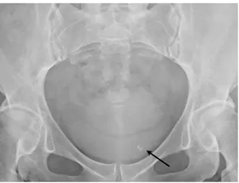
KUB showed an amorphous hyperdense material in left lower pelvis (Figure-1). Computed tomography (CT) study revealed a 3 cm curvilin-ear hyperdense lesion at left anterolateral wall of urinary bladder (Figure-2). Under the impression of intravesical foreign body, cystoscopy was per-formed which revealed the misplacement mesh with stone encrustation (Figure-3). It was removed by Thulium Laser. There was no pyuria in the fol-low up urianalysis (WBC 0-2/ high power field) and her symptoms were improved.
Mesh repair has been used for surgi-cal treatment of SUI since 1990s and has gained popularity. Urinary tract injury is a known com-plication of the procedure, which may be due to iatrogenic injury during the surgery or subsequent erosion of the synthetic mesh through the mucosa (1). The incidence of urinary tract injuries from
mesh repair is not exactly known, with higher re-ported rate in synthetic material (2-4). The definite diagnosis of urinary bladder foreign body is made by cystoscopy, and endoscopic or surgical removal of the foreign body effectively cure the symptom (1). However the diagnosis is usually delayed with the median of 24 months after the original surgery, because the presenting symptoms are variable and confused with recurrent SUI or post-sling obstruc-tion symptoms (1-3,5-7). This is the first report of the imaging findings of urinary tract injury follow-ing SUI surgery. In patient with previous history of pelvic surgery, calcified urinary tract foreign body on CT should alert the present of iatrogenic injury.
Figure 1 - The plain KUB at postoperative 29 months showed a small irregular round radiopaque material (black arrow) at left lower pelvis which was not seen on the postoperative 1 week film (not shown).
564
IBJU |RADIOLOGY PAGE
REFERENCES
1. Frenkl TL, Rackley RR, Vasavada SP, Goldman HB: Manage-ment of iatrogenic foreign bodies of the bladder and urethra fol-lowing pelvic floor surgery. Neurourol Urodyn. 2008; 27: 491-5. 2. Dmochowski RR, Blaivas JM, Gormley EA, Juma S, Karram
MM, Lightner DJ, et al.: Update of AUA guideline on the sur-gical management of female stress urinary incontinence. J Urol. 2010; 183: 1906-14.
3. Clemens JQ, DeLancey JO, Faerber GJ, Westney OL, Mc-guire EJ: Urinary tract erosions after synthetic pubovaginal slings: diagnosis and management strategy. Urology. 2000; 56: 589-94.
4. Atherton MJ, Daborn JP, Tsokos N, Jeffery JT, Yin MJ: Com-plications associated with tissue anchor migration after vagi-nal surgery using the tissue fixation system - a case series. Aust N Z J Obstet Gynaecol. 2012; 52: 83-6.
Figure 2 - Axial scan of pelvic CT, (a) noncontrast and (b) contrast enhanced, bone window. A curvilinear hyperdense material (black arrow) was demonstrated at left anterolateral wall of urinary bladder.
A B
Figure 3 - Cystoscopy demonstrated misplaced suture (long arrow) with stone encrustation (short arrows).
5. Baessler K: Do we need meshes in pelvic floor reconstruc-tion? World J Urol. 2012; 30: 479-86.
6. Deval B, Haab F: Management of the complications of the synthetic slings. Curr Opin Urol. 2006; 16: 240-3.
7. Tse V, Chan L: Outlet obstruction after sling surgery. BJU Int. 2011; 108(Suppl 2): 24-8.
Correspondence address: