Pan American Health Organization
Regional Office of theWorld Health Organization
VISUAL INSPECTION
OF THE UTERINE CERVIX
WITH ACETIC ACID (VIA)
A CRITICAL REVIEW
PAHO HQ Library Cat aloguing-in-Publicat ion
Visual Inspect ion of t he Ut erine Cervix w it h Acet ic Acid (VIA): A Crit ical Review and Select ed Art icles Washingt on, D.C.: PAHO, © 2003.
ISBN 92 75 12444 2
Preparat ion of t his publicat ion w as support ed by t he Bill & M elinda Gat es Foundat ion t hrough t he Alliance f or Cervical Cancer Prevent ion (ACCP). This report is part of t he project Prevent ion of Cervical Cancer of t he Pan American Healt h Organizat ion, a member of t he Alliance f or Cervical Cancer Prevent ion (ACCP).
Prepared under t he supervision of Dr. Sylvia Robles
Aut hors of t he review w ere Dr. Cat t erina Ferreccio and M s. Julia Gage
ACKNOWLEDGMENTS
The aut hors w ish t o t hank Dr. Lynne Gaff ikin, JHPIEGO Corporat ion, Dr. R. Sankaranarayanan, Int ernat ional Agency f or Research on Cancer, and Dr. M ichael St ot o, The George Washingt on Universit y School of Public Healt h and Healt h Services, f or t heir comment s and cont ribut ions.
The Noncommunicable Diseases Unit w elcomes comment s about t his mat erial. Please direct all comment s t o your local PAHO Represent at ive or direct ly t o [email protected].
This document is not a f ormal publicat ion of t he Pan American Healt h Organizat ion (PAHO).
The ment ion of specif ic companies or of cert ain manuf act urers’ product s does not imply t hat t hey are endorsed or recommended by t he Pan American Healt h Organizat ion in pref erence t o ot hers of a similar nat ure t hat are not men-t ioned. Errors and omissions excepmen-t ed, men-t he names of propriemen-t ary producmen-t s are dismen-t inguished by inimen-t ial capimen-t al lemen-t men-t ers.
Pan American Health Organization
Area of Disease Prevention and Control Non Communicable Diseases UnitVISUAL INSPECTION
OF THE UTERINE CERVIX
WITH ACETIC ACID (VIA)
A CRITICAL REVIEW
AND SELECTED ARTICLES
Pan American Health Organization
Pan American Sanitary Bureau,
PERMISSIONS
1. Denny et al. “Evaluation of alternative methods of cervical cancer screening for resource-poor settings”
Cancer2000; 89: 826-833 © John Wiley & Sons, Inc. Jan. 17, 2001
2. University of Zimbabwe/JHPIEGO Cervical Cancer Project
“Visual inspection with acetic acid for cervical-cancer screening: test qualities in primary-care settings”
The Lancet 1999; 353: 869-873 © The Lancet Publishing Group Dec. 7, 2000
3. Sankaranarayanan et al. “Visual inspection with acetic acid in the early detection of cervical cancer and precursors” Letter, Intl. J. of Cancer. 1999; 80: 161-163
© John Wiley and Sons, Inc. Sept. 21,2001
4. Sankaranarayanan et al. “Visual inspection of the uterine cervix after the application of acetic acid in the detection of cervical carcinoma and its precursors”
Cancer1998; 83: 2150-2156 © John Wiley & Sons, Inc Sept. 11, 2000
5. Megevand et al. “Acetic acid visualization of the cervix: an alternative to cytologic screening”
Obstetrics and Gynecology1996; 88: 383-386 © Elsevier Science Ltd.
Jan. 31, 2001
6. Cecchini et al. “Testing cervicography and cervicoscopy as screening tests for cervical cancer”
Tumori1993; 79: 22-25 © Tumori
Nov. 13, 2000
Slawson et al “Are Papanicolaou smears enough? Acetic acid washes of the cervix as adjunctive therapy: a HARNET study”
J. of Family Practice1992; 35: 271-277 © Dowden Health Media
Dec. 1, 2000
Ottaviano et al. “Examination of the cervix with the naked eye using acetic acid test”
Am. J. of Obstetrics and Gynecology1982; 143: 139-142 © Mosby, Inc
CONTENTS
LIST OF TABLES … … … 4
LIST OF FIGURES … … … 4
PREFACE … … … 5
PART 1:CRITICAL REVIEW … … … 7
Background … … … 8
Methodology followed and studies selected… … … 9
Time period, location, purpose of the studies … … … 10
Sample size and characteristics of study subjects… … … 13
VIA providers and their training … … … 14
Characteristics of VIA, other screening tests used … … … 16
Sequence of tests used for screening and diagnosis … … … 19
Reference standard … … … 19
Positivity results of VIA and conventional cytology … … … 19
Accuracy of VIA … … … 21
Detection of moderate or worse dysplasia: VIA and conventional cytology … … 23
Conclusions of study authors … … … 23
Discussion and unresolved questions … … … 23
Epilogue … … … 25
REFERENCES … … … 26
GLOSSARY … … … 26
ANNEX: Studies not selected … … … 28
PARTE 2:FULL-TEXT ARTICLES … … … 29
Denny L, Kuhn L, Pollack A, Wainwright H, Wright T. Evaluation of alternative methods of cervical cancer screening for resource-poor settings. Cancer2000; 89: 826-833. … … … 30
University of Zimbabwe / JHPIEGO Cervical Cancer Project. Visual inspection with acetic acid for cervical-cancer screening: test qualities in a primary-care setting. The Lancet1999; 353: (869-873). … … … 39
Sankaranarayanan R, Shyamalakumary B, Wesley R, Sreedevi Amma N, Parkin DM, Krishnan Nair M. Visual inspection with acetic acid in the early detection of cervical cancer and precursors. (Letter) Int J Cancer1999; 80: 161-163. … … … 47
Sankaranarayanan R, Wesley R, Somanathan N, Dhakad N, Shyamalakumary B, Amma NS, Parkin DM, Nair MK. Visual inspection of the uterine cervix after the application of acetic acid in the detection of cervical carcinoma and its precursors. Cancer1998; 83: 2150-2156. … … … 51
Megevand E, Denny L, Dehaeck K, Soeters R, Bloch B. Acetic-acid visualization of the cervix: an alternative to cytologic screening . Obstet Gynecol 1996; 88: 383-386. … … … 58
Cecchini S, Bonardi R, Mazzotta A, Grazzini G, Iossa A, Clatto S. Testing cervicography and cervicoscopy as screening tests for cervical cancer. Tumori1993; 79: 22-25. … … … 62
Slawson D, Bennett J, Herman, J. Are Papanicolaou smears enough? Acetic acid washes of the cervix as adjunctive therapy: a HARNET study. J Fam Pract1992; 35: 271-277. … … … 67
LIST OF TABLES
1 Articles selected and time period and location of each study, by year
of publication … … … 10
2 Purpose of the studies included in this review … … … 13
3 Sample size and selected other characteristics of the studies and the subjects … … 14
4 Providers of VIA and the training they received … … … 15
5a VIA characteristics, other screening tests used … … … 16
5b Criteria for positive or abnormal VIA test results … … … 16
6 Sequence of actions in the conduct of screening and follow-up … … … 17
7 Criteria for application of the reference standard and percent of study group tested with it … … … 18
8 Comparison of positivity results, conventional cytology and VIA … … … 19
9 Estimated VIA accuracy, sample size, and coverage with the reference standard… … 20
10 Cases of CIN II or worse that were positive to either VIA or the Pap test and were negative to the other … … … 21
11 Conclusions of the studies, as stated by the authors … … … 22
LIST OF FIGURES
1 Percent of women with abnormal conventional cytology or abnormal VIA, by study … … … 20The significantly limited impact of cytology based cervi-cal cancer screening programs in developing countries is now widely recognized. There are several reasons for these limits, ranging from the nature of participation of women in screening programs to the access and timely completion of treatment when necessary. Much of the discussion on how to improve the effectiveness of screening programs has been centered on analysis of the sensitivity and specificity of screening tests. As a result, new potential screening tests are being proposed, among them visual inspection with acetic acid, which is quite appealing for low resource settings.
In this publication we review the available evidence which speaks to the accuracy of visual inspection with acetic acid. The information and findings provided are directed to health professionals and managers of health services as an aid in the decision making process. As with any new technology, there must be a process of evalua-tion for this screening method. Ideally, evidence from randomized trials would provide the basis for policy changes. In such trials women are randomly assigned to be screened with either VIA or another method, and then both groups are followed up and compared. Currently, at least two studies of this nature are on-going. In the meantime, evidence from cross-sectional studies seems to point out that VIA may be equally or more sensitive than cytology; this would signify a reduc-tion in the rate of women with false negative results. The overarching question is how much and what level of evidence is necessary in order to incorporate this tech-nology into public health programs.
An important factor for consideration is that the results presented in these papers were obtained under research conditions, with meticulous attention to per-formance of health care providers and training. In this
way, sensitivity and specificity were estimated under the best of circumstances and conditions. By contrast, a 'real life' screening program entails a much more varied and complex set of circumstances. Therefore, the ultimate accuracy of the test must be evaluated under conditions, which are less than ideal; in other words, conditions which fit with the various realities of developing coun-tries. This is important because in the end the feasibility and appropriateness of implementing VIA programs may not depend on the test itself but on the program-matic conditions surrounding the test.
No comprehensive consideration of costs is made throughout the studies contained in this report, but sev-eral of the papers assume that the cost must be relatively low since VIA relies on trained human resources and low cost materials, such as vinegar, etc. It should be noted, however, that costs may increase through high referral rates which might be required with this test, through the intensive and continuous training that a subjective tech-nique usually requires, and most importantly, through quality control measures that a program must have.
With this report the Pan American Health Organization, as a member of the Alliance for Cervical Cancer Prevention, intends to encourage and facilitate discussion about this test in developing countries. We hope, especially, that as health professionals consider the evidence in light of their own knowledge of the sit-uation in their respective countries, a more confident assessment of VIA may be made. This document does not constitute a formal recommendation, since impor-tant results of new studies will be forthcoming. However, it is important at this preliminary stage to pay attention to the issues of methodology and implemen-tation that merit consideration in the design of public health programs.
PART 1.
Visual inspection with acetic acid (VIA), also called cervi-coscopy, consists of naked-eye visualization of the uter-ine cervix (without magnification) after the application of diluted acetic acid, to screen for cervical abnormali-ties. A solution of 3% to 5% acetic acid is used, and the cervix is illuminated with a light source (See pictures). If low-power magnification is used, the technique is called
VIA with magnification (VIAM). The purpose is to iden-tify acetowhite areas, which may indicate tissue under-going precancerous changes. Either ablation (destroy-ing them) or excision (cutt(destroy-ing them out) can then elimi-nate these acetowhite areas.
Early studies of visual inspection involved simply looking at the cervix (unaided visual inspection, or UVI) to identify and treat pre-cancerous lesions as early as possible, a strategy referred to as downstaging. This was the main tool used before conventional cytology and, together with improved treatment and increased public and professional awareness of cervical cancer, con-tributed to the decline of cervical cancer deaths in
Northern Europe(1). The major drawback of this
approach is that lesions are not detected early enough to prevent invasion, because a large proportion of the cancers detected are relatively advanced, requiring com-plex medical therapy that is difficult to provide in many settings. By contrast, visual inspection after swabbing the cervix with acetic acid causes pre-cancerous cells to turn white, enabling much earlier detection, and treat-ment, of pre-cancerous lesions.
Other visual inspection approaches are:
Cervicography, which entails photographing the cervix with a patented, uniquely-designed camera. The photo-graphs, called cervigrams, are viewed as projected slides by colposcopists trained in their interpretation.
Speculoscopy,where acetic acid is applied to the cervix, but a chemical, or chemiluminescent, light–source and magnifying lens are used to visualize the acetowhite lesions of the cervix(2).
VILI: Visual Inspection with Lugol Iodine, which involves visualizing the cervix after applying Lugol’s iodine to detect lesions. This technique is under evaluation as an independent primary screening test(3).
After conventional cytology became the standard test for cervical cancer screening, increased utilization of the colposcope ensued in order to confirm the findings. Years later, given the expense and inconvenience of col-poscopy services, clinicians began to explore whether unmagnified visualization of the cervix, aided by acetic acid, could be used as an adjunct to conventional cytol-ogy to identify those patients in need of colposcopy and thereby use resources more efficiently. However, few studies were conducted which examined the value of unmagnified inspection of the cervix after the applica-tion of acetic acid for purposes of identifying a normal “transformation zone” or for detecting pre-cancerous lesions of the cervix (i.e., primary screening)(4).
In 1982, Ottaviano and La Torre published the results of a study involving 2,400 women who were examined visually and colposcopically after the applica-tion of a cervical wash with acetic acid(5). A key result
was that naked-eye (unmagnified) inspection detected an abnormality in 98.4% of the 312 patients assessed colposcopically as having an abnormal transformation zone. In addition, (unmagnified) visual inspection with acetic acid identified as normal, 98.9% of the 1,584 women diagnosed as normal by colposcopy. That land-mark study, as well as others conducted since, is among those included in this document.
Most of the scientific discussion on screening for pre-cancerous lesions of the cervix has focused on iden-tifying the technique that will provide the best balance between true and false positives. Efforts have been devoted to evaluating the effectiveness of combining screening tests in order to improve the overall sensitivi-ty attained by the screening program. However, in
addi-tion to performing one or more screening tests, the prompt availability of results is at times critical in guar-anteeing the effectiveness of a screening program. Of all the above-described screening techniques, only VIA provides instant results, allowing for immediate treat-ment if necessary.
METHODOLOGY FOLLOWED
AND STUDIES SELECTED
Studies pertaining to visual inspection of the cervix, pub-lished from 1955 through December 2000, were retrieved by searching the PubMed database available in the U.S. National Library of Medicine. The reference lists provided in the articles retrieved were reviewed as well, and a number of researchers in the field were also con-tacted.
The OVID version 4.1.0 search and retrieval software was used for accessing the PubMed database. Initially, the search identified all studies with at least one of the following MeSH subject headings (both exploded and limited to a MeSH major topic):
- cervix dysplasia
- cervical intraepithelial neoplasia - cervix neoplasms, and
- cervix uteri.
Next, the search was narrowed to those publications that also included the MeSH keyword acetic acid in the title, abstract, registry number word, or MeSH subject heading; 67 such articles were finally identified. The search through the reference lists given in the articles retrieved produced another 12 publications, and six more studies were identified at conferences or through contacts with other researchers. Thus, a total of 85 sci-entific articles or studies were identified. Only 20 of these met our main selection criterion that the article or study must present original data from a clinical study of the accuracy of VIA in detecting pre-invasive cervical lesions.
The following secondary criteria were then applied: (1) The paper should have been published in a
peer-reviewed journal or should be in press.
(2) The study should have included a diagnostic evalua-tion for all or a sample of negative, and all VIA-positive, women.
(3) The thresholds used in determining VIA- and PAP-positives should be specified in the paper.
(4) The sample size should have been large enough to have the statistical power to detect a VIA sensitivity of approximately 60%-80%. This implies that, con-sidering various prevalences of CIN, the sample size should have been between 500 and 3,000 for confi-dence intervals of 80% to 90%.
Seven scientific articles met the above criteria and are included in this document; an eighth, by Ottaviano and La Torre, was selected for inclusion as well because of its landmark status and also because it was conducted in a clinical setting. This was, in fact, the first study conduct-ed on VIA. The list of articles chosen is presentconduct-ed in
Table 1.
The lack, or insufficient use, of a reference standard was common to the majority of studies identified in the search. Therefore, it proved necessary to omit a selec-tion criterion concerning a reference standard.
It was found that some of the published studies applied a reference standard in which VIA was com-pared to all methods, some had no reference standard at all, and others used the Pap smear test as a reference standard screening test. In this review, the Pap smear test is considered to yield insufficiently definitive results to allow prudent use as a reference standard.
Time period, location,
purpose of the studies
Five of the studies selected were conducted in develop-ing countries: two in South Africa, one in Zimbabwe, two in India*. Three other studies were carried out in developed countries: two in Italy, one in the United States of America. None were conducted in Latin America. The first VIA study, by Ottaviano and La Torre, was published in 1982 and conducted in a clinical setting in Italy, while the other seven were published between 1992 and 2000 and primarily correspond to reports of community-based studies.
All the studies were cross-sectional, and aimed to evaluate VIA’s ability to detect pre-cancerous lesions as compared to a reference standard. They did not investi-gate the overall effectiveness of a VIA-based screening program for the purpose of lowering cervical cancer inci-dence or mortality, which would have required a longi-tudinal study design.
The authors had various objectives in carrying out a study on VIA (Table 2). Broadly, these objectives may be classified as follows:
1. Measuring the accuracy** of VIA in screening for cervical dysp lasia.
2. Comparing the accuracy of VIA to that of other screening tests.
3. Measuring the added accuracy when using VIA as an adjunct to other screening tests.
4. Assessing the concordance of VIA with colposcopy (colposcopy being the reference standard).
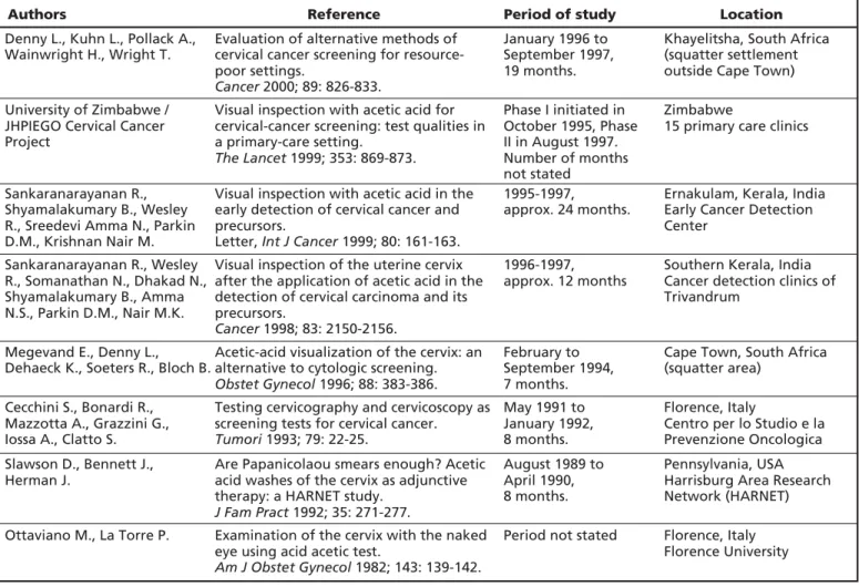
Authors Reference Period of study Location
Denny L., Kuhn L., Pollack A., Wainwright H., Wright T.
Evaluation of alternative methods of cervical cancer screening for resource-poor settings.
Cancer 2000; 89: 826-833.
January 1996 to September 1997, 19 months.
Khayelitsha, South Africa (squatter settlement outside Cape Town)
University of Zimbabwe / JHPIEGO Cervical Cancer Project
Visual inspection with acetic acid for cervical-cancer screening: test qualities in a primary-care setting.
The Lancet 1999; 353: 869-873.
Phase I initiated in October 1995, Phase II in August 1997. Number of months not stated
Zimbabwe
15 primary care clinics
Sankaranarayanan R., Shyamalakumary B., Wesley R., Sreedevi Amma N., Parkin D.M., Krishnan Nair M.
Visual inspection with acetic acid in the early detection of cervical cancer and precursors.
Letter, Int J Cancer 1999; 80: 161-163.
1995-1997,
approx. 24 months.
Ernakulam, Kerala, India Early Cancer Detection Center
Sankaranarayanan R., Wesley R., Somanathan N., Dhakad N., Shyamalakumary B., Amma N.S., Parkin D.M., Nair M.K.
Visual inspection of the uterine cervix after the application of acetic acid in the detection of cervical carcinoma and its precursors.
Cancer 1998; 83: 2150-2156.
1996-1997, approx. 12 months
Southern Kerala, India Cancer detection clinics of Trivandrum
Megevand E., Denny L., Dehaeck K., Soeters R., Bloch B.
Acetic-acid visualization of the cervix: an alternative to cytologic screening.
Obstet Gynecol 1996; 88: 383-386.
February to September 1994, 7 months.
Cape Town, South Africa (squatter area)
Cecchini S., Bonardi R., Mazzotta A., Grazzini G., Iossa A., Clatto S.
Testing cervicography and cervicoscopy as screening tests for cervical cancer.
Tumori 1993; 79: 22-25.
May 1991 to January 1992, 8 months.
Florence, Italy
Centro per lo Studio e la Prevenzione Oncologica
Slawson D., Bennett J., Herman J.
Are Papanicolaou smears enough? Acetic acid washes of the cervix as adjunctive therapy: a HARNET study.
J Fam Pract 1992; 35: 271-277.
August 1989 to April 1990, 8 months.
Pennsylvania, USA Harrisburg Area Research Network (HARNET)
Ottaviano M., La Torre P. Examination of the cervix with the naked eye using acid acetic test.
Am J Obstet Gynecol 1982; 143: 139-142.
Period not stated Florence, Italy Florence University
TABLE 1. Articles selected and time period and location of each study, by year of publication
_____________________________
* The first of the two Sankaranarayanan studies presented here began in 1995, ended in 1997, and was published in 1999; it will be referred to as “Sankaranarayanan (1995-97).” The second began in 1996, ended in 1997, and was published in 1998; it will be referred to as “Sankaranarayanan (1996-97).”
The objectives of six of the eight studies selected fall into the first two categories. Those that aimed to assess the accuracy of VIA as a screening test, such as the University of Zimbabwe / JHPIEGO+and Cecchini studies,
often applied a reference standard (such as histology) to all subjects (i.e., regardless of whether they tested posi-tive or negaposi-tive to VIA), in order to be able to measure accuracy by means of indicators such as sensitivity, speci-ficity, and positive and negative predictive values++. In
these studies, the ultimate objective was to determine whether VIA can act as an exclusive, stand-alone screen-ing method.
In the studies by Denny, Sankaranarayanan, and Megevand, which aimed to compare the accuracy of VIA with that of other screening tests, the reference stan-dard was not applied to all subjects, but only to those who tested positive to any of the initial screening tests. Although in principle all study subjects should be tested with the reference standard, when several tests are used and all subjects having any positive result are then
test-ed by means of a reference standard, the sampling frac-tion of people receiving the reference test will be high-er, thereby decreasing the verification bias ‡.
The study by Slawson aimed to determine the increased accuracy obtained by using VIA in combina-tion with convencombina-tional cytology. The main focus was on the number of cases that would have been missed with-out VIA as an adjunct, rather than on indicators of screening accuracy. For the authors of this study, the question was whether combining conventional cytology with VIA significantly improves the results of a screening approach.
_____________________________
+ The Zimbabwe study had two phases. In the first phase not all subjects received the reference standard, whereas in the second phase included a standard test for all study subjects. In this publication, when analyzing results only the second phase is taken into consideration.
++ See first box in next page for definitions.
‡ Verification bias: results when only those people with a positive screening test result receive the confirmatory diagnostic test. See article in page 67.
Measures of data quality:
accuracy and precision
Accuracy: A measure of how closely the results of the test correspond to the true state of affairs. It is measured by the cal-culation of sensitivity and specificity. A test with perfect accuracy will have a sensitivity of 100% (identifies every single case in the population under study) and a specificity of 100% (recognizes every normal subject as such in the population under study).
Precision: A measure of how closely replicate observation of the same thing produces the same results. Measures can be highly precise but inaccurate. Measured by the standard deviation of a series of replicate determinations. The Kappa coef-ficient, a measure of the degree of nonrandom agreement between observers or measurements of the same categorical variable, is also used. In the conventional cytology reading the degree of dysplasia is highly variable among pathologists,i.e. there is low agreement. However, for distinguishing between cancer and no cancer, conventional cytology is highly precise, i.e. there is a high agreement rate among pathologists.
Effectiveness of screening
Effectiveness: Reduction of incidence or mortality due to the particular disease (cancer). Most studies do not attempt to measure these long-term outcomes. Instead, they use the test sensitivity to evaluate the detection capacity of the screening and the pre-cancerous lesions treated as a proxy for invasive cancers prevented.
Biases in the interpretation of a screening program’s effectiveness
Several different biases might cause overestimation of a program's benefits. Although not relevant to the studies under review, for reference purposes they are mentioned below.
1. Lead time bias: Although screening only speeds up diagnosis, it does not necessarily delay death. Upon analyzing the study group, screening appears to increase post-diagnostic lifespan. To measure lead time bias a controlled longitudinal design would be required, with one group screened and another unscreened, both followed until death.
2. Length bias: Asymptomatic cases that are detected by screening have a better prognosis, or more favorable progression of the disease, than those symptomatic cases that result in spontaneous consultation. This improved disease progression and longer life expectancy is then erroneously attributed as a benefit of the screening program. Measurement of this bias would require following asymptomatic cases without treatment, which is ethically unacceptable.
The options for incorporating VIA into a screening and treatment program are:
• As an adjunct to conventional cytology. Both tests are carried out, and a positive VIA result is followed either by colposcopy or by immediate treatment.
• As a complement to conventional cytology.
Identification of low grade lesions by conventional cytology is followed by VIA to decide whether to send the woman for colposcopy.
• As the sole screening technique. A positive VIA
result is followed by colposcopic examination or immediate treatment.
In all studies, the outcome that determines accuracy is the actual detection of cervical lesions (squamous cell intraepithelial lesions). Detection and treatment of these precancerous lesions serve as a proxy for the reduction of cervical cancer incidence, which ultimately is the true measure of effectiveness for a cervical cancer screening program.
Sensitivity is the proportion of truly diseased persons in the screened population who are identified as diseased by the screening test. Sensitivity is a measure of the proba-bility of correctly diagnosing a case, or the probaproba-bility that any given case will be identified by the test (Syn: true pos-itive rate).
Specificity is the proportion of truly non-diseased persons who are so identified by the screening test. It is a measure of the probability of correctly identifying a non-diseased person with a screening test (Syn: true negative rate).
Positive predictive valueis the proportion of people with a positive test who have the disease in question. It is a measure of the probability that a patient with a positive screening result has the disease. The prevalence of a dis-ease in a population, as well as the sensitivity of the test being used when the disease is infrequent easily affects the positive predictive value.
Negative predictive value is the proportion of people with a negative test who do not have the disease, thereby measuring the probability that a patient with a negative screening result does not have the disease.
The above listed relationships are shown in the following table, in which the letters a, b, c, and d represent the quantities specified below the table.
Screening Reference Standard Total test Diseased Not diseased results
Positive a b a+b
Negative c d c+d
Total a+c b+d a+b+c+d
a = Diseased individuals detected by the test (true positives)
b = Non-diseased individuals positive by the test (false positives)
c = iseased individuals not detectable by the test (false negatives)
d = Non-diseased individuals negative by the test (true negatives)
Sensitivity = a / (a+c) Specificity = d / (b+d)
Positive predictive value (positive test result) = a / (a+b) Negative predictive value (negative test result) = d / (c+d)
_____________________________ Sources:
Last JM. A dictionary of epidemiology. Third edition, 1995. Oxford University Press. (p. 154)
Gordis L. Epidemiology 1996 W.B. Saunders Company. (pp 55-65)
Properties of
screening tests
Reference standard
In order to determine the accuracy of a screening test, it is usually compared with a reference or gold standard - an external source of “truth” that can determine the true dis-ease status of an individual resulting from a more defini-tive and often more invasive test. Accuracy is therefore described using various indicators that explain this com-parison: sensitivity, specificity, positive and negative pre-dictive values. Currently some authors are beginning to use a score that, in one single measurement, simultane-ously synthesizes the specificity and sensitivity of a test. The score indicates how close each study is to the ideal of 100% sensitivity and 100% specificity; it is called accuracy score (see Table 9).
Two other factors may be distinguished:
a) Concurrent validity: The ability of a test to detect exist-ing cases. Its measurement requires that a reference test be applied simultaneously to the entire study group. This is the detection capacity of the test to identify those cases that the reference also identifies as cases. The studies reviewed focus on concurrent validity of VIA to detect CIN and cancer.
It should be noted that, in order to obtain the true sensitivity or specificity of a test, it is necessary to apply a reference standard‡ test to all the study subjects, regardless of their screening results. Otherwise, only a relative comparison of the screening tests can be made. Denny and Sankaranarayanan calculated the ratio of the sensitivity of one test to that of another (ratio of the number of true cases detected by each test) and used McNemar’s test of statistical significance of the differ-ence between them. Relative comparisons of screening outputs (number of cases detected) are valid and useful in regard to cost-effectiveness analyses, since they pro-vide information that is helpful in choosing which screening tests to implement.
Since most of the studies did not intend to measure the sensitivity and specificity of VIA, estimating accuracy indicators will incur some degree of verification bias.
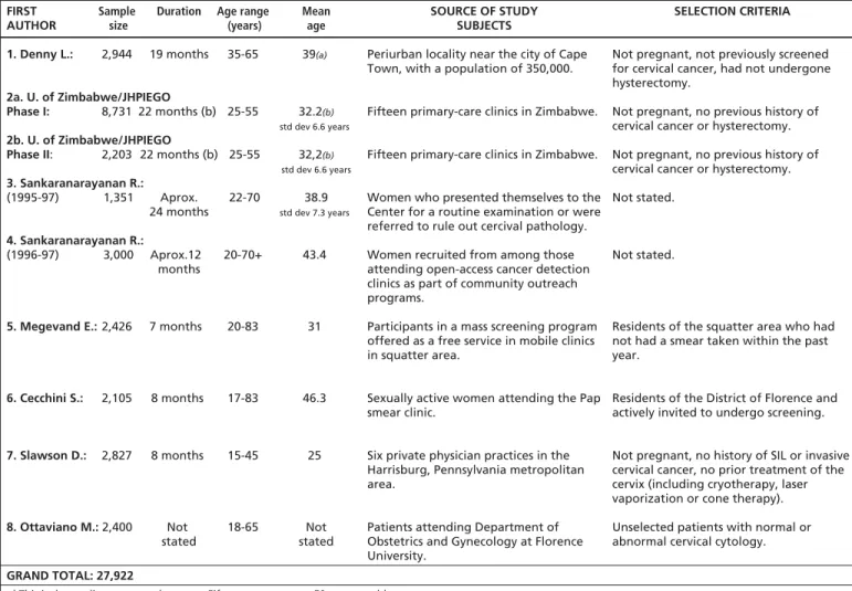
Sample size and
characteristics of study
subjects
A combined total of 27,922 women were screened with VIA in the eight studies (Table 3). Sample sizes ranged between 2,000 and 3,000 subjects, except for the Sankaranarayanan (1995-97) and University of Zimbabwe / JHPIEGO Phase I studies, with 1,351 and 8,731 subjects respectively. Except for Ottaviano, whose study subjects were patients subjected to both VIA and colposcopy, all other studies were conducted among women who had come for a screening test without rea-son to suspect disease
_____________________________
‡ See second box in the opposite page for definitions.
First author Purpose (as stated in the paper)
General objective: To measure the accuracy of VIA in screening
To assess the specificity and sensitivity of VIA carried out by non-physicians in a primary-care setting. A secondary objective was to compare the qualities of VIA with those of the Pap smear test
To evaluate the possible advantages of combining cervicography with vaginal cytology in a screened population. The study also investigated the diagnostic accuracy of direct
examination of the cervix after acid lavage.
General objective: To compare the accuracy of VIA relative to other tests
To evaluate the use of alternative screening methods in a resource-poor setting
To compare the performance of VIA and cervical cytology in detecting cervical lesions in a clinic-based study of women attending for routine examination or because of referral to rule out cervical pathology.
To evaluate the performance of unmagnified, naked-eye visual examination of the uterine cervix after application of 3-4% acetic acid (VIA) in detecting cervical lesions.
To determine if direct visualization of the cervix after application of acetic acid would be an adequate alternative to cytology in the detection of premalignant lesions of the cervix.
General objective: To measure the added value of VIA as an adjunct to other tests
To determine whether acetic acid wash and the cytology smear, if used together, would identify more cases of cervical disease than the cytology smear alone.
General objective: To assess the concordance of VIA and colposcopy
To draw attention to the importance of naked-eye inspection of the cervix, employing a good light source, after preparing the cervix with a 3% acetic acid solution
Ottaviano M. Slawson D. Megevand E.
Sankaranarayanan R. (1995-97)
Sankaranarayanan R. (1995-97)
Denny L. Cecchini S.
U. of Zimbabwe / JHPIEGO, Phases I & II
Duration of the studies was between seven months and two years. The mean age of patients ranged from 25 to 46.3 years.
The exclusion criteria most commonly applied were: • previous treatment of pre-cancerous lesions of the
cervix;
• pregnancy;
• gross distortion of the cervical anatomy.
VIA providers
and their training
In five of the studies VIA was performed by nurses, while in one (Sankaranarayanan 1996-1997) it was performed by cyto-technicians and, in two others (Slawson, Ottaviano), by physicians (Table 4). Not all the authors mention how many providers conducted VIA, yet those that do (Sankaranarayanan, Megevand, Cecchini, Ottaviano) indicate that only one or two providers per-formed all of the VIA exams. In the University of Zimbabwe / JHPIEGO study, six nurses were used (per-sonal communication with one of the authors, LG).
The duration of the training of providers was usual-ly three to six days, however, little or no information was given as to the number of hours of training and clinical practice, the number of patients seen during the train-ing, or the criteria for competency of the trainees.
FIRST Sample Duration Age range Mean
AUTHOR size (years) age
1. Denny L.: 2,944 19 months 35-65 39(a)
2a. U. of Zimbabwe/JHPIEGO
Phase I: 8,731 22 months (b) 25-55 32.2(b)
std dev 6.6 years
2b. U. of Zimbabwe/JHPIEGO
Phase II: 2,203 22 months (b) 25-55 32,2(b)
std dev 6.6 years
3. Sankaranarayanan R.:
(1995-97) 1,351 Aprox. 22-70 38.9 24 months std dev 7.3 years
4. Sankaranarayanan R.:
(1996-97) 3,000 Aprox.12 20-70+ 43.4
months
5. Megevand E.: 2,426 7 months 20-83 31
6. Cecchini S.: 2,105 8 months 17-83 46.3
7. Slawson D.: 2,827 8 months 15-45 25
8. Ottaviano M.: 2,400 Not 18-65 Not stated stated
SELECTION CRITERIA
Not pregnant, not previously screened for cervical cancer, had not undergone hysterectomy.
Not pregnant, no previous history of cervical cancer or hysterectomy.
Not pregnant, no previous history of cervical cancer or hysterectomy.
Not stated.
Not stated.
Residents of the squatter area who had not had a smear taken within the past year.
Residents of the District of Florence and actively invited to undergo screening.
Not pregnant, no history of SIL or invasive cervical cancer, no prior treatment of the cervix (including cryotherapy, laser vaporization or cone therapy).
Unselected patients with normal or abnormal cervical cytology.
SOURCE OF STUDY SUBJECTS
Periurban locality near the city of Cape Town, with a population of 350,000.
Fifteen primary-care clinics in Zimbabwe.
Fifteen primary-care clinics in Zimbabwe.
Women who presented themselves to the Center for a routine examination or were referred to rule out cercival pathology.
Women recruited from among those attending open-access cancer detection clinics as part of community outreach programs.
Participants in a mass screening program offered as a free service in mobile clinics in squatter area.
Sexually active women attending the Pap smear clinic.
Six private physician practices in the Harrisburg, Pennsylvania metropolitan area.
Patients attending Department of Obstetrics and Gynecology at Florence University.
GRAND TOTAL: 27,922
a) This is the median age, not the mean. Fifteen percent were 50 years or older. b) These figures are fro phases I and II combined.
1. Denny L.: 1 trained nurse
Nurse conducted gynecological examination, VIA, Cervicography, and obtained a Pap smear and a sample for HPV DNA testing.
PAP smears were read at local cytopathology laboratory; not masked. Cervigrams were evaluated in the United States of America, masked. HPV DNA test assayed at local University.
Gynecological oncologist conducted colposcopy, biopsy, LEEP and endocervical curettage.
All histology exams were evaluated in the United States of America, masked.
2a. U. of Zimbabwe/JHPIEGO, Phase I: 6 trained nurse-midwives
Nurse-midwives obtained a Pap smear and conducted VIA. Pap smears were analyzed by cytotechnicians in Harare, masked. All positive smears and a 10% random sample of negative smears were re-assessed by a local pathologist and also by a cytopathologist in the United States of America.
Colposcopy was conducted in Harare by three faculty members, masked.
Biopsies were read by the local pathologist, masked. HPV samples were analyzed in the United States of America.
2b. U. of Zimbabwe/JHPIEGO, Phase II: 6 trained nurse-midwives
Same as above, except:
• All slides were reviewed, masked, by the local pathologist and by a cytopathologist in the United States of America
• All patients received a masked colposcopy in a Harare clinic
3. Sankaranarayanan R. (1995-97): 1 nurse
Nurse did a speculum examination, VIA exam and obtained a Pap smear.
PAP smears were examined by a cytopathologist. Colposcopy was conducted by the same cytopathologist
4. Sankaranarayanan R. (1996-97): 2 cytotechnicians
Two cytotechnicians conducted speculum examinations, VIA, and obtained cervical smears.
Smears were examined by cytopathologists and cytotechnologists. Providers of colposcopy and biopsies were not mentioned.
5. Megevand E.: 1 nurse
Una enfermera adiestrada realizó IVAA y obtuvo frotis para prueba de A trained nurse conducted VIA and obtained a Pap smear. Cervical smears were screened and processed by a masked cytotechnologist.
No information is given on providers of colposcopy, LEEP and biopsies conducted in mobile clinic.
6. Cecchini S.: 2 “smear-takers”
Two “smear-takers,” midwives without any training in colposcopy, conducted cervicoscopy (VIA).
Cervicography was performed by the "smear-taker."
An expert colposcopist, masked to the cytologic report, interpreted cervigrams.
No information is given on providers of colposcopy nor on Pap smears.
7. Slawson D.: Clinicians; number not stated
Clinicians (medical practitioners or family practice residents) obtained Pap smear and conducted VIA.
A qualified cytotechnologist performed cytology, masked. Smears found to be abnormal were reviewed, masked, by a board-certified pathologist who also reviewed biopsies.
Physicians performed colposcopies and biopsies. Colposcopist was unaware of which area of the cervix was abnormally acetowhite in VIA.
8. Ottaviano M.: 1 postgraduate MD student and 1 colposcopist
A postgraduate MD student conducted a naked-eye examination before and after application of acetic acid solution.
An experienced colposcopist conducted colposcopic examination before and after application of acetic acid solution.
Naked-eye and colposcopic findings, before and after acetic acid application, were comparedimmediately after both persons conducted the examinations.
Nurses received training on all four testing methods in a four-day course involving clinical examinations and extensive review of photographs of normal and abnormal cervices.
Nurse-midwives received:
Refresher training in speculum insertion and PAP collection. Three-day training on VIA, where trainees were
familiarized with the naked-eye appearance of the cervix in various states of health and disease. Pictorial atlas was used during training and service delivery. All were assessed as competent.
Over five days, the study cytotechnicians took part in a review course.
Training as above, plus, before initiation of Phase II, a job refresher training of nurses in VIA was held, as well as refresher training of cytotechnicians in reading PAP smears.
The nurse was trained in recognizing cervical abnormalities and acetowhite lesions after acetic acid application.
Training for speculum examinations was provided by a gynecologist and a pathologist during a two-month period before the studies began, on the following competencies: VIA without magnification, recognition of acetowhite lesions, and identification of macroscopic abnormalities.
Education was provided on-site, under the supervision of a qualified gynecologist trained in oncology and colposcopy, starting one week before the arrival of the mobile clinic.
"Smear -takers" underwent "short" training to recognize acetowhite areas.
All clinicians received "standard instruction" on the identification of abnormal results of acetic acid washes. This included observation of photographs demonstrating normal and abnormal cervices. No specific instruction in colposcopic technique was given.
Colposcopy and directed biopsies were performed by physicians with training and certification in colposcopic techniques.
“Adequately instructed” postgraduate MD student. First author/Number, characteristics
of providers of screening tests
Training received
The information given in Table 4includes whether or not the health personnel examining the patient knew the results of previous tests. This is particularly impor-tant since knowledge of initial VIA results could poten-tially affect subsequent interpretation of Cervigrams or colposcopic evaluations. Most of the authors report that the complementary screening and diagnostic tests were conducted in a masked fashion with regard to VIA results. Unless independently assessed, serious bias may occur which could undermine the validity of the study.
Characteristics of VIA, other
screening tests used
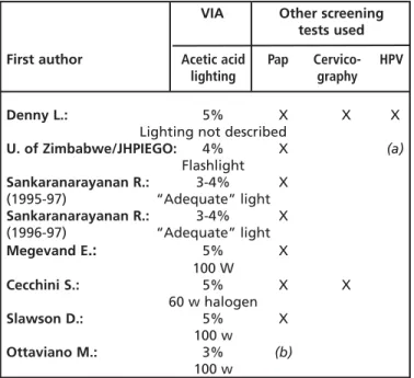
Given that VIA is still under evaluation as a cervical can-cer screening tool, while conventional cytology is a widely accepted screening test, the latter was used con-currently with VIA in all the studies considered here. In the study by Denny a third screening test was also used: HPV testing. In Phase II of the University of Zimbabwe / JHPIEGO study, samples were also collected for HPV test-ing, but the results were not reported in the paper. Two other studies (those by Denny and Cecchini) also incor-porated cervicography as a screening test.
When the reference standard screening test was not systematically applied to all study subjects, the use of additional screening tests often played a major role in determining the coverage with the reference standard diagnostic tool (colposcopy with histology in all studies). The specifics of the application of the VIA method varied across studies, for example with respect to the acetic acid concentration used, which ranged from 3% to5%. None of the authors report how, or whether, acetic acid concentration was controlled during the intervention. Recent studies conducted under field con-ditions have shown that the acidity of acetic acid solu-tion drops dramatically after exposure to air for one week.
VIA Other screening tests used
First author Acetic acid Pap Cervico- HPV lighting graphy
Denny L.: 5% X X X
Lighting not described
U. of Zimbabwe/JHPIEGO: 4% X (a)
Flashlight Sankaranarayanan R.: 3-4% X (1995-97) “Adequate” light Sankaranarayanan R.: 3-4% X (1996-97) “Adequate” light
Megevand E.: 5% X
100 W
Cecchini S.: 5% X X
60 w halogen
Slawson D.: 5% X
100 w
Ottaviano M.: 3% (b)
100 w a. Phase II only; findings not presented.
b. The study subjects had had cervical cytology carried out prior to the study, with normal or abnormal results. The cytologic data were avail-able for comparison to VIA and colposcopic findings.
TABLE 5a. VIA characteristics, other screening tests used
Criteria for positive
or abnormal VIA test results
Denny L.*:Acetowhite lesions, cervical ulcers or cervical growths. Did not grade the severity of lesions. U. de Zimbabwe*/JHPIEGO:Normal: Smooth, pink,
uni-form, featureless
Atypical:Cervicitis (inflammation, red spots), dis-charge, ectropion, polyp, nabothian cyst
Abnormal (positive): White plaques, ulcer, acetowhite epithelium
Cervical cancer (positive):Cauliflower-like growths, fungating mass
Sankaranarayanan R.*: Positivo:Any distinct acetowhite area(s) detected on cervix
Abnormalities: Cervicitis, cervical warts, polyps. Nabothian cysts, bleeeding, erosions, stippled cervix, irregular edematous elongated cervix, growths and ulcers
Negative: No acetowhite area, whitish appearance doubtful or faint, polyps took faint acetowhite stain-ing, nabothian follicles (which are whitish before acetic acid application) became more prominent after acetic acid application, grape-like glands or erosions in the endocervix are slightly paler than ectocervix. Megevand E.*: Positivo:Presence of acetowhite areas on
the cervix
Cecchini S.*: Negativo:No acetowhite areas
Positive:Evidence of acetowhite areas
Slawson D.*: Positivo (abnormal):Acetowhite areas detect-ed outside the transformation zone
Ottaviano M.*: Normal:Original columnar epithelium; physiologic transformation zone (TZ)
Abnormal or atypical transformation zone: White epithelium; other findings (metaplasia, cervicitis, atro-phy, true erosions, polyps)
• Firts author
First Author Sequence of actions
Denny L.: Day 0 1. Pap smear
2. HPV DNA 3. VIA and VIAM 4. Cervicography
Days 2-6 5. If positive VIA or HPV referred for immediate colposcopy
Day 14 If positive Pap and not previously referred for colposcopy tracked and referred for colposcopy
Day 56-76 If positive cervigram and not previously referred for colposcopy tracked and referred for colposcopy
Day 2-76 6. If significant grade lesions at colposcopy lesions electrosurgically excised using loop electrodes
If minor grade lesions at colposcopy biopsy
If no visible lesions endocervical curettage performed U. of Zimbabwe/JHPIEGO Fase I: Day 0 1. Pap smear
2. VIA
Day not specified 3. If VIA showed abnormal result scheduled for colposcopy and biopsy when indicated
Every 10th woman with normal or atypical VIA scheduled for colposcopy and biopsy when indicated
U. of Zimbabwe/JHPIEGO Fase II : Day 0 1. Pap smear 2. VIA 3. HPV DNA
4. Colposcopy and biopsy when indicated
Sankaranarayanan R. (1995-97): Day 0 1. Unaided visual inspection 2. Pap smear
3. VIA
Day not specified 4. If positive VIA or Pap referred for colposcopy
If grossly abnormal-looking cervix referred for colposcopy Day not specified 5. If abnormal colposcopic findings performed directed biopsy Sankaranarayanan R. (1996-97): Day 0 1. Unaided visual inspection
2. Pap smear 3. VIA
Day 3-80 4. If positive VIA or Pap referred for colposcopy
If grossly abnormal-looking cervix referred for colposcopy
Day 3-80 5. If abnormal colposcopic diagnosis directed biopsy obtained from ace-towhite and suspicious areas
If no lesions or features of reparative and reactive changes found on col-poscopy no biopsy
Megevand E.: Day 0 1. Speculum examination
2. VIA 3. Pap smear
Day 0-3 4. If positive VIA or Pap referred for colposcopy
5. If features consistent with HGSIL patient treated with large loop excision of transformation zone
If features consistent with LGSIL punch biopsy of most abnormal area taken
If no abnormal colposcopy histology exam (not defined, but probably endocervical curettage)
Cecchini S.: Day 0 1. Papsmear
2. Cervicoscopy (VIA) 3. Cervicography
Day not specified 4. If abnormal Pap, cervicoscopy or cervicography colposcopically-guided biopsies of all acetowhite areas
Slawson D.: Day 0 1. Pap smear
2. VIA
Day not specified 3. If positive Pap immediate colposcopy
Day 120-180 3. If abnormal VIA or atypical, inflammatory, or negative Pap colposcopy 4. Endocervical curettage performed on all patients with colposcopy
Vaginal sidewalls and vulvar areas also examined and biopsied when indicated
Ottaviano M.:. Day not specified 1. Pap (performed prior to the study)
Day 0 2. VIA and colposcopic exam performed on all women 3. When appropriate, punch biopsy carried out
In addition, the type of lighting used was not con-sistent across studies, nor was it concon-sistently described by the authors. In some studies a halogen light was used, in others a handheld flashlight; in some, a 100-watt light source was used, while in some cases only “adequate” lighting is mentioned (Table 5a). Both the acetic acid concentration and the type of lighting could play a role in identifying acetowhite lesions. Table 5bcomplements
Table 5a,and presents the criteria for a positive test as stated by the authors in their reports. All of the studies mention an acetowhite lesion as a criterion, while only the University of Zimbawe / JHPIEGO and both Sankaranarayanan studies provide additional details. All studies distinguish VIA positive from negative (nor-mal) test results, although the acetowhite lesions are not graded
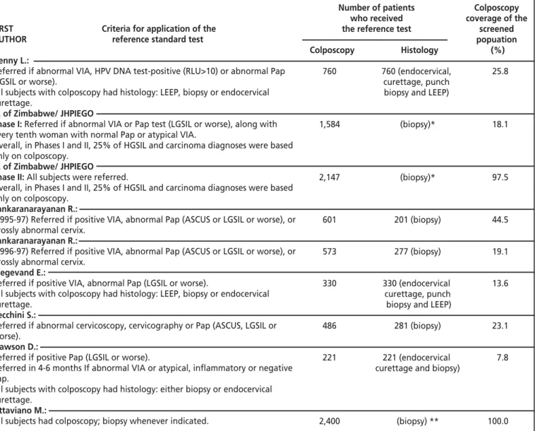
FIRST Criteria for application of the AUTHOR reference standard test
Denny L.:
Referred if abnormal VIA, HPV DNA test-positive (RLU>10) or abnormal Pap (LGSIL or worse).
All subjects with colposcopy had histology: LEEP, biopsy or endocervical curettage.
U. of Zimbabwe/ JHPIEGO
Phase I: Referred if abnormal VIA or Pap test (LGSIL or worse), along with every tenth woman with normal Pap or atypical VIA.
Overall, in Phases I and II, 25% of HGSIL and carcinoma diagnoses were based only on colposcopy.
U. of Zimbabwe/ JHPIEGO Phase II: All subjects were referred.
Overall, in Phases I and II, 25% of HGSIL and carcinoma diagnoses were based only on colposcopy.
Sankaranarayanan R.:
(1995-97) Referred if positive VIA, abnormal Pap (ASCUS or LGSIL or worse), or grossly abnormal cervix.
Sankaranarayanan R.:
(1996-97) Referred if positive VIA, abnormal Pap (ASCUS or LGSIL or worse), or grossly abnormal cervix.
Megevand E.:
Referred if positive VIA, abnormal Pap (LGSIL or worse).
All subjects with colposcopy had histology: LEEP, biopsy or endocervical curettage.
Cecchini S.:
Referred if abnormal cervicoscopy, cervicography or Pap (ASCUS, LGSIL or worse).
Slawson D.:
Referred if positive Pap (LGSIL or worse).
Referred in 4-6 months If abnormal VIA or atypical, inflammatory or negative Pap.
All subjects with colposcopy had histology: either biopsy or endocervical curettage.
Ottaviano M.:
All subjects had colposcopy; biopsy whenever indicated.
Number of patients Colposcopy who received coverage of the the reference test screened
popuation
Colposcopy Histology (%)
760 760 (endocervical, 25.8 curettage, punch
biopsy and LEEP)
1,584 (biopsy)* 18.1
2,147 (biopsy)* 97.5
601 201 (biopsy) 44.5
573 277 (biopsy) 19.1
330 330 (endocervical 13.6 curettage, punch
biopsy and LEEP)
486 281 (biopsy) 23.1
221 221 (endocervical 7.8 curettage and biopsy)
2,400 (biopsy) ** 100.0
* Although all patients positive on colposcopy had a biopsy, the results for all patients were not available at the time of publication. Among 624 positive final diagnoses of HGSIL or cancer in Phases I and II, 74.8% were confirmed by biopsy.
** A punch biopsy was obtained when appropriate; however, the number of patients is not given.
Sequence of tests used for
screening and diagnosis
With the single exception of the Megevand study, the Pap smear was always obtained prior to the application of acetic acid (Table 6).
The time interval between VIA and confirmatory tests varied greatly, ranging from zero days to 6 months. It should be noted that, when a confirmatory test is per-formed months after the initial screening has taken place, it is possible to obtain a result which is discordant with that obtained with the original VIA, because many lesions (particularly, those that are low-grade) may have regressed. (This would have been a false-positive VIA screening test result.) On the other hand, new lesions may also have become evident during the time interval between the tests.
Although VIA provides the opportunity for immedi-ate diagnosis and treatment, only Ottaviano offered immediate diagnoses, and conization was provided to patients with severe dysplasia or carcinoma in situ; the other researchers offered neither immediate diagnosis nor immediate treatment.
Reference standard
Colposcopy was used as a reference standard in all of the studies. Several authors did not conduct any further tests when the colposcopy result was negative; others per-formed a biopsy (endocervical curettage) for histologic examination, even when the colposcopy result was neg-ative.
Regardless of whether colposcopy was conducted alone or was accompanied by a histologic exam, in order to determine the number of true positives in the study group the reference standard test should ideally have been applied to all patients referred for evaluation. However, with the exception of Phase II of the Zimbabwe study, all the other studies only referred for application of the reference standard, the 10%-50% of patients who had abnormal results in at least one of the screening tests. Therefore, all of the studies, except Zimbabwe Phase II, carry some verification bias (6). This
bias skews the estimated sensitivity and specificity of VIA. However, a correction for this can be made by eval-uating a random sample of the screen-negative subjects with the reference standard.
It is also necessary to track how many of the subjects who were intended to receive the reference standard test, actually did. In the present group of studies, between 78% and 100% of the subjects referred for it, received the reference test.
In summary, the proportion of all women studied who actually received the reference standard test depended not only on the criteria used for applying the standard, but also on the rate of compliance with these criteria (Table 7). The last column of Table 7shows the final figure for coverage with a reference standard test in each of the studies considered; it varied from as low as 8.1% (Slawson) to 97.5% (University of Zimbabwe / JHPIEGO Phase II).
Positivity results of VIA and
conventional cytology
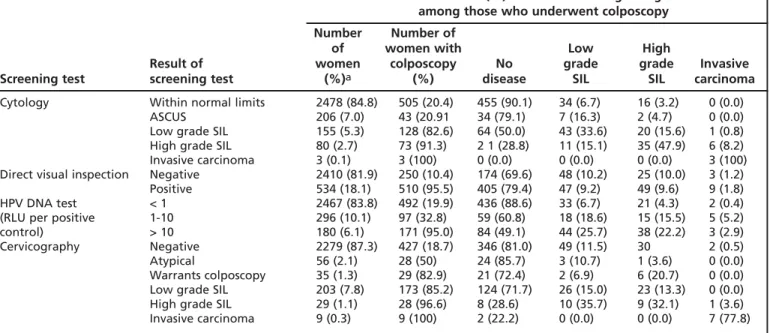
In five of the studies (Denny, both phases of the University of Zimbabwe / JHPIEGO study, Sankaran-arayanan 1995-97, Cecchini), VIA resulted in more posi-tives than conventional cytology, while in two studies (Megevand, Slawson), positivity was higher with con-ventional cytology. In Sankaranarayanan’s second study (1996-1997), both techniques gave similar results (Table 8, Figure 1).
Positivity rates for conventional cytology varied between 1.0% and 15.2% among the eight studies, with a weighted average of 11.1 %. VIA positivity rates var-ied from 3.1% to 39.8%, with a weighted average of 18.2%.
There was a large difference between the two Sankaranarayanan studies as to the VIA positivity rates observed: 37.7% in the study that began in 1995 and
Pap LGSIL Abnormal
and worse VIA
First author No. % No. %
Denny L 238 8.2 534 18.1
U. of Zimbabwe/
I JHPIEGO Phase I 1,218 14.6 1,762 20.2 U. of Zimbabwe/
JHPIEGO Phase II 269 12.6 868 39.8 Sankaranarayanan R.
(1995-97) 205 15.2* 509 37.7
Sankaranarayanan R.
(1996-97) 307 10.2* 298 9.9
Megevand E. 315 13.0 76 3.1
Cecchini S. 21 1.0 341 25.4
Slawson D. 196 7.1* 113 4.2
Average positivity rate, weighted according to
sample size 11,1 18,2
* ASCUS or worse
9.9% in the one that began in 1996. At the same time, the differences in positivity rates (ASCUS or worse) for conventional cytology were not as great: 15.2% and 10.2%, respectively. The lower VIA positivity found in the 1996-97 study may represent a change in the appli-cation of the positivity criteria as a result of experience gained by the provider. On the other hand, in the University of Zimbabwe / JHPIEGO study the positivity rate of VIA doubled (from 20% to 40%) between Phases
I and II while the positivity of conventional cytology was stable (15% and 13%, repectively). The authors attrib-uted this change to the application of less stringent pos-itivity criteria in Phase II, in the interest of maximizing sensitivity.
Positivity by age
The paper by Cecchini is the only one that provides pos-itivity results for VIA, Pap and cervicography classified by
7.1 1.0
13.0 10.2
15.2 12.6
14.6 8.2
4.2
25.4 3.1
9.9
37.7 39.8 20.2
18.1
Slawson D. Cecchini S. Megevand E. Sankaranarayanan R. (1995-97) Sankaranarayanan R. (1996-97) U. of Zimbabwe / JHPIEGO Phase II U. of Zimbabwe / JHPIEGO Phase I Denny L.
Percent
VIA
Papanicolaou
0 10 20 30 40 50
Figure 1. Percent of women with abnormal conventional cytology or abnormal VIA, by study TABLE 9. Estimated(A)VIA accuracy, sample size, and coverage with the reference standard
CINII threshold for positive final diagnosis
Estimated Estimated Accuracy N Coverage with the sensitivity % specificity % score (B) reference standard (%)
Sankaranarayanan R. (1996-97): 90.2 92.2 87.5 3000 19.1
Cecchini S.: 87.5 82.3 78.3 2105 23.1
Sankaranarayanan R. (1995-97): 95.8 67.9 67.6 1351 44.5
Megevand E.: 64.5 97.7 64.4 2426 13.6
U. of Zimbabwe/JHPIEGO Phase I: 65.5 88.7 64.2 8731 18.1
Denny L.: 67.4 83.7 63.6 2944 25.8
U. of Zimbabwe/JHPIEGO Phase II 76.7 64.1 57.2 2203 97.5
Slawson D.: 29.0 97.1 28.9 2827 7.8
Studies are shown in descending accuracy score sequence
A. Sensitivity and specificity can only be estimated, given that most of the studies suffer from verification bias.
B. The accuracy score shows how close to the ideal each study is, where “ideal” is defined as 100% specificity and 100% sensitivity. The formula to obtain the score is: 100 - (SQR ([100-Sensitivity%]2+ [100 –Specificity%]2))
age. Two age groups were used: over 50 years and under 50. Statistically significant lower positivity rates were found for women in the older age group, for all the screening tests. The positivity rates found were: cytology, 5.1% among women under 50 vs. 2.5% among
women over 50; cervicography, 24.3% vs. 3.2%, respec-tively, for those two age groups; and for VIA, 29.0% vs. 20.5%.
In that same study, approximately 50% of the women who had a positive result to any of the tests received a biopsy if colposcopic examination confirmed a lesion. The biopsy confirmation rate for high-grade lesions was similar for both age groups: 1.7% biopsy-confirmed CINII/III among women under age 50 and 1.3% among women over 50. No significant difference was found, with regard to age, in the positive predictive values of the tests.
Accuracy of VIA
Estimates of the sensitivity and specificity of VIA for detecting CINII and worse lesions were calculated for all those studies that provided the data (Table 9, Figure 2). Caution is advisable in comparing the results because, due to differences in study design, from 8% to 98% of the screened population was actually tested with the reference standard. When calculating estimated sensi-tivity and specificity it was assumed that all those women who were not tested with the reference stan-dard were true negatives, which introduced a bias into the estimates.
TABLE 10. Confirmed cases of CIN II or worse that were positive to either VIA or the Pap test and were negative to the other
Pap + Pap
-and VIA- and VIA + First author No. % of all No. % of all
cases cases
Sankaranarayanan R.*
(1996-97): 2 3.9 5 9.8
Sankaranarayanan R.‡
(1996-97): 3 5.9 5 9.8
Cecchini S.*‡: 1 12.5 3 37.5
Sankaranarayanan R.‡
(1995-97) 1 1.4 25 34.5
Megevand E.*: 11 35.5 0 0.0
U. of Zimbabwe/
JHPIEGO Phase I*: 99 24.9 95 23.9 U. of Zimbabwe/
JHPIEGO Phase II*: 14 7.0 80 40.0
Slawson D.‡: 22 71.0 4 12.9
Denny L.: Not mentioned Not mentioned
* Positive Pap: LSIL and above ‡ Pap result: ASCUS and higher
Slawson D. Cecchini S.
Megevand E. Sankaranarayanan R. (1995-97) Sankaranarayanan R. (1996-97)
U. of Zimbabwe / JHPIEGO Phase II U. of Zimbabwe / JHPIEGO Phase I
Denny L.
Percent
Sensitivity
Specificity
0
90,2 92,2
87,5 82,3
95,8 67,9
64,5
97,7
65,5
88,7
67,4
83,7
76,7 64,1
29,0
97,1
20 40 60 80 100
Studies are shown in descending accuracy score sequence
To measure how well VIA distinguishes true nega-tives from true posinega-tives, an “accuracy score” was also calculated for each study (Table 9). This score, which combines sensitivity and specificity to produce a single figure, attempts to facilitate comparison of the results obtained in the different studies. It summarizes the trade-off between, on the one hand, the need for high
sensitivity in a screening test and, on the other, the need for a sufficiently high level of specificity.
Any interpretation of the accuracy scores shown on the table must reflect the fact that those women who were not tested with the reference standard were assumed to be true negatives. This assumption resulted in inflated accuracy scores for VIA, for all the studies
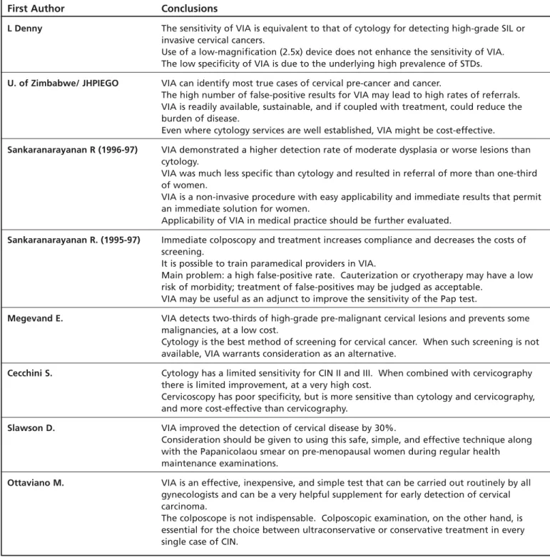
First Author Conclusions
L Denny The sensitivity of VIA is equivalent to that of cytology for detecting high-grade SIL or invasive cervical cancers.
Use of a low-magnification (2.5x) device does not enhance the sensitivity of VIA. The low specificity of VIA is due to the underlying high prevalence of STDs.
U. of Zimbabwe/ JHPIEGO VIA can identify most true cases of cervical pre-cancer and cancer.
The high number of false-positive results for VIA may lead to high rates of referrals. VIA is readily available, sustainable, and if coupled with treatment, could reduce the burden of disease.
Even where cytology services are well established, VIA might be cost-effective.
Sankaranarayanan R (1996-97) VIA demonstrated a higher detection rate of moderate dysplasia or worse lesions than cytology.
VIA was much less specific than cytology and resulted in referral of more than one-third of women.
VIA is a non-invasive procedure with easy applicability and immediate results that permit an immediate solution for women.
Applicability of VIA in medical practice should be further evaluated.
Sankaranarayanan R. (1995-97) Immediate colposcopy and treatment increases compliance and decreases the costs of screening.
It is possible to train paramedical providers in VIA.
Main problem: a high false-positive rate. Cauterization or cryotherapy may have a low risk of morbidity; treatment of false-positives may be judged as acceptable.
VIA may be useful as an adjunct to improve the sensitivity of the Pap test.
Megevand E. VIA detects two-thirds of high-grade pre-malignant cervical lesions and prevents some malignancies, at a low cost.
Cytology is the best method of screening for cervical cancer. When such screening is not available, VIA warrants consideration as an alternative.
Cecchini S. Cytology has a limited sensitivity for CIN II and III. When combined with cervicography there is limited improvement, at a very high cost.
Cervicoscopy has poor specificity, but is more sensitive than cytology and cervicography, and more cost-effective than cervicography.
Slawson D. VIA improved the detection of cervical disease by 30%.
Consideration should be given to using this safe, simple, and effective technique along with the Papanicolaou smear on pre-menopausal women during regular health maintenance examinations.
Ottaviano M. VIA is an effective, inexpensive, and simple test that can be carried out routinely by all gynecologists and can be a very helpful supplement for early detection of cervical carcinoma.
The colposcope is not indispensable. Colposcopic examination, on the other hand, is essential for the choice between ultraconservative or conservative treatment in every single case of CIN.