Effect of angiotensin-(1-7) on
reperfusion arrhythmias in
isolated rat hearts
1Laboratório de Hipertensão, Departamento de Fisiologia e Biofísica,
Instituto de Ciências Biológicas, Universidade Federal de Minas Gerais, 31270-901 Belo Horizonte, MG, Brasil
2Cleveland Clinic Foundation, Cleveland, OH 44122, USA
L.A.A. Neves1,
A.P. Almeida1,
M.C. Khosla2,
M.J. Campagnole-Santos1
and R.A.S. Santos1
Abstract
There is increasing evidence that angiotensin-(1-7) (Ang-(1-7)) is an endogenous biologically active component of the renin-angiotensin system (RAS). In the present study, we investigated the effects of Ang-(1-7) on reperfusion arrhythmias in isolated rat hearts. Isolated rat hearts were perfused with two different media, i.e., Krebs-Ringer (2.52 mM CaCl2) and low-Ca2+ Krebs-Ringer (1.12 mM CaCl2). In hearts perfused with Krebs-Ringer, Ang-(1-7) produced a concentra-tion-dependent (27-210 nM) reduction in coronary flow (25% reduc-tion at highest concentrareduc-tion), while only slight and variable changes in contraction force and heart rate were observed. Under the same conditions, angiotensin II (Ang II; 27 and 70 nM) produced a signifi-cant reduction in coronary flow (39% and 48%, respectively) associ-ated with a significant increase in force. A decrease in heart rate was also observed. In low-Ca2+ Krebs-Ringer solution, perfusion with Ang-(1-7) or Ang II at 27 nM concentration produced similar changes in coronary flow, contraction force and heart rate. In isolated hearts perfused with normal Krebs-Ringer, Ang-(1-7) produced a significant enhancement of reperfusion arrhythmias revealed by an increase in the incidence and duration of ventricular tachycardia and ventricular fibrillation (more than 30-min duration). The facilitation of reperfu-sion arrhythmias by Ang-(1-7) was associated with an increase in the magnitude of the decreased force usually observed during the post-ischemic period. The effects of Ang-(1-7) were abolished in isolated rat hearts perfused with low-Ca2+ Krebs-Ringer. The effect of Ang II (27 nM) was similar but less pronounced than that of Ang-(1-7) at the same concentration. These results indicate that the heart is a site of action for Ang-(1-7) and suggest that this heptapeptide may be in-volved in the mediation of the cardiac effects of the RAS.
Correspondence R.A.S. Santos
Laboratório de Hipertensão Departamento de Fisiologia e Biofísica, ICB, UFMG Av. Antonio Carlos, 6627 31270-901 Belo Horizonte, MG Brasil
Fax: 55 (031) 441-0835/448-1291 E-mail: marrob@oraculo.lcc.ufmg.br
Research supported by FAPEMIG and CNPq. L.A.A. Neves was the recipient of a CNPq fellowship.
Received September 4, 1996 Accepted April 15, 1997
Key words
•Renin-angiotensin system
•Reperfusion arrhythmias
•Angiotensin-(1-7)
•Coronary flow
•Isolated heart
Introduction
A large body of data has been accumu-lated regarding the existence of a local, inde-pendently regulated, myocardial renin-an-giotensin system (RAS) (1,2). Besides the direct modulation of cardiac function through
effects on coronary vascular tone, myocar-dial contractility, neurotransmitter release from sympathetic nerve endings and cardiac growth, the cardiac RAS appears to be in-volved in post-ischemic reperfusion arrhyth-mias (3).
ef-fects of the RAS were attributed solely to the interaction of the octapeptide angiotensin II (Ang II) with its receptors (1). Over the past few years, however, other endogenous bio-logically active products of the RAS have been identified including angiotensin-(1-7) (1-7)) (4,5) and angiotensin-(3-8) (Ang-(3-8)) (6). Among the biologically active end-products of the RAS, the heptapeptide Ang-(1-7) is particularly interesting because it can be formed directly from angiotensin I by an angiotensin converting enzyme (ACE) independent pathway (7-9) and is essentially devoid of effects exerted by Ang II through AT1 receptors including vasoconstriction and
induction of drinking (4,10).
ACE inhibitors have beneficial effects on reperfusion arrhythmias which have been attributed to a reduction of both local Ang II generation and bradykinin degradation (3). However, plasma Ang-(1-7) concentration increases several-fold during treatment with ACE inhibitors (11,12). Furthermore, Ang-(1-7) has been recently reported to produce vasodilation in canine coronary artery rings pre-constricted with the thromboxane A2
analogue U46619 (13) and to increase the release of [3H]norepinephrine from isolated
rat atria (14). These observations suggest that Ang-(1-7) can participate in the pharma-cological effects of ACE inhibitors in the reperfusion arrhythmias. However, there are no data regarding the effects of Ang-(1-7) on ischemic hearts. In this study we evaluated the role of Ang-(1-7) in post-ischemic reper-fusion arrhythmias by determining its ef-fects on isolated rat hearts.
Material and Methods
Male Wistar rats (200-300 g body weight) were decapitated 15 min after intraperito-neal injection of 200 IU heparin. The thorax was opened and the heart was carefully dis-sected and perfused with Krebs-Ringer solu-tion or a low-Ca2+ Krebs-Ringer solution
through a 1.0 ± 0.3 cm aortic stump. The
perfusion fluid was maintained at 37 ± 0.1oC,
with a pressure of 65 mmHg and constant oxygenation (5% CO2 and 95% O2). A force
transducer (model BG-25 g, Gould, Valley View, OH) was attached through a heart clip to the apex of the ventricles to record the contractile force (developed tension) on a Gould recorder (model RS 3200). A dia-stolic tension of 1.0 g was applied to the heart. Electrical activity was recorded with an electrocardiograph (Nihon Kohden, To-kyo, Japan) with the aid of two cotton wicks placed directly on the surface of the right atrium and left ventricle (bipolar lead). Cor-onary flow was measured by collecting the perfusate over a period of 30 s at regular intervals. The composition of the perfusion solutions was as follows: 1) Krebs-Ringer solution: 118.6 mM NaCl, 4.75 mM KCl, 2.52 mM CaCl2, 2.52 mM KH2PO4, 1.17
mM MgSO4.7 H2O, 25.0 mM NaHCO3 and
11.0 mM glucose, and 2) low-Ca2+
Krebs-Ringer solution: 118.6 mM NaCl, 4.75 mM KCl, 1.12 mM CaCl2, 2.52 mM KH2PO4,
1.17 mM MgSO4.7 H2O, 25.0 mM NaHCO3
and 11.0 mM glucose.
The hearts were allowed to equilibrate for 20-30 min and then perfused for an addi-tional 20 min with Krebs-Ringer solution (control group) or Krebs-Ringer solution containing Ang-(1-7) (27, 70 or 210 nM) or Ang II (27 or 70 nM). Each isolated heart was perfused with only one concentration of peptide (N = 3-11 for each concentration). To study the effect of Ang-(1-7) and Ang II on the reperfusion arrhythmias isolated hearts were perfused for an initial 20-min period with 1) Krebs-Ringer solution (control, N = 11), 2) Krebs-Ringer solution containing Ang-(1-7) (27 nM, N = 8) and 3) Krebs-Ringer solution containing Ang II (27 nM, N = 6). Since the biological effects of Ang-(1-7) are considered to be independent of cal-cium, additional hearts were perfused with 4) low-Ca2+ Krebs-Ringer solution (N = 5),
5) low-Ca2+ Krebs-Ringer solution
low-Ca2+ Krebs-Ringer solution containing Ang
II (27 nM, N = 2). Immediately after the 20-min perfusion period, the left anterior de-scending coronary artery was ligated by the method described by Lubbe et al. (15) be-neath the left auricular appendage together with the adjacent veins. The ligature was released after 15 min and reperfusion with different Krebs-Ringer solutions (above) was performed for an additional 30 min. Cardiac arrhythmias were defined as the presence of ventricular tachycardia and/or ventricular fi-brillation after the ligature of the coronary artery was released. In order to obtain a quantitative measurement, the arrhythmias were graded arbitrarily according to their duration considering a duration of 30 min as irreversible arrhythmia. Therefore, the oc-currence of cardiac arrhythmias for up to 5 min was assigned the factor 1, 10 min was assigned the factor 2, 15 min was assigned the factor 3, 20 min was assigned the factor 6, 25 min was assigned the factor 9, and 30 min was assigned the factor 12. A value of 0-12 was thus obtained in each experiment and is denoted “arrhythmia severity index” (ASI) (16,17). Data are reported as means ± SEM. Statistical analysis was performed by the Student t-test or ANOVA followed by the Least Significant Difference test or the Newman-Keuls test, when appropriate. The level of significance was set at P<0.05.
Results
The averaged basal values for contrac-tion force, heart rate and coronary flow in isolated rat hearts (N = 58) perfused with normal Krebs-Ringer solution were 8.35 ± 0.21 g, 264 ± 7 bpm and 7.92 ± 0.35 ml/min, respectively. When the isolated rat hearts (N = 11) were perfused with low-Ca2+
Krebs-Ringer there was, as expected, a decrease in contraction force to 6.61 ± 0.39 g that was not accompanied by changes in heart rate (260 ± 9 bpm) or coronary flow (7.80 ± 0.68 ml/min).
Figure 1 shows the percentage of change in contraction force, heart rate and coronary flow in isolated hearts perfused with normal Ringer solution or normal Krebs-Ringer solution containing different concen-trations of Ang-(1-7) and Ang II. No signifi-cant differences were observed in contrac-tion force (7.60 ± 0.47 g vs 7.76 ± 0.73 g at the end of the equilibration period), heart rate (278 ± 14 bpm vs 260 ± 21 bpm at the end of the equilibration period), or coronary flow (9.03 ± 1.10 ml/min vs 9.12 ± 1.04 ml/ min at the end of the equilibration period) in isolated hearts perfused with normal Krebs-Ringer solution. Perfusion of isolated hearts with normal Krebs-Ringer solution contain-ing Ang-(1-7) did not consistently change
Percent
120
110
100
90
80
70
60
50 Contraction force
Heart rate
Coronary flow Ang-(1-7)
Krebs-Ringer
Krebs-Ringer + 27 nM Ang-(1-7)
Krebs-Ringer + 70 nM Ang-(1-7)
Krebs-Ringer + 210 nM Ang-(1-7)
Krebs-Ringer
Krebs-Ringer + 27 nM Ang II
Krebs-Ringer + 70 nM Ang II Ang II
Percent
120
110
100
90
80
70
60
50
Figure 1 - Contraction force, heart rate and coronary flow in isolated rat hearts perfused with 1) Krebs-Ringer solution (control, N = 9); 2) Krebs-Ringer solution containing Ang-(1-7) concentrations of 27 nM (N = 9), 70 nM (N = 9), or 210 nM (N = 3) and 3) Krebs-Ringer solution containing Ang II concentrations of 27 nM (N = 11), or 70 nM (N = 6). *P<0.05 compared to the end of the equilibration period (Student t-test for paired observations). **P<0.05 compared to the end of the equilibration period and the control group (Student t -test for paired observations and ANOVA followed by the Newman-Keuls -test).
Contraction force
Heart rate
Coronary flow
*
* **
** **
** ** *
* *
In contrast, Ang II produced a dose-de-pendent increase in contraction force (9% and 12% at 27 nM and 70 nM concentra-tions, respectively, Figure 1) and a decrease in heart rate (approximately 20% at both concentrations, Figure 1). In addition, Ang II markedly reduced the coronary flow by 39% and 48% at concentrations of 27 nM and 70 nM, respectively (Figure 1). Under basal conditions no changes in diastolic tension were produced by Ang-(1-7) or Ang II.
Occlusion of the coronary artery in other rat hearts resulted in a comparable flow re-duction (approximately 50%), which was sustained throughout the ischemic period. The flow reduction was associated with a marked decrease in contractility (60%) with-out major changes in diastolic tension. The changes in coronary flow in the hearts per-fused with Ang II were greater than those of the control group since a significant decrease (35%) in flow was already observed before occlusion. A slight decrease in heart rate was also observed during ischemia in all groups (data not shown). Figures 2 and 3 graphi-cally show the percentage of change in coro-nary flow (before, after 15 min of corocoro-nary occlusion and after 30 min of reperfusion) and the percentage of change in contraction force and diastolic tension (before, after 15 min of coronary occlusion and after 5, 10, 20 and 30 min of reperfusion).
Upon reperfusion, coronary flow in-creased to values corresponding to approxi-mately 80% of the values before occlusion. However, upon reperfusion there was an initial further decrease (35%) in contraction force and a significant increase (250%) in diastolic tension in all groups (Figure 2). In addition, following the initial increase in diastolic tension a small decrease was ob-served for all groups (Figure 2). The initial decrease in force was followed by a small gradual increase in the control and Ang II-perfused hearts (Figure 2). In contrast, in the hearts perfused with Ang-(1-7) no apparent recovery was observed (Figure 2).
Figure 2 - Coronary flow (upper), contraction force (middle) and di-astolic tension (lower) in isolated rat hearts perfused with Krebs-Ringer solution (control, N = 11), Krebs-Ringer solution containing 27 nM Ang-(1-7) (N = 8) or Krebs-Ringer solution containing 27 nM Ang II (N = 6) before and after (reperfusion) coronary oc-clusion. Percentage was calcu-lated in relation to the last value of the equilibration period. *P <0.05 compared to the control group (ANOVA followed by the Least Significant Difference test).
Coronary flow (%)
140
Coronary occlusion
min 120
100
80
60
40
20
0
0 10 20 30 40 50 60 70 -10
Before Reperfusion
*
* *
Contraction force (%)
Coronary occlusion
min 120
100
80
60
40
20
0
0 10 20 30 40 50 60 70 -10
Before Reperfusion
* *
Diastolic tension (%)
Coronary occlusion
min 500
400
300
200
100
0
0 10 20 30 40 50 60 70 -10
Before Reperfusion
contraction force or heart rate. However, a concentration-dependent decrease in coro-nary flow of 10%, 18% and 25% was ob-served with Ang-(1-7) concentrations of 27 nM (N = 9), 70 nM (N = 9) and 210 nM (N = 3), respectively (Figure 1).
When the isolated hearts were perfused with low-Ca2+ Krebs-Ringer solution,
addi-tion of Ang-(1-7) or Ang II produced results in basal heart rate (data not shown) and contraction force and coronary flow (Figure 3) similar to those observed with normal Krebs-Ringer. Coronary occlusion in low Ca2+
-perfused hearts also produced a significant decrease in coronary flow associated with a decrease in contraction force without changes in diastolic tension (Figure 3). Upon reper-fusion, the initial changes were also similar to those of normal Krebs-Ringer-perfused hearts except for the changes in diastolic tension that were more modest (Figures 2 and 3). However, different from the data ob-served with normal Krebs-Ringer, after these initial changes there was an increase in con-traction force accompanied by a complete re-covery of diastolic tension for control, Ang II-and Ang-(1-7)-perfused hearts (Figure 3).
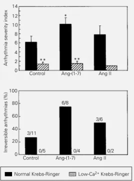
Ventricular tachycardia and/or ventricu-lar fibrillation upon reperfusion were ob-served in all groups. A facilitatory effect of Ang-(1-7) on reperfusion arrhythmias in iso-lated hearts perfused with normal Krebs-Ringer solution was evidenced by a signifi-cant increase of about 60% in ASI (10.62 ± 1.32 vs 6.64 ± 1.29 for the control group; Figure 4). On the other hand, a tendency to an increased ASI was observed in Ang II-perfused hearts (8.00 ± 1.84 vs 6.64 ± 1.29
for the control group, P>0.05; Figure 4). In addition, there was an increase in the occur-rence of irreversible arrhythmias ranging from 27% in control hearts to 75% and 50% in the hearts perfused with Krebs-Ringer solution containing Ang-(1-7) and Ang II, respectively (Figure 4). Perfusion of isolated hearts with Krebs-Ringer containing low Ca2+
reduced the ASI from a value of 6.64 ± 1.29 observed in normal Krebs-Ringer solution to 1.40 ± 0.40, and abolished the occurrence of irreversible arrhythmias. In addition, low-Ca2+ Krebs-Ringer prevented the
ar-rhythmogenic effect of Ang-(1-7) and Ang II (Figure 4).
Coronary flow (%)
140
120
100
80
60
40
20
*
Contraction force (%)
120
100
80
60
40
20
Diastolic tension (%)
Coronary occlusion 500
400
300
200
100
0
Control Ang-(1-7) Coronary occlusion
min 0
0 10 20 30 40 50 60 70 -10
Before Reperfusion
Coronary occlusion
min 0
0 10 20 30 40 50 60 70 -10
Before Reperfusion
min 0 10 20 30 40 50 60 70 -10
Before Reperfusion
Discussion
In the present study we observed that Ang-(1-7) produced a concentration-depend-ent reduction in coronary flow in isolated rat hearts. More important, Ang-(1-7) enhanced
Figure 3 - Coronary flow (upper), contraction force (middle) and diastolic tension (lower) in iso-lated rat hearts perfused with low-Ca2+ Krebs-Ringer solution
(control, N = 5), low-Ca2+
Krebs-Ringer solution containing 27 nM Ang-(1-7) (N = 4) or low-Ca2+
Krebs-Ringer solution containing 27 nM Ang II (N = 2) before and after (reperfusion) coronary oc-clusion. Percentage was calcu-lated in relation to the last value at the equilibration period. *P<0.05 compared to the con-trol group (ANOVA followed by the Least Significant Difference test).
reperfusion arrhythmias. The increase in di-astolic tension and the decrease in contrac-tion force observed during the post-ischemic period in isolated rat hearts were also poten-tiated by Ang-(1-7). These effects resembled those produced by Ang II. However, these two peptides differed significantly in their action on basal force and frequency. Ang II produced a significant increase in force and a decrease in frequency while Ang-(1-7) produced only slight and inconsistent changes in these parameters.
Our data confirm and extend previous observations showing that the heptapeptide Ang-(1-7) is a biologically active end-prod-uct of the renin-angiotensin system (4,5,18, 19). The selectivity of the biological action of Ang-(1-7) reported previously (4,18,20) is apparently also true for isolated rat hearts. Although Ang-(1-7) and Ang II decreased coronary flow, Ang-(1-7) did not signifi-cantly change the contraction force or heart rate, which were significantly modified by Ang II. In addition, Ang-(1-7) was more potent in inducing arrhythmogenesis under our experimental conditions. As pointed out earlier, the selectivity of Ang-(1-7) is
prob-ably related to the absence of phenylalanine at the carboxyl terminus (4).
A vasoconstrictor effect of Ang-(1-7) on the coronary vessels was first reported by Kumagai et al. (21) in isolated hamster hearts. However, it has been recently reported that Ang-(1-7) has a vasodilator effect on canine coronary arteries (13). These contrasting observations may be related to species dif-ferences. In this regard, the vasodilator ef-fect of Ang-(1-7) on the feline hindquarter vascular bed is mainly dependent on the release of nitric oxide (22), while in pithed rats Ang-(1-7) produced systemic vasodila-tation dependent on prostaglandin release (23). Although the mechanism of the vaso-constrictor effect of Ang-(1-7) in the iso-lated rat heart was not investigated a likely explanation is that it depends, at least in part, on the release of coronary vasoconstrictors (14).
The arrhythmogenic effect of Ang II has been attributed to its vasoconstrictor action and to its facilitatory effect on sympathetic neurotransmission (3). Alternatively, Ang II may promote reperfusion arrhythmias by stimulating phospholipase C and/or A2, thus
enhancing the formation of free radicals via the arachidonic acid pathway (24). Our ob-servation that the arrhythmogenic effect of Ang II was absent in the two isolated hearts perfused with low-Ca2+ Krebs-Ringer also
suggests an important role for Ca2+ in this
mechanism. However, these data should be interpreted with caution due to the small number of isolated hearts used. Although we have no data regarding the mechanism of the Ang-(1-7) arrhythmogenic effect, it appears that Ca2+ also plays an important role. This
finding contrasts with a previous study on CRTG3 cells showing that Ang-(1-7) in-duced the release of prostaglandins inde-pendent of Ca2+ mobilization (25). It should
be pointed out, however, that Fura 2, the compound used to study Ca2+ transport in
the study by Tallant et al. (25), could inhibit some types of calcium mobilization such as
Figure 4 - Averaged arrhythmia severity index (upper panel) and percentage of irreversible ar-rhythmias (more than 30 min) (lower panel) upon reperfusion after 15 min of occlusion of the left anterior descending coro-nary artery in isolated rat hearts perfused with normal or low-Ca2+ Krebs-Ringer solution
(con-trol), normal or low-Ca2+
Krebs-Ringer solution containing 27 nM Ang-(1-7) or normal or low-Ca2+ Krebs-Ringer solution
con-taining 27 nM Ang II. Numbers above the bars indicate the inci-dence of irreversible arrhythmias during the reperfusion period. *P<0.05 in comparison to the control group (Student t-test). **P<0.01 compared to the re-spective group submitted to nor-mal Krebs-Ringer solution (Stu-dent t-test).
Arrhythmia severity index
12 10 8 6
4 2 0
100 14
Irreversible arrhythmias (%)
80
60
40
20
0
Control Ang-(1-7) Ang II
Control Ang-(1-7) Ang II 3/11
0/5 6/8
3/6
0/4 0/2
Normal Krebs-Ringer Low-Ca2+ Krebs-Ringer *
that induced by ATP, through IP3 receptors
in megakaryocytes (26). Activation of phos-pholipase A2, as demonstrated in rat
proxi-mal tubular cells (27), and facilitation of sympathetic neurotransmission (14) are also likely mechanisms involved in the Ang-(1-7) arrhythmogenic effects in isolated rat hearts.
We have found that in isolated hearts perfused with normal Krebs-Ringer solution Ang-(1-7) facilitated the decrease in force observed during the post-ischemic period. The absence of this effect in isolated hearts perfused with a low-Ca2+ Krebs-Ringer
so-lution suggests that Ang-(1-7) could be act-ing by facilitatact-ing the increase in cytosolic Ca2+ usually observed during the
reperfu-sion period (28). This increased cytosolic Ca2+ may impair myocardial function by
several mechanisms including impairment of mitochondrial function and activation of Ca2+-ATPases leading to a reduction in
in-tracellular ATP, or by activation of Ca2+
-lipases (28).
We have observed that Ang II produced a dose-dependent positive inotropic effect in Krebs-Ringer-perfused hearts. This finding is in contrast to previous studies showing that Ang II has no inotropic effect on the heart of adult rats (29). Ang II, however, has been reported to stimulate contractility in isolated adult rat ventricular myocytes (30). In addition, Ang II has been reported to produce a direct positive chronotropic effect in cultured neonatal rat heart cells (31). How-ever, we found a negative chronotropic ef-fect, not dose dependent, in rat hearts per-fused with normal Krebs-Ringer solution. No chronotropic effect was observed in hearts perfused with low-Ca2+ Krebs-Ringer
solu-tion.
Similarly to Ang II, Ang-(1-7) produced a reduction in coronary blood flow and fa-cilitated reperfusion arrhythmias whereas its effect on cardiac force under basal condi-tions was not consistent, in contrast to the significant increase in force produced by
Ang II. This selectivity indicates that differ-ent angiotensin receptor subtypes are in-volved in the mediation of the Ang-(1-7) and Ang II effects. All the known actions of Ang II in the heart appear to be mediated mainly by the AT1 receptor subtype (32,33). The
receptor(s) mediating the Ang-(1-7) effects remains to be determined. Although no di-rect data were obtained in this study, recent reports strongly indicate that a selective Ang-(1-7) receptor may exist (4,13). We have recently found that central and peripheral actions of Ang-(1-7) can be blocked by its analog, compound A-779, which does not change the pressor, myotropic or dipsogenic effects of Ang II (20). On the other hand, the cardiovascular effects produced by microin-jection of Ang-(1-7) into the rostral ventro-lateral medulla (4) or its vasodilator effect on canine coronary arteries (13) could not be blocked by AT1 or AT2 angiotensin receptor
antagonists. In contrast, Gironacci et al. (14) have recently reported that the facilitatory effect of Ang-(1-7) on the release of [3H]
norepinephrine in isolated rat atria could be blocked by either DUP 753 or the AT2
an-tagonist PD 123177. This observation sug-gests that the Ang-(1-7) receptor in the rat heart resembles the AT1 receptor subtype
described by Ernsberger et al. (34) in rat mesangial cells.
studies are necessary to elucidate whether the results obtained in our study for isolated rat hearts are also demonstrable in vivo and/
or in other species.
Acknowledgments
We wish to thank Paulo Cesar Nogueira and Kátia Marques for technical assistance.
References
1. Dzau VJ (1988). Cardiac renin-angiotensin system: molecular and functional aspects. American Journal of Medicine, 84: 22-27. 2. Lindpaintner K & Ganten D (1991). The cardiac renin-angiotensin system: an ap-praisal of present experimental and clini-cal evidence. Circulation Research, 68: 905-921.
3. Grinstead WC & Young JB (1992). The myocardial renin-angiotensin system: ex-istence, importance, and clinical implica-tions. American Heart Journal, 123: 1039-1045.
4. Santos RAS & Campagnole-Santos MJ (1994). Central and peripheral actions of angiotensin-(1-7). Brazilian Journal of Medical and Biological Research, 27: 1033-1047.
5. Schiavone MT, Santos RAS, Brosnihan KB, Khosla MC & Ferrario CM (1988). Re-lease of vasopressin from the hypothal-amo-neurohypophyseal system by angio-tensin-(1-7) heptapeptide. Proceedings of the National Academy of Sciences, USA, 85: 4095-4098.
6. Sardinia MF, Hanesworth JM, Krebs LT & Harding JW (1993). AT4 receptor binding characteristics: D-amino acid- and glycine-substituted peptides. Peptides, 14: 949-954.
7. Santos RAS, Brosnihan KB, Chappell MC, Pesquero JL, Chernicky CL, Greene LJ & Ferrario CM (1988). Converting enzyme activity and angiotensin metabolism in the dog brainstem. Hypertension, 11: I.230-I.238.
8. Santos RAS, Brum JM, Brosnihan KB & Ferrario CM (1990). The renin-angiotensin system during acute myocardial ischemia in dogs. Hypertension, 15: I.121-I.127. 9. Santos RAS, Brosnihan KB, Jacobsen
DW, DiCorleto PE & Ferrario CM (1992). Production of angiotensin-(1-7) by human vascular endothelium. Hypertension, 19: II.56-II.61.
10. Timmermans PBMWM, Wong PC, Chiu AT, Herblin WF, Benfield P, Carini DJ, Lee RJ, Wexler RR, Saye JAM & Smith RD (1993). Angiotensin II receptors and an-giotensin II receptor antagonists. Pharma-cological Reviews, 45: 205-251.
11. Campbell DJ, Lawrence AC, Towrie A, Kladis A & Valentijn AJ (1991). Differential regulation of angiotensin peptide levels in plasma and kidney of the rat. Hyperten-sion, 18: 763-773.
12. Lawrence AC, Evin G, Kladis A & Campbell DJ (1990). An alternative strat-egy for the radioimmunoassay of angio-tensin peptides using amino-terminal-di-rected antisera: measurement of eight an-giotensin peptides in human plasma. Jour-nal of Hypertension, 8: 715-724. 13. Brosnihan KB, Li P & Ferrario CM (1996).
Angiotensin-(1-7) dilates canine coronary arteries through kinins and nitric oxide. Hypertension, 27: 523-528.
14. Gironacci MM, Adler-Graschinsky E, Peña C & Enero MA (1994). Effects of angio-tensin II and angioangio-tensin-(1-7) on the re-lease of [3H]norepinephrine from rat atria.
Hypertension, 24: 457-460.
15. Lubbe WF, Daries PS & Opie LH (1978). Ventricular arrhythmias associated with coronary artery occlusion and reperfusion in the isolated perfused rat heart: a model for assessment of antifibrillatory action of antiarrhythmic agents. Cardiovascular Re-search, 12: 212-220.
16. Bernauer W (1986). The effect of ethanol on arrhythmias and myocardial necrosis in rats with coronary occlusion and reperfu-sion. European Journal of Pharmacology, 126: 179-187.
17. Bernauer W & Ernenputsch I (1988). An-tagonistic effects o f α-adrenoceptor blocking agents on arrhythmias, enzyme released and myocardial necrosis in iso-lated rat hearts with coronary occlusion and reperfusion. Naunyn-Schmiedebergs Archives of Pharmacology, 338: 88-95. 18. Campagnole-Santos MJ, Heringer SB,
Batista EN, Khosla MC & Santos RAS (1992). Differential baroreceptor reflex modulation by centrally infused angio-tensin peptides. American Journal of Physiology, 263: R89-R94.
19. Ferrario CM, Barnes KL, Block CH, Brosnihan KB, Diz DI, Khosla MC & Santos RAS (1990). Pathways of angiotensin for-mation and function in the brain. Hyper-tension, 15: I.13-I.19.
20. Santos RAS, Campagnole-Santos MJ, Baracho NCV, Fontes MAP, Silva LCS, Neves LAA, Oliveira DR, Caligiorne SM, Rodrigues ARV, Gropen Jr C, Carvalho WS, Simoes e Silva AC & Khosla MC (1994). Characterization of a new tensin antagonist selective for angio-tensin-(1-7): Evidence that the actions of angiotensin-(1-7) are mediated by specific angiotensin receptors. Brain Research Bulletin, 35: 293-298.
21. Kumagai H, Khosla MC, Ferrario C & Fouad-Tarazi FM (1990). Biological activ-ity of angiotensin-(1-7) heptapeptide in the hamster heart. Hypertension, 15: I.29-I.33. 22. Osei SY, Ahima RS, Minkes RK, Weaver JP, Khosla MC & Kadowitz PJ (1993). Dif-ferential responses to angiotensin-(1-7) in the feline mesenteric and hindquarters vascular beds. European Journal of Phar-macology, 234: 35-42.
23. Benter IF, Diz DI & Ferrario CM (1993). Cardiovascular actions of angiotensin-(1-7). Peptides, 14: 679-684.
24. Linz W, Scholkens BA & Han YF (1986). Beneficial effects of the converting en-zyme inhibitor, ramipril, in ischemic rat hearts. Journal of Cardiovascular Pharma-cology, 8: S91-S99.
25. Tallant EA, Jaiswal N, Diz DI & Ferrario CM (1991). Human astrocytes contain two distinct angiotensin receptor subtypes. Hypertension, 18: 32-39.
26. Akaike N & Uneyama H (1994). ATP-in-duced K+ current oscillation in
megakaryo-cytes: a unique purinoceptor. News in Physiological Sciences, 9: 49-53. 27. Andreatta-Van Leyen S, Romero MF,
Khosla MC & Douglas JG (1993). Modula-tion of phospholipase A2 activity and
so-dium transport by angiotensin-(1-7). Kid-ney International, 44: 932-936.
28. Nayler WG (1981). The role of calcium in the ischemic myocardium. American Jour-nal of Physiology, 102: 262-270. 29. Doggrell SA (1989). The effects of
30. Neyses L & Vetter H (1989). Action of atrial natriuretic peptide and angiotensin II on the myocardium: studies in isolated rat ventricular cardiomyocytes. Biochemical and Biophysical Research Communica-tions, 163: 1435-1443.
31. Allen IS, Cohen NM, Dhallan RS, Gaa ST, Lederer WJ & Rogers TB (1988). Angio-tensin II increases spontaneous contrac-tile frequency and stimulates calcium cur-rent in cultured neonatal rat heart myo-cytes: Insight into the underlying bio-chemical mechanisms. Circulation Re-search, 62: 524-534.
32. Feolde E, Vigne P & Frelin C (1993). An-giotensin II receptor subtypes and biologi-cal responses in the rat heart. Journal of Molecular and Cellular Cardiology, 25: 1359-1367.
33. Wiemer G, Scholkens BA, Wagner A, Heitsch H & Linz W (1993). The possible role of angiotensin II subtype AT2 recep-tors in endothelial cells and isolated i-schemic rat hearts. Journal of Hyperten-sion, 11: S234-S235.