Arq Neuropsiquiatr 2010;68(3):396-399
396
Article
Intramedullary tumors in children
Analysis of 24 operated cases
Ricardo de Amoreira Gepp1, Jose Mauro Cardoso Couto1,
Maria Dorvalina da Silva2, Régis Tavares da Silva1, Eidmar Augusto Neri1
ABSTRACT
Intramedullary tumors are rare. The authors reviewed 24 cases operated between 1996 and 2006. The study assessed the clinical characteristics and surgical results based upon the neurological function. Method: Medical records of patients with intramedullary astrocytoma and ependymoma were reviewed. The minimal follow up time was 6 months and, at the end of this period, a comparative analysis of the neurological function was performed based using the McCormick scale score. Results: Most patients had astrocytoma (75%). Male gender was more prevalent (58.3%). The most common type of tumor was graded as I or II, and in three cases these were malignant. The total resection of the tumor was achieved in 20.8% of the cases. The statistical analysis did not show a statistically significant difference between preoperative and postoperative grades at McCormick scale. Conclusion: The authors concluded that microsurgery to intramedullary tumors did not significantly alter the neurological function after six months.
Key words: astrocytoma, ependymoma, microsurgery, spinal cord.
Tumores intramedulares em crianças: análise de 24 casos operados
RESUMO
Os tumores intramedulares são doenças raras. Os autores analisaram 24 casos operados entre 1996 e 2006. O estudo analisou as características clínicas e o resultado da cirurgia quanto à função neurológica. Método: Foram analisados pacientes com astrocitomas e ependimomas intramedulares. O tempo mínimo de acompanhamento foi de 6 meses e ao final deste período foi realizada a avaliação comparativa da variação do estado neurológico baseado na escala de McCormick. Resultados: A maioria dos pacientes era de astrocitoma (75%). O gênero masculino foi mais prevalente (58,3%). A maioria dos tumores era de grau I ou II, 3 casos eram malignos. A ressecção total do tumor ocorreu em 20,8% dos casos. A avaliação estatística demonstrou que não houve diferença significativa entre o estado neurológico na escala de McCormick pré-operatória e pós-operatória. Conclusões: Os autores concluem que a microcirurgia para ressecção dos tumores intramedulares não ocasionou variação funcional significativa nos pacientes após seis meses da cirurgia.
Palavras-chave: astrocitoma, ependimoma, microcirurgia, medula espinhal.
Correspondence
Ricardo de Amoreira Gepp
SQSW 300 bloco M / apt 204 Sudoeste 70673-014 Brasília DF - Brasil E-mail: [email protected] [email protected]
Received 12 June 2009
Received in final form 19 September 2009
Accepted 19 October 2009 Sarah Network of Rehabilitation Hospitals, Brasília DF, Brazil: 1Neurosurgeon; 2Neurophysiologist.
Spinal cord tumors represent 4 to 8% of central nervous system tumors in the pe-diatric population1. Despite this low
prev-alence, there are doubts about the clini-cal management of these cases. In chil-dren, astrocytomas are 3 times more fre-quent than ependymomas. Due to the slow growth in the natural history of many of these tumors, neurosurgeons usually do not make a deinitive statement regarding
treatment2. In the pediatric population, at
the time of diagnosis, some patients have few symptoms, leading to medical con-cerns regarding radical surgery3.
Advances in surgical techniques and the widespread availability of improved imaging techniques have enhanced the ability of neurosurgeons to perform rad-ical resections of these tumors4.
neuro-Arq Neuropsiquiatr 2010;68(3)
397
Intramedullary tumors in children Gepp et al.
logical outcomes after surgery performed to treat spinal cord tumors in children.
Method
A consecutive series of 24 children, aged 1 to 17 years old at the time of diagnosis, with pathologically conirmed astrocytomas or ependymomas were studied. hese chil-dren were treated at the Sarah Network of Rehabilitation Hospitals, from 1996 until December 2006. Inclusion cri-teria were: age below eighteen years old, diagnosis of in-tramedullary astrocytoma or ependymoma, and a follow up of six months or more. All of these cases were operat-ed by the neurosurgical team at Sarah Hospitals. Patients who were operated at other hospitals, but followed in the rehabilitation spinal cord program, were evaluated by the neurosurgical team at Sarah Hospitals but were excluded from this study. Patients with other types of intramedul-lary tumors or those who had less than six months follow up after the surgical procedure were excluded from this assessment. Prior to surgery, all cases underwent magnet-ic resonance imaging (MRI). Following the surgmagnet-ical proce-dure, all patients underwent MRI scans to assess the vol-ume of the resected tumor. he pictures were studied by independent group of radiologists of Sarah Hospitals.
he children were assessed using the following vari-ables: age at diagnosis, type of the tumor, histological grade (World Health Organization classiication), time of diagno-sis, volume of tumor resection, McCormick grade before and after surgery. he preoperative functional status was assessed by McCormick classic scale. he postoperative outcome grades were performed at one and six months5.
Patients underwent laminectomy and, more recent-ly, laminotomies, whenever the tumor extended to more than two vertebral segments. Both the dorsal myeloto-my and the surgical resection were made using micro-surgical techniques. he tumor dissection was carried out along the cleavage plane with the spinal cord for as far as it could be identiied. Ultrasonic aspiration was used to excavate the tumor from inside out, until its interface with the white matter was reached. Sensory evoked and, recently motor evoked potentials were used routinely for on-line feedback to the surgeon.
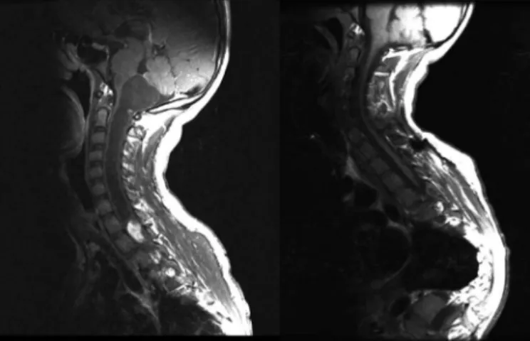
Volume of surgical resection was classiied in biopsy when only 50% of the tumor was resected, or when more than 50% was resected. When less than 90% of the tumor was removed, it was considered to have been a partial re-section. A subtotal resection meant that more 90% of the tumor was resected, but the MRI showed some residual tumor. Total resection was achieved only when MRI did not show any residual tumor (Fig 1).
he statistical analyses were performed by the soft-ware SPSS version 13.0, with the assistance of the statis-tics group of Sarah Hospitals. he Wilcoxon test was used
Fig 1. Preoperative and postoperative MRI showing total resection of the tumor in the cervical spinal cord.
Table. Clinical and surgical characteristics of patients. Clinical features Number Percentage
Astrocytoma Grade I Grade II Grade III
18 8 8 2
75.0% 33.3% 33.3% 8.3% Ependymomas
Grade I Grade II Grade III
6 3 2 1
25.0% 12.5% 8.3% 4.2% Tumor location
Cervical Thoracic Lumbar
8 13 3
33.3% 54.2% 12.5% Age
0 to 3 years 4 to 10 years 11 to 18 years
4 5 15
16.7% 20.8% 62.5% Scoliosis
Yes No
15 9
62.5% 37.5% Sex
Female
Male 1014 41.7%58.3%
Motor deicit Yes
No 195 79.2%20.8%
Tumor resection Biopsy Partial Subtotal Total
0 10 9 5
0% 41.7% 37.5% 20.8%
for analysis of variation in the clinical status after surgery, at one and six months.
Results
Preoperative clinical status
pa-Arq Neuropsiquiatr 2010;68(3)
398
Intramedullary tumors in children Gepp et al.
tients were divided into 18 cases of astrocytomas and 6 cases of ependymomas. he average age of these patients was 11 years old. We divided the ages of patients in three groups. he population in the irst group (0 to 3 years) consisted of only four cases, and most of patients were older than eleven years old (Table). he median time pri-or to the procedure was 14 months, varying from 28 days to 26 months. he Table provides a summary of the main clinical features in our patients. he most common histo-logical type of tumor was astrocytoma grade I. No cases of anaplastic multiform glioblastoma were found in our series. Ependymomas were found only in patients older than 10 years. he most common location for tumors was the thoracic region (Table). Scoliosis was diagnosed in 15 radiological ilms. Motor deicit was identiied in 19 cases at the time of admission. McCormick classiication5 was
performed in all of cases to deine the clinical preopera-tive status and it was used again for comparison with the postoperative status.
Postoperative clinical status
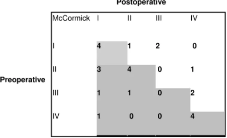
All of these 24 patients were submitted to surgical treatment. Biopsies were not performed in 10 cases in which only a partial resection was achieved. Total resec-tion was done in 5 cases and subtotal in 9 children (Table). he McCormick scale was used to assess the surgical results and the impact of the procedure in the clinical sta-tus at one and six months after surgery (Fig 2). Most the patients maintained the same neurological function as in the preoperative assessment. For the group of children with preoperative grade 1 or 2 on the McCormick scale, three cases presented a worse grading in the postopera-tive assessments. Patients with preoperapostopera-tive grades 3 or 4 kept the same clinical status, with exception of three cas-es that showed improvement. In one case, a child with paraplegia, the ability to walk was recovered after sur-gery (Fig 2)
he statistical analysis performed by non parametric test showed no diference in the preoperative and post-operative groups (p=0.773), six months after the surgery. his result showed that the patients kept the same neu-rological grade after the surgical procedure, and the sur-gery in itself was not responsible for any change in neu-rological function.
discussion
Intramedullary tumors are rare in children, with an annual incidence of about one case per million. Astro-cytomas and ependymomas are the most frequent in-tramedullary tumors, accounting for about 80% of all tu-mors in this area1,6. For the pediatric population, there is
an increased number of spinal cord tumors in compari-son with other spinal neoplasms7.
Surgical treatment is the initial approach for treat-ing spinal cord tumors, with the objective of resecttreat-ing most of the tumor, while attempting to keep the patient in the same neurological function grade8. Epstein published
his work in spinal cord tumors showing the possibility of complete resection in most of 75% of his cases7-9.
Nad-Kani and Rekate performed a systematic review of this subject, and no evidence was found that total resection of spinal cord tumors could improve de survival rates1.
he French multicenter study showed two prognos-tic factors: time to the diagnosis (relative risk – 4.93) and histological type of tumor (relative risk – 7.69)10. The
other variables analyzed in that study did not show sta-tistical diference, and gross total removal of intramed-ullary tumors did not correlate with increased survival rates10. More recently, another study from England with
453 adults patients showed that extension of tumor re-section did not correlate with improvement in survival rates11. We believe that the aim of surgery in
intramed-ullary tumors should be radical removal, although this is possible only in cases where there is cleavage plane. A clear cleavage plane is usually found in ependymomas, where radical tumor removal should be the objective of surgery5. Total removal is rarely possible in astrocytomas
which, even when viewed under a microscope, rarely have a clear cleavage plane5. his is the reason for the better
surgical outcome from spinal ependymomas in compar-ison with spinal astrocytomas.
he limitations on the use of radiotherapy and chemo-therapy, as well as the fact that there is no clear evidence that these treatments may improve outcomes in children, have made surgery the best option for treatment of spinal cord tumors1,12,13. Concerns regarding neurological function
are a very important issue. Most cases are astrocytomas grade 1 or 2, and the survival rate in this type of tumor is very good, so the clinical status after surgery in these pa-tients will be of great importance to their quality of life10,14.
Arq Neuropsiquiatr 2010;68(3)
399
Intramedullary tumors in children Gepp et al.
In the present study, it was observed that, after 6 months, the patients still maintained the same function-al neurologicfunction-al grade on the McCormick scfunction-ale. No statis-tical diference was noticed in the preoperative and post-operative groups. he present study used the McCormick scale to compare the neurological status, since this is the most common method used in the literature to assess clinical function after surgical resection5,15.
One hundred cases of spinal tumors were studied by Ferreira et al16. In this series of cases from Porto Alegre,
ependymomas (4%) and astrocytomas (2%) were report-ed16. he pediatric population difers from the adult cases.
Taricco et al published results from a series of forty-eight patients with primary spinal cord tumors. he thoracic cervical cord was most common site involved, and mi-crosurgical removal was achieved in 71% of the patients without added neurological morbidity17. Ependymoma
was the most common tumor found in their paper. his is an important diference, since the pediatric population presented astrocytoma as the most frequent tumor, and resection of these lesions is more complex. Koerbel et al studied 35 patients with spinal cord tumors18. Total
re-section was more often obtained among patients with ependymomas (13 out of 17) than with astrocytomas (5 out of 12). However, the degree of resection and tumor histology did not interfere in the postoperative clinical morbidity, although the authors found that the tumors located at the thoracic level were associated with higher morbidity (p=0.021)18.
his study collected 24 cases with spinal cord tumors in pediatric patients. here is no comparable series of cases in the Brazilian literature, and the present results showed the importance of surgery, which did not alter the neurological function of the patients.
In conclusion, the surgical resection did not alter the neurological function six months after surgery. Early diag-nosis and treatment improved the surgical morbidity rate. he strongest predicting factor of functional outcome was the preoperative clinical status, as assessed by the McCor-mick scale. he neurosurgical team aimed to remove most of the tumor, while maintaining neurological function.
he larger numbers of patients followed for longer periods is helping to clarify the results from surgery for spinal cord tumors. It is important to emphasize that the natural history of these tumors is variable, afecting even patients who were considered to have had total tumor re-moval. With longer follow-up periods, the natural histo-ry and the efect of the surgical treatment may become clearer in the future.
RefeRences
1. Nadkarni TD, Rekate HL. Pediatric intramedullary spinal cord tumors. Critical review of the literature. Childs Nerv Syst 1999;15:17-28.
2. Kothbauer KF. Neurosurgical management of intramedullary spinal cord tu-mors in children. Pediatr Neurosurg 2007;43:222-235.
3. Miller DC. Surgical pathology of intramedullary spinal cord neoplasms. J Neu-rooncol 2000;47:189-194.
4. Steinbok P, Cochrane DD, Poskitt K. Intramedullary spinal cord tumors in chil-dren. Neurosurg Clin N Am 1992;3:931-945.
5. McCormick PC, Torres R, Post KD, Stein BM. Intramedullary ependymoma of the spinal cord. J Neurosurg 1990;72:523-532.
6. Constantini S, Houten J, Miller DC, et al. Intramedullary spinal cord tumors in children under the age of 3 years. J Neurosurg 1996;85:1036-1043. 7. Epstein FJ. Spinal cord tumors in children. J Neurosurg 1995;82:516-517. 8. Constantini S, Miller DC, Allen JC, Rorke LB, Freed D, Epstein FJ. Radical
exci-sion of intramedullary spinal cord tumors: surgical morbidity and long-term follow-up evaluation in 164 children and young adults. J Neurosurg 2000; 93(Suppl 2):S183-S193.
9. Epstein F, Epstein N. Surgical treatment of spinal cord astrocytomas of child-hood: a series of 19 patients. J Neurosurg 1982;57:685-689.
10. Boufet E, Pierre-Kahn A, Marchal JC, et al. Prognostic factors in pediatric spi-nal cord astrocytoma. Cancer 1998;83:2391-2399.
11. Tseng JH, Tseng MY. Survival analysis of 459 adult patients with primary spi-nal cancer in England and Wales: a population-based study. Surg Neurol 2007;67:53-58.
12. Robinson CG, Prayson RA, Hahn JF, et al. Long-term survival and function-al status of patients with low-grade astrocytoma of spinfunction-al cord. Int J Radiat Oncol Biol Phys 2005;63:91-100.
13. Lowis SP, Pizer BL, Coakham H, Nelson RJ, Boufet E. Chemotherapy for spi-nal cord astrocytoma: can natural history be modiied? Childs Nerv Syst 1998; 14:317-321.
14. Goh KY, Velasquez L, Epstein FJ. Pediatric intramedullary spinal cord tumors: is surgery alone enough? Pediatr Neurosurg 1997;27:34-39.
15. Tihan T, Chi JH, McCormick PC, Ames CP, Parsa AT. Pathologic and epidemi-ologic indings of intramedullary spinal cord tumors. Neurosurg Clin N Am 2006;17:7-11.
16. Ferreira NP, Chaves DL, Moraes AC, de Oliveira LM. Spinal tumor: apropos of 100 cases. Arq Neuropsiquiatr 1981;39:25-31.
17. Taricco MA, Guirado VM, Fontes RB, Plese JP. Surgical treatment of prima-ry intramedullaprima-ry spinal Cord tumors in adult patients. Arq Neuropsiquiatr 2008;66:59-63.