Arq Neuropsiquiatr 2008;66(1):90-92
90
Median (third) occipital condyle causing
atlantoaxial instability and Myelopathy
Nicandro Figueiredo
1, Laryssa Brito Moraes
2, Alexandre Serra
3,
Sérgio Castelo
4, Douglas Gonsales
2, Roger Rotta Medeiros
3terceiro côndilo occipital causando instabilidade atlantoaxial e Mielopatia
1 MD, MSc, Professor of Neuroanatomy and Neurosurgery, Federal University of Mato Grosso, Cuiaba MT, Brazil; 2Medical-Student, University of Cuiaba,
Cuiaba MT, Brazil; 3 MD, Professor of Neuroanatomy and Neurosurgery, University of Cuiaba, Cuiaba MT, Brazil; 4 MD, Professor of Neuroradiology,
University of Cuiaba, Cuiaba MT, Brazil.
Received 9 August 2007, received in inal form 15 October 2007. Accepted 22 November 2007.
Dr. Nicandro Figueiredo – Avenida das Flores 941 / 1st loor / Instituto Neurológico e da Coluna Vertebral - 78043-172 Cuiaba MT - Brasil. E-mail:
Abnormalities of the craniovertebral junction are of great interest not only to anatomists but also to clinicians, because many of these malformations can produce neu-rological symptoms and death. Median occipital condyle is presented in postmortem anatomic studies, but there is a lack of clinical study regarding this anomaly in the literature1-3. Considering the segmental complexities
in-volved in the phyletic and developmental establishment of the normal human craniovertebral articulations, the ocasional ocurrence of anomalous separations, fusions, and intercalated ossicles should not be surprising4. One
of these manifestations of variant segmentation is the third condyle (basilar tubercle). This structure occurs as a projection on basion (anterior central point) of the fo-ramen magnum. Some incidences are expressed as a sim-ple rounded tubercle, but in more developed cases there is actually an articular facet that receives the tip of the odontoid process forming a true diarthrosis. In a series of 600 skulls, some suggestions of craniovertebral malfor-mations, including a third condyle, was present in 14 per cent5. The mamalian apical odontoid element is the
phy-logenetic equivalent of the proatlas of reptiles. In most members of this vertebrate class this proatlas fuses to the occiput of the skull, whereas in mammals it becomes fused to the atlantal contribution of the odontoid6.
We therefore report a case of a patient with median occipital condyle, associated to transverse ligament laxi-ty and atlantoaxial instabililaxi-ty (AAI), who had mild symp-toms few months before a sudden tetraplegia.
case
A 45-year-old man, Japanese descendant, had a sudden tet-raplegia unleashed after a fast ride down a roller coaster. At that instant, the patient stated that he felt that he had a “neck cracking” and at the moment the car stopped he felt syncope for some seconds; faintness; scotoma; neck pain; dyspnea and
quadriplegia. He was immediately taken to a local hospital by the paramedics, where he was evaluated, submitted to some ex-ams, like X-ray, CT scan and MRI of the cervical spine. His cervi-cal motion was partially restricted by a rigid cervicervi-cal brace, he received initial medical care, and started to recover his neuro-logical functions. The family contacted us, and the patient was transferred by airplane to our hospital, two days later. The pa-tient had a history of some episodes of paresthesias affecting his four limbs, about eight months earlier. He also had some previ-ous neck and lumbar pain, restriction of neck movements, con-stipation and sleeplessness.
Physical examination made in our hospital revealed: tetrapa-resis (classiied as ASIA D), worse on his left side, increased pro-found relex on four limbs; hypoesthesia below the neck (about C3 cord level); feet clonus, and bilaterally Babinski’s sign.
We immediately installed a halo vest to restrict his cervical motion, and then we completed the study with the CT scan in-volving the craniovertebral junction.
Arq Neuropsiquiatr 2008;66(1)
91
Median occipital condyle Figueiredo et al.
Image study
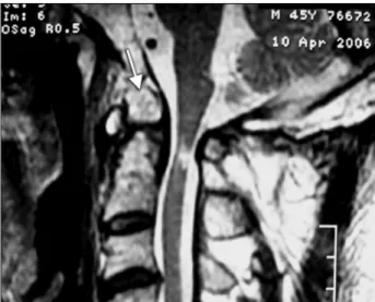
The initial X-ray was inconclusive, and the CT scan he brought, including only the levels below C3, showed no severe abnormality. His MRI showed an anomalous bone element, sug-gesting initially an odontoid fracture, but with the lack of bone edema on T2 (fat supression image), it could not be an acute fracture line. The dens was relatively short, with a thick and lac-cid transverse ligament, compatible with atlanto-axial chronic instability. The spinal cord was narrow at the C1-2 level, with al-tered signal, compatible with chronic edema or myelomalacia, and not with an acute contusion (Figs 1 and 2).
After the external immobilization, we repeated the CT scan, including the upper cervical levels (C1-2), when we saw the anomalous bone, connected with the occipital bone, conirming the presence of the third occipital condyle, together with odon-toid hypoplasia and atlanto-axial chronic instability (Fig 3).
We also ordered a CT scan with angiogram, to better study and plan the surgery, according to the osseous-vascular anatomy.
The patient was transferred to the intensive care ward of our hospital, and we immediately installed a halo vest to restrict his
cervical motion, and then we completed the study with the CT scan involving the craniovertebral junction. Then, we analyzed carefully all the images and when we conirmed the diagnosis of the third condyle and AAI, we discussed the case with the pa-tient, and proposed the surgery, mainly because of the cranio-vertebral malformation and the ligamentous instability.
One month later, we performed a C1-2 posterior screw-rod ixation7 and removed the central part of the posterior arc of C1 to decompress the canal, with the patient in prone position, under general anesthesia, and intraoperative neuromonitoring (SSEP). We introduced two screws at the lateral mass of atlas, and two more at the pedicle of the axis. Because the C2 root was too close to the lateral mass of C1, and the space between C1 and C2 was too narrow, we had to cut the nerve bilaterally. We took autograft from his iliac bone, and we added it at the C1-2 posterior area for bone fusion.
The patient had excellent recovery, and was discharged from the hospital three days later, wearing a cervico-thoracic brace (Minerva) for 20 days. He resumed his previous usual activities about 45 days after surgery, and he wore a Philadelphia collar for 3 months after surgery. He was followed clinically and radiologi-cally for 6 months, with X-ray and CT scan of the cervical region, and he had excellent clinical and radiological results. He still has some numbness in his right hand, increased profound relex of four limbs, but he’s working and driving by himself.
discussion
Atlantoaxial instability may result for many reasons, including hypoplasia of the odontoid process and from laxity of the transverse ligament8. The patient we
de-scribed, had a large third occipital condyle, associated with a hypoplastic odontoid process and laxity of the transverse ligament, according to the exams. These ab-normalities resulted in AAI, which caused a mild chronic spinal cord impingement, and a sudden cord lesion when he was going down fast on the roller coaster.
Atlantoaxial subluxation (AAS) is deined as the situ-ation in which the atlas is unstable relative to the axis due to the disruption or insuficiency of the ligamentous complex of the atlantoaxial joint. This pathogenesis has many causes, including congenital diseases. The cause of congenital craniovertebral junction varies. In patients with C1-2 subluxation, what probably transiently happened to our patient, the condition is likely related to ligamentous laxity. Osseous malformation of the dens region may be accounted for abnormal ligamentous laxity due to dis-turbances in the blood supply to the developing bone as a result of inordinate mobility9,10. In contrast, trauma is a
relatively uncommon cause of such instability, and the pathomechanics are reported to be due to lexion, dis-traction and rotation11-15. Miyamoto et al described a rare
case of AAS that occurred with a professional rugby ath-lete whose mechanism was in a hiperlexion manner16.
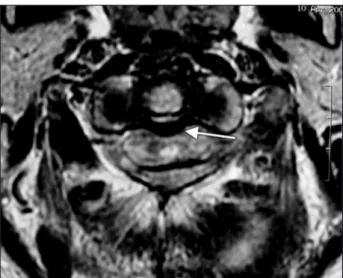
Fig 2. Axial MRI on T2 of C1-C2 level:, show a thick and laccid trans-verse ligament, compatible with atlanto-axial chronic instability, the spinal cord was narrowed at C1-2 level, with altered signal, compati-ble with chronic edema or myelomalacia.
Arq Neuropsiquiatr 2008;66(1)
92
Median occipital condyle Figueiredo et al.
Kotil et al.1 also reported a rare case of a 40-year-old
woman presenting myelopathy related to condylus oc-cipitalis located in the anterior foramen magnum region. They performed a posterior decompression without fu-sion, which they concluded not to be effective.
The C1-2 posterior screw for AAI has several advan-tages include the following: it produces high fusion rates; it allows intraoperative direct reduction, if necessary; C2 pedicle screws provide a higher margin of safety and in-creased lexibility in altered anatomy; it does not require halo immmobilization; it requires only a small incision with minimal dissection; it also avoids dangerous sublami-nar wire passage7,17,18.
In conclusion, we have reported a rare case of an adult who had a sudden tetraplegia during a roller coaster ride, probably related to an AAI due to transverse ligamentous laxity, associated to a congenital malformation of the cra-niovertebral junction, a median occipital condyle. The pa-tient had a good neurological recovery. He was initially immobilized in a halo device, and submitted to C1-2 pos-terior screw-rod ixation, with an excellent result.
Acknowledgments – We would like to thank CEDIC (Cen-tro de Medicina Diagnóstica), which provided the main images for this paper, as well as Mr. John Hall for his trans-lation of the text.
reFerences
1. Kotil K, Kalayci M. Ventral cervicomedullary junction compression sec-ondary to condylus occipitalis (median occipital condyle), a rare entity. J Spinal Disord Tech 2005;18:382-384.
2. Prasada Rao PVV. Median (third) occipital condyle: case report. Clin Anat 2002;15:148-151.
3. Ludinghausen M, Schindler G, Kageyama I, Pomaroli A. The third oc-cipital condyle, a constituent part of a median occipito-atlanto-odon-toid joint: a case report. Surg Radiol Anat 2002;24:71-76.
4. Parke WW. Development of the spine. In Rothman-Simeone (Ed). The Spine. Philadelphia: Saunders 1999:3-27.
5. Lang J. Clinical anatomy of the head. Berlin: Springer-Verlag, 1983.
6. Sensing EC. The origin of the vertebral column in deermouse,
Peromys-cus maniculatus ruinus. Anat Rec 1943;86:123-141.
7. Harms J, Melcher RP. Posterior C1-2 fusion with polyaxial screw and rod ixation. Spine 2001;26:2467-2471.
8. Shaffrey CI, Chenelle AG, Abel MF, Menezes AH, Wiggins GC. Anat-omy and physiology of congenital spinal lesions. In Benzel EC (Ed). Spine surgery: techniques, complication avoidance, and management.
2nd Ed. Amsterdam: Elsevier 2005:61-87.
9. Crockard HA, Stevens JM. Craniovertebral junction anomalies in in-herited disorders: part of the syndrome or caused by the disorder? Eur J Pediatr 1995;154:504-512.
10. Stevens JM, Kendall BE, Crockard HA, Ransford A. The odontoid pro-cess in Morquio-Brailsford’s disease: the effects of occipitocervical fu-suion. J Bone Joint Surg Br 1991;73:851-858.
11. Huang CI, Chen IH, Lee LS. Traumatic atlantoaxial distractive instabil-ity: case report. J Trauma 1994;36:599-600.
12. Carrol EA, Gordon B, Sweeney CA, et al. Traumatic atlantoaxial dis-tractive injury: case report. Spine 2001;26:454-457.
13. Fielding JW, Hawkins RJ. Atlanto-axial rotatory ixation. J Bone Joint Surg Am 1977;59:37-44.
14. Moore KR, Frank EH. Traumatic atlantoaxial rotatory subluxation and dislocation. Spine 1995;20:1928-1930.
15. Wise JJ, Cheney R, Fishgrund J. Traumatic bilateral rotatory dislocation of the atlanto-axial joints: a case report and review of the literature. J Spinal Disord 1997;10:451-453.
16. Miyamoto H, Doita M, Kotaro N et al. Traumatic anterior atlantoaxial subluxation occurring in a professional rugby athlete: case report and review of literature related to atlantoaxial injuries in sports activities. Spine 2004;29:61-64.
17. Melcher RP, Harms J. C1-2 posterior screw-rod ixation. In Bradford DS,
Zdeblick TA (Eds). The spine. 2nd Ed. Lippincott Williams & Wilkins
2004:129-145.