Arq Neuropsiquiatr 2006;64(1):149-152
1Chefe do Serviço de Neurocirurgia do Hospital Municipal Souza Aguiar, Rio de Janeiro RJ, Brasil. Ex-neurocirurgião da Policlínica
de Botafogo, Rio de Janeiro RJ, Brasil;2Chefe do Serviço de Clínica Médica e Cardiologia da Policlínica de Botafogo, Rio de Janeiro
RJ, Brasil;3Estudante de Graduação da Escola de Medicina e Ciru rgia da Universidade Federal do Estado do Rio de Janeiro (UNIRIO),
Rio de Janeiro RJ, Brasil; 4Serviço de Patologia da Policlínica de Botafogo, Rio de Janeiro RJ, Brasil.
Received 6 May 2005, received in final form 17 August 2005. Accepted 7 October 2005.
Dr. José Fernando Guedes Corrêa - Rua Santa Clara 303/404 - 22041-010 Rio de Janeiro RJ - Brasil.
INTRAMEDULLARY SPINAL CYSTICERCOSIS
SIMULATING A CONUS MEDULLARIS TUMOR
Case report
José Fernando Guedes-Corrêa
1, Ricardo Caratta Macedo
2, Rafael Pereira Vaitsman
3,
Jorge Gomes de Mattos
4, Jovita Marques Agra
4ABSTRACT - Cysticercosis is an endemic condition in many developing countries. Although it is the most common parasitic disease of the central nervous system, cysticercal involvement of the spinal cord is rare. It may occur as intradural extramedullary, intramedullary, intramedullary associated with intradural-e x t r a m intradural-e d u l l a ry or as thintradural-e vintradural-ertintradural-ebral printradural-esintradural-entation. Wintradural-e rintradural-e p o rt thintradural-e casintradural-e of a 53-yintradural-ear-old woman who printradural-e s intradural-e n t-ed with low back pain of acute onset and no other symptoms. Magnetic resonance imaging (MRI) showt-ed an intramedullary cyst of the conus medullaris region which, at pathological examination, was diagnosed as a cysticercal cyst. She refused anticysticercal agents and steroids postoperatively. After an eight-year fol-low-up, the patient performs the activities of her daily living with no difficulties, and annual spinal MRIs show no residual signs of the disease. Clinical, pathofisiological, diagnostic and therapeutic aspects of spinal cord intramedullary cysticercosis are discussed.
KEY WORDS: conus medullaris, cysticercosis, intramedullary cysticercosis, spinal cord.
Cisticercose intramedular simulando tumor do cone medular: relato de caso
RESUMO - Cisticercose é uma doença endêmica em vários países em desenvolvimento. Embora seja a doença parasitária mais freqüente do sistema nervoso central, o acometimento medular por cisticercos é raro. Pode o c o rrer nas formas intradural extramedular, intramedular isolada, intramedular em associação com intradu-ral extramedular, além da forma vertebintradu-ral. Relatamos o caso de mulher de 53 anos de idade que se apre-sentou com dor lombar de início agudo, sem outros sintomas. Ressonância magnética (RM) identificou imagem cística na região do cone medular que, no estudo histopatológico, foi diagnosticada como cisticer-co. A paciente recusou tratamento pós-operatório com anti-helmínticos e corticosteróides. Após oito anos de seguimento, a paciente exerce suas atividades quotidianas sem dificuldades, e estudos de RM a n u a i s não mostram sinais de doença residual. Aspectos clínicos, fisiopatológicos, diagnósticos e terapêuticos da cisticercose intramedular são discutidos.
PALAVRAS-CHAVE: cone medular, cisticercose, cisticercose intramedular, medula espinhal.
C y s t i c e rcosis is the infection caused byC y s t i c e rc u s cellulosae,the larvae of the tapewormTaenia soli -um,which affects humans mainly by accidental inges-tion of eggs containing infective oncosfere s1 - 4. It is
the most common parasitic disease of the central ner-vous system1 - 3 , 5 - 1 3, and its related mortality rates range
f rom 6 to 50%1 2. It is an endemic condition to
Bra-z i l8 , 1 2, Peru1 4, Mexico1 , 1 2, Korea, India1 1 , 1 2and other
South American, Tropical African, and Southeast A-sian countries1 , 6. Its frequency in developed countries
is increasing, as migration rates increase from ende-mic are a s3 , 6 , 1 2 , 1 5. Cysticercal involvement of the spinal
c o rdis rare1 , 4 - 8 , 1 1 , 1 5 - 2 1, even in endemic are a s1, and
ac-counts for 0.7 to 5.85% of all cases2 , 8 , 1 0 - 1 3 , 1 5 , 1 9 , 2 1 - 2 3. Its
prevalence may be underestimated, since brain cys-t i c e rcosis, which is a more common condicys-tion, fre-quently occours concomitantly5 , 8 , 1 5 , 1 8 , 2 1 , 2 4, spinal canal
is not routinely examinated in necro p s i e s1 , 8 , 2 0 , 2 1 , 2 4a n d
small asymptomatic cysts may be easily overlooked1.
o-150 Arq Neuropsiquiatr 2006;64(1)
sis is more prevalent than intramedullary cysticerc o-sis (IC)5 , 6 , 1 1 , 2 0 , 2 1 , 2 3and than the association of
intrame-d u l l a ry anintrame-d intraintrame-dural-extrameintrame-dullary pre s e n t a t i o n s (54% vs. 17% vs. 17%, re s p e c t i v e l y )2 5. Extradural
loca-tion is even more rare1 , 5 , 6 , 8 , 1 5 , 1 6 , 2 0 , 2 1 , 2 5. The vertebral
ty-pe has also been reported2,22.
We report the case of a 53-year-old woman who p resented with low back pain of acute onset and no other symptoms. Magnetic resonance imaging (MRI) revealed an intraparenchymal cyst of the conus medu-llaris region which, at pathological examination, was diagnosed as a cysticercal cyst. Based on a brief re v i e w of the literature, clinical, pathofisiological, diagnos-tic and therapeudiagnos-tic aspects of IC are discussed.
CASE
An otherwise healthy 53-year-old woman wakes up in the early morning with intense incapacitating low back pain with lower limbs irradiation. No predisposing condtion for cysticercal infeccondtion was present, and she is re s i-dent in a non-endemic region of Rio de Janeiro, at the
Sou-theast of Brazil. At the Emergency Department she pre-sented afebrile with stable vital signs and normal mental status. General, ophthalmologic and neurologic examina-tions, routine blood, urine analysis, plain x-ray films of her lumbosacral spinal cord and chest were normal. There were no signs of subcutaneous nodules, nor meningeal irr i t a-tion. As pain relieved after non-steroidal anti-inflammato-ry drugs administration, she was discharged for ambulato-ry investigation and follow-up. Twelve hours after hospi-tal discharge, she re t u rned with re c u rrence of the symp-toms. Again, no abnormallity was noted on physical and laboratory examinations.
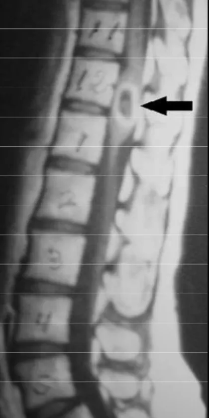
The patient underwent lumbosacral spine MRI, which showed a cystic lesion in the conus medullaris. On sagittal T1-weighted image (Fig 1), cyst fluid was isointense to cere-b rospinal fluid (CSF) and there was a hyper- to isointense nodule attached to the interior of the cyst wall. Further ima-ges were not available for publication. As definitive diag-nosis was missing, pain rapidly worsened and did not re s o l v e with pro g ressive more potent analgesic agents, an ord i n a ry T11 to T12 laminectomy was perfomed 36 hours after the first episode. Dural sac was tough and tense (Fig 2A). After longitudinal dural opening at posterior midline, an enlarg e d
Fig 1. Sagittal T1-weighted MRI of the spine showing an intramedullary well delimitating cyst at the conus medul laris region (arrow). Cyst fluid is isoin tense to CSF and the scolex is visual -ized as a hyper- to isointense nodule attached to the interior of the cyst wall.
Arq Neuropsiquiatr 2006;64(1) 151
spinal cord was visualized (Fig 2B). Five milliliters of a xantho-c h romixantho-c fluid were then aspirated with punxantho-cture at spinal posterior midline, leaving some fluid inside the cyst. Fluid examination did not reveal malignant cells. Under micro-scope vision and with micro s u rgical techniques, myelotomy was made at the posterior median sulcus (Fig 2C), followed by extirpation of the cyst (Fig 2D). Histological findings at the operating room showed a non-tumoral, inflammatory p rocess. Postoperative histological diagnosis of cysticerc o-sis was made by means of hematoxylin and eosin-stained samples of the surgical specimen (Fig 3).
T h e re was no evidence of cysticercal infection elsewhere . The patient, a mathematics teacher married to a doctor and mother of two other ones, refused postoperative tre a t-ment with anticysticercal agents and steroids. After an eight-year follow -up, she presents with hypesthesia over the S1 dermatome and absent Achilles tendon reflex bilat-e r a l l y, prbilat-e s bilat-e rvation of anal sphinctbilat-er and bladdbilat-er function, no motor deficit and no difficulty on deambulation, with-out compromise of the activities of her daily living. Annual spinal MRIs show no residual signs of the disease.
DISCUSSION
I n t r a m e d u l l a ry cysticercal involvement, usually s o l i t a ry2 , 2 3, most probably occurs trough arterial blood
c i rc u l a t i o n2 , 5 , 8 , 9 , 1 1 , 1 2 , 1 5 , 1 6 , 1 8 , 2 0 , 2 1 , 2 4 , 2 5. The site of infection
could be pro p o rtional to regional blood flow2 , 8 , 1 0 -1 2 , -1 5 , -1 6 , 2 0 - 2 2, and this would help explain why the most
common region of IC is thoracic, followed by cerv i-cal, lumbar and sacral regions2,8,12,15,20,22,23,25. The
cys-ticercus can cause direct mass effect, induce region-al or distant inflammatory reaction, and medullar degeneration due to meningitis or vascular compre s-sion and insuff i c i e n c y5 , 8 , 1 0 , 1 2 , 1 4 , 1 6 , 2 6. Inflammatory re a
c-tion against the dead parasite is associated with per-ilesional edema, which can damage medullar pare n-c h y m a1 5 , 1 8and, there f o re, worsen symptoms and
pre-dict a poorer outcome1. We believe that mass eff e c t ,
inflammation and, mainly, ischemia of the sacral
re-gion, an area of low blood flow, would explain the onset of symptoms in the present case.
Most re p o rted cases of IC ranged between 20-45 years of age, and symptoms duration varied fro m one week to 10 years1 0 , 1 2. Common symptoms include
pain, para- or quadriparesis, spasticity, bowel and bladder incontinence8and sexual dysfunction. They
can be accompanied by a variety of sensory defi-c i t s1 5 , 2 3. These symptoms may be secondary to mass
effect lesions, and their occurrence, as happened in the present case, should not be primarily attributed to IC. This can be the cause of delayed diagnosis, mainly in non-endemic areas, unless there is evidence of concomitant cysticercal infection elsewhere. Our patient presented with pain of acute onset and pro-g ressive worseninpro-g, without other sipro-gns and symp-toms of neurologic disease. As she is resident in a non-endemic area of the country, has good educa-tional and economical status and no evident risk fac-tors for cysticercosis, the presumptive diagnosis was an intramedullary tumor of the conus.
D i f e rential diagnosis of an intramedullary cystic lesion in a patient presenting with back pain is exten-se and includes neoplastic2 , 1 2 , 1 5 , 1 8 , 1 9, infectious (e.g.
a b s c e s s )2 7, inflammatory (e.g. multiple sclerosis),
post-traumatic spinal cord changes (e.g. syringomyelic cav-i t a t cav-i o n )1 2 , 1 5and parasitic infestations (e.g. IC and
hydat-ic cysts)1 5. MRI can identify characteristic features of
I C9, and this would be of great diagnostic import a n c e .
It is the pre f e rred image method for intramedullary lesions visualization1and correlates with cysticerc a l
pathological stages (vesicular, colloidal vesicular, gran-ular nodgran-ular and calcific nodule)1 0, but visualization
of the scolex is sometimes not possible. Although a nodule was present inside the cyst, clinical and epi-demiological history did not lead us to IC as the pri-m a ry diagnostic hypothesis in the present case.
152 Arq Neuropsiquiatr 2006;64(1)
The optimal treatment for IC remains controver-s e2 3. As surgical excision can give definitive
diagno-sis and alleviate compressive symptoms5,6,12, it is
rec-ommended by the majority of authors as the tre a t-ment of choice5 , 6 , 1 0 , 1 2 , 1 3 , 1 8 , 2 0. As cysts usually have a
s u p e rficial location (3 mm deep or less)1 2, adhere
wea-kly to medullar parenchyma even in the degenera-tive stage5and their walls are made of a dense
lay-er of fibrous tissue4,10,15, total resection is possible in
most cases6 , 1 7. In our case, aspiration of part of the
cyst fluid was necessary in order to reduce its ten-sion, preventing the medullary cavity from closing above a completely evacuated cyst. Then, microsur-gical techniques and gentle manipulation made total resection possible.
According to the American Society for Microbio-logy Current Consensus Guidelines for Treatment of N e u ro c y s t i c e rc o s i s3, the treatment of spinal
cysticer-cosis, intra- or extramedullary, is primarily surg i c a l (evidence III: opinions of respected authorities, based on clinical experience; descriptive studies and case reports; or reports of expert committees). Some au-thors state that postoperative treatment with anti-c y s t i anti-c e ranti-cal agents would be warranted, sinanti-ce anti-cystianti-cer- cysticer-cosis is a generalized disease with focal symp-t o m s1 , 1 1 , 1 2 , 2 2. Our patient was operated on in an
emer-gency basis, 36 hours after the onset of the symp-toms and, despite postoperative anticysticercal and s t e roids refusal, her postoperative deficits were non-significant.
Total resection, nevertheless, is not always feasi-ble. Adhesions to nervous tissue and vessels can make i n t r a m e d u l l a ry degenerating cysts resection techni-cally difficult in some cases1 , 5 , 6 , 1 0. Besides, while surg
e-ry has high related mortality and morbidity rates (15% and 85%, re s p e c t i v e l y )2 , 1 2 , 1 8 , 2 8, good results after
8- and 30-day regimens of albendazole (15 mg/kg/ day) and praziquantel (50 mg/kg/day) have been re p o rt e d9 , 1 8 , 2 3 , 2 7. It seems that albendazole is more
e ffective for IC than praziquantel, as it is for cere b r a l c y s t i c e rc o s i s1 8. Dexamethasone is also used because
it increases albendazole blood levels and attenuates treatment-associated inflammatory reactions23.
IC is a rare condition, mainly at the sacral re g i o n , and the difficulties in making a definitive diagnosis a re greater in non-endemic areas. Once spread to medullar parenchyma, most probably by arterial em-bolization, cysticercus may cause mass effect, inflam-m a t o ry reaction and inflam-medullar degeneration. The majority of symptoms are non-specific, and MRI is the pre f e rred diagnostic method. The optimal tre a t-ment for IC (surgical exeresis versus anticysticerc a l d rugs) remains controverse. High surg e ry - re l a t e d
m o rtality and morbidity have been re p o rted, while medical regimens have achieved good results in re-cently re p o rted cases. Clinical studies on this issue are still lacking.
REFERENCES
1. Alsina GA, Johnson JP, McBride DQ, Rhoten PRL, Mehringer CM, Stokes JK. Spinal neurocysticercosis. Neurosurg Focus 2002;12:1-7. 2. Dantas FLR, Fagundes-Pereyra WJ, Souza CT, Veja MG, Souza AA.
In-tramedullar cysticercosis: case report. A rq Neuropsiquiatr 1999;57: 301-305.
3. G a rcía HH, Evans CAW, Nash TE, et al. Current consensus guidelines for treatment of neuro c y s t i c e rcosis. Clin Microbiol Rev 2002;15: 747-756.
4. Hesket KT. Cysticercosis of the dorsal cord. J Neurol Neuro s u rg Psychiatry 1965;28:445-448.
5. Colli BO, Assirati JA J r, Machado HR, Santos F, Takayanagui OM. C y s t i c e rcosis of the central nervous system: II. Spinal cysticercosis. A rq Neuropsiquiatr 1994;52:187-199.
6. Colli BO, Valença MM, Carlotti Jr CG, Machado HR, Assirati JA J r. Spinal cord cysticercosis: neuro s u rgical aspects. Neuro s u rg Focus 2002; 12:1-7.
7. Egberts JH, van der Horst C, Bannowsky A, Junemann KP, Braun PM. Micturition dysfunction triggered by spinal intramedullary neuro c y s-ticercosis. Aktuelle Urol 2004;35:58-61.
8. Gallani NR, Zambelli HJL, Roth-Va rgas AA, Limoli C Jr. Cysticerc o s i s of the spinal cord: report of two cases, literature review and comments on pathogenesis. Arq Neuropsiquiatr 1992;50:343-350.
9. Gaur V, Gupta RK, Dev R, Kathuria MK, Husain M. MR imaging of in-tramedullary spinal cysticercosis: a report of two cases. Clin Radiol 2000;89:311-314.
10. Mathuriya SN, Khosla VK, Vasishta RK, Tewari MK, Pathak A, Pra-bhakar S. Intramedullary cysticercosis: MRI diagnosis. Neurol India 2001;49:71-74.
11. Mohanty A, Venkatrama SK, Das S, Das BS, Rao BR, Vasudev MK. Spinal intramedullary cysticercosis. Neurosurgery 1997;40:82-87. 12. Sheehan JP, Sheehan J, Lopes MB, Jane JA. Intramedullary spinal
cys-t i c e rcosis: case reporcys-t and review of cys-the licys-teracys-ture. Neuro s u rg Focus 2002;12:1-4.
13. Singh NN, Verma R, Pankaj BK, Misra S. Cauda-conus syndrome re s u l t-ing from neurocysticercosis. Neurol India 2003;51:118-120.
14. Trelles JO, Caceres A, Palomino L. La cysticercose médullaire. Rev Neurol (Paris) 1970;123:187-202.
15. Leite CC, Jinkins JR, Escobar BE, et al. MR imaging of intramedullary and intradural-extramedullary spinal cysticercosis. Am J Roentgenol 1997;169:1713-1717.
16. Akiguchi I, Fujiwara T, Matsuyama H, Muranaka H, Kameyama M. Intramedullary spinal cysticercosis. Neurology 1979;29:1531-1534. 17. Barini O. Cisticerco macrocístico intramedular: extirpação cirúrg i c a .
Arq Neuropsiquiatr 1954;12:264-266.
18. Corral I, Quereda C, Moreno A, et al. Intramedullary cysticercosis cure d with drug treatment: a case report. Spine 1996;21:2284-2287. 19. Vázquez MAS, Carachure IJ, Maltos URC, Herrera FS. Cisticerc o s i s
intramedular. Arch Neuroci 2001;6:36-38.
20. Siqueira MC, Koury LS, Boer CAA, Rezende-Filho CP. Cisticerc o solitário intramedular: relato de caso e revisão da literatura. Arq Bras Neurocir 1987;38:131-139.
21. Canelas HM, Ricciard i - C ruz O, Escalante OAD. Cysticercosis of the nervous system: less frequent clinical forms; III - Spinal cord forms. Arq Neuropsiquiatr 1963;21(2):77-86.
22. Singh P, Sahai K. Intramedullary cysticercosis. Neurol India 2004; 52: 264-265.
23. Torabi AM, Quiceno M, Mendelsohn DB, Powell CM. Multilevel in-tramedullary spinal neuro c y s t i c e rcosis with eosinophilic meningitis. Arch Neurol 2004;61:770-772.
24. Sperlescu A, Balbo RJ, Rossitti SL. Brief comments on the pathogene-sis of spinal cysticercopathogene-sis. Arq Neuropsiquiatr 1989;47:105-109. 25. Amaral L, Maschietto M, Maschietto R, et al. Unusual manifestations
of neuro c y s t i c e rcosis in MR imaging: analysis of 172 cases. A rq Neu-ropsiquiatr 2003;61:533-541.
26. Yamashita S, Mesquita MVG, Machado JCM, Miranda AH, Morceli J. Intramedullary spinal cysticercosis: a case report and review of the lit-erature. Radiol Bras 2003;36:255-257.
27. Robertson HJ, Watson J. Case 4: Neuro c y s t i c e rcosis with cervical menin-geal involvement. Am J Roentgenol 1998;171:879-880.