225 Pediatric Cardiovascular Surgery, São José do Rio Preto – Hospital de
Base – Medical School, São José do Rio Preto.
Correspondence address: Ulisses Alexandre Croti
Hospital de Base – FAMERP – Avenida Brigadeiro Faria Lima, 5544. São José do Rio Preto, SP. CEP 15090-000
Phone (Fax): (17) 3201-5025 / 9772-6560. E-mail: [email protected]
Ulisses Alexandre CROTI, Domingo Marcolino BRAILE, Carlos Henrique DE MARCHI, Lilian BEANI
Braz J Cardiovasc Surg 2006; 21(2): 225-226
CLINICAL-SURGICAL CORRELATION
RBCCV 44205-822
Lactente com endocardite fúngica na via de saída do ventrículo direito
Breastfeeding baby with fungal endocarditis at the
right ventricle outflow tract
CLINICAL DATA
A female Caucasian infant of one month and seventeen days old living in São João da Boa Vista, São Paulo had been born prematurely after a normal delivery. Amniotic membrane rupture had occurred more than 24 hours previously, the meconium was thick at birth and the infant presented with whimpering and cyanosis followed by a convulsive crisis and hypoglycemia. The infant evolved with changes in the chest radiograph and was treated with crystalline penicillin and amicacin for pulmonary infection, which became systemic. Thus, prolonged broad spectrum antibiotic therapy was necessary. During breastfeeding dyspnea was noticed. The child was evaluated by a cardiologist who requested an echocardiogram that demonstrated a large mass in the right ventricle outflow tract (RVOT) as far as the pulmonary trunk (PT) with fungal characteristics. Treatment with amphotericin was initiated and there was an improvement in the clinical condition, but no changes in the echocardiographic image.
Article received in Abril, 2006 Article accepted in May, 2006
Case 3/2006
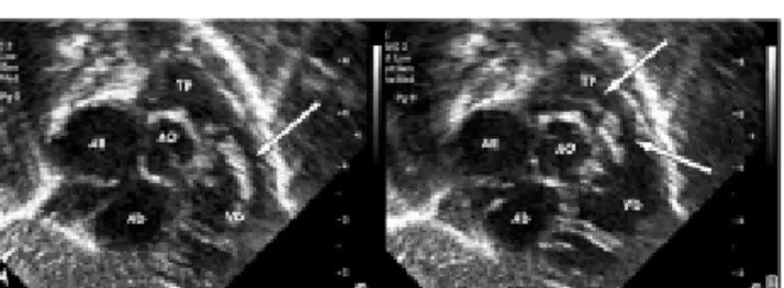
Fig. 1 - Coronal subcostal echocardiographic slices demonstrating: A – In diastole, a large vegetation in the right ventricle outflow tract (arrow). B – In systole, vegetation through the pulmonary valve extending as far as the bifurcation of the pulmonary arteries (arrows).
AE: Left atrium; AD: right atrium; VD: right ventricle; AO: aorta; TP: pulmonary trunk
226
CROTI, UA ET AL - Breastfeeding baby with fungal endocarditis at the right ventricle outflow tract - Case 3/2006
Braz J Cardiovasc Surg 2006; 21(2): 225-226
of the lungs was symmetrical without adventitious noises. The abdomen was swollen with the liver 4 cm from the right costal border. The pulses were symmetrical and the peripheral oxygen saturation was 96%.
ELECTROCARDIOGRAM
The electrocardiogram evidenced sinusal rhythm with a frequency of 125 bpm. The QRS angle was + 60º and the PR interval was 0.16 s. The QRS was 0.08s without direct signs of right ventricular overload.
RADIOGRAM
The radiogram demonstrated visceral situs solitus. The heart area and lungs were normal.
ECHOCARDIOGRAPH
The echocardiograph showed situs solitus and levocardia with normal venoatrial, atrioventricular and ventriculo-arterial connections. The longilinea image was irregular projecting from the RVOT through the pulmonary valve to the pulmonary trunk with an irregular aspect suggestive of fungal vegetation. The infant had a good ventricular function without pericardial effusions (Figure 1).
DIAGNOSIS
The clinical history and the long period using a central nervous catheter to administer endovenous antibiotics due to neonatal sepsis, the echocardiographic and the clinical improvement with antifungal agents made the diagnosis of fungal endocarditis caused by vegetation obvious; there was a high possibility of thrombogenic events.
OPERATION
A median transsternal thoracotomy was performed and a cardiopulmonary bypass was established with hypothermia at 34 ºC. Intermittent anterograde sanguineous cardioplegia at 4 ºC was also necessary. After opening the pulmonary
Fig. 2 - Pulmonary trunk opened lengthwise showing the large vegetation fixed in the right ventricle outflow tract extending through the pulmonary valve as far as the bifurcation of the pulmonary arteries