Hospital de Clínicas da Universidade Federal de Uberlândia
Correspondência: Renato E. S. Achá – Rua Guaicurus, 270 – 38408-394 – Uberlândia, MG – E-mail: [email protected]
Objective - To evaluate cardiac arrhythmias during and after pregnancy in women with Chagas’ disease wi-thout apparent heart disease using dynamic electrocar-diography.
Methods - Twenty pregnant women with Chagas’ di-sease without apparent heart didi-sease aged 19 to 42 years (26.96 ± 3.6) and a control group of 20 non-chagasic pregnant patients aged 16 to 34 years (22.5 ± 4.8). The pa-tients were submitted to passive hemagglutination and in-direct immunofluorescence for the detection of Trypano-soma cruzi evaluation, and electrocardiography, echocar-diography and 24-h dynamic electrocarechocar-diography.
Results - Supraventricular premature depolarizations were observed in 18 (90%) patients and ventricular prema-ture depolarization in 11 (55%) patients of both groups du-ring pregnancy. After delivery, supraventricular premature depolarizations were present in 13 (60%) chagasic patients and in 16 (89.4%) control patients (P≤0.05). Ventricular premature depolarization were observed in 9 (45%) cha-gasic patients and 11 (57.8%) control patients.
Conclusion - The prevalence of ventricular premature depolarization was similar for the chagasic and control groups during and after pregnancy. The incidence of su-praventricular premature depolarizations was similar in the two groups during pregnancy, while after delivery a predominance was observed in the control group compa-red to the chagasic group.
Key words: Chagas’disease, pregnancy, arrhythmia
Arq Bras Cardiol, volume 79 (nº 1), 5-9, 2002
Renato Enrique Sologuren Achá, Marco Túlio Oliveira Rezende,
Rimmel Amador Guzmán Heredia, Aguinaldo Coelho da Silva, Elmiro Santos Rezende,
Cleber Augusto Oliveira Souza
Uberlândia, MG - Brazil
Prevalence of Cardiac Arrhythmias During and After
Pregnancy in Women with Chagas’ Disease without
Apparent Heart Disease
The presence of arrhythmias as well as palpitations, dizziness, presyncope and, eventually, syncope 1 are events known to occur during pregnancy and are the main reason for the consultation of a cardiologist 2. Palpitations are related to premature depolarization and/or benign tachyarr-hythmias, which are well tolerated from a clinical point of view. However, patients with underlying heart disease and significant left ventricular dysfunction are more susceptible to malignant arrhythmias, which are poorly tolerated and may place the pregnant woman’s life at risk due to sudden death2. An increase in the incidence of cardiac arrhythmias during pregnancy, including ventricular tachycardia, has been reported in patients with and without heart disease 3,4.
Many young, healthy women present supraventricular and ventricular premature depolarizations with and without symptoms. Sobotka et al5, studying 50 non-pregnant women without apparent heart disease by 24-h dynamic electrocardiography, found isolated (supraventricular or ventricular) premature depolarizationss in 44 (88%) of the patients.
The expression “Chagas’ disease without apparent heart disease” refers to patients who present positive sero-logy for Chagas’ disease and who have a normal electrocar-diogram and chest X-ray, but who cannot be assigned to the so-called indeterminate form of Chagas’ disease since they were not submitted to radiologic analysis of the digestive tract6.
Studies in the literature on cardiac rhythm disturban-ces in pregnant patients with Chagas’ disease are scarce. This fact led us to determine the prevalence of cardiac arr-hythmias in pregnant women with Chagas’ disease without apparent heart disease.
Methods
seen at the Heart Disease and Pregnancy Outpatient Clinic of the University Hospital, Universidade Federal de Uberlândia (HC-UFU), were studied. All patients were initially submit-ted to clinical evaluation and then classified according to the criteria established by the New York Heart Association (NYHA). For the diagnosis of chagasic infection two sero-logic tests for the detection of Trypanosoma cruzi (passive hemagglutination and indirect immunofluorescence) were carried out. The patients were submitted to conventional 12-lead electrocardiography using a simultaneous 6-channel Ecafix apparatus, with the exam being considered to be nor-mal when the sinus rhythm was present and no rhythmic disturbances or any other electrocardiographic alterations were observed, and to echocardiographic examination using a Siemens model Sonoline CD apparatus. The diameters of the left ventricle (LV), left atrium, ascending aorta, interven-tricular septum and posterior wall of the LV, and the ejection fraction of the LV were determined, with values considered to be normal according to pre-established criteria. A dy-namic electrocardiogram (24-h Holter monitoring) was ob-tained using a Dynamis 3000 recorder, with the records being analyzed by the Cardiosystems program, version ALT V5 08C. All 24-h Holter exams were analyzed by a single observer. Cardiac rhythm, heart rate (maximum, minimum and mean) and the presence of cardiac arrhythmias were determined and analyzed with respect to the site of origin (supraventricular and ventricular), frequency and complexity (isolated, poly-morphic, coupled, and supraventricular or ventricular ta-chycardia) when exceeding 50 beats per 24 h, i.e., more than 10 beats per h. The presence of atrioventricular and/or intra-ventricular block and pauses or alterations in the ST segment were also recorded.
The control group consisted of 20 patients aged 16 to 34 years (22.5 ± 4.8), selected at the Prenatal Outpatient Clinic of HC-UFU, with negative serology for Chagas’ disea-se, asymptomatic in terms of the cardiovascular apparatus, non-smokers, and with electrocardiographic and echocar-diographic exams within normal parameters. Dynamic elec-trocardiography (24-h Holter monitoring) was carried out in both groups between 25 and 30 weeks of gestation.
All patients were again submitted to clinical evalua-tion, electrocardiography, echocardiography and 24-h dyna-mic electrocardiography during the second postpartum month. Only one patient from the control group refused to perform the exams. The study was approved by the Medical Ethics Committee of HC-UFU.
Statistical analysis. The nonparametric Wilcoxon test was applied to related variables to compare each group du-ring and after pregnancy. The Mann-Whitney U test was ap-plied to independent variables to compare the two groups during each period analyzed. The Student t-test was used for paired or dependent samples when the complexity of ar-rhythmias was compared between groups. A P value <0.05 was considered to be significant for all tests.
Results
The mean age of the group of pregnant women with Chagas’ disease was 26 years and 11 months (26.91 ± 3.6 years) and the mean age of the control group was 22 years and 6 months (22.5 ± 4.8 years), with this difference being statistically significant (P<0.002).
The mean ejection fraction was 74.55 ± 4.67 for the chagasic group and 75.5 ± 4.55 for the control group, with no significant difference between groups. Twenty-four-hour Holter monitoring was carried out in both groups bet-ween 24 and 30 weeks of gestation, with the mean value being 27.75 ± 1.52 weeks for the chagasic group and 27.41 ± 1.27 weeks for the control group (not significant). The mean heart rate was 88.8 ± 10.85 bpm for the chagasic group and 87.85 ± 6.47 bpm for the control group, with no significant difference between groups.
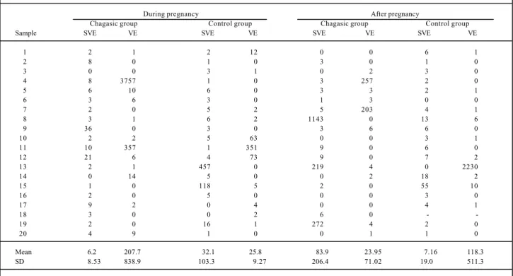
Table I shows the distribution of the number of prema-ture depolarizationss during and after pregnancy for the chagasic and control groups. During pregnancy, the number of supraventricular premature depolarizations was 6.2 ± 8.53 for the chagasic group and 32.1 ± 103.3 for the control group, and the mean number of 24-h ventricular premature depolarizations was 207.7 ± 839.9 for the chagasic group and 25.8 ± 79.27 for the control group, with no significant difference between groups. After pregnancy, the mean number of supraventricular premature depolarizations was 83.9 ± 260.4 for the chagasic group and 7.16 ± 19 for the control group. The number of 24-h ventricular premature depolarization was 23.95 ± 71.02 for the chagasic group and 118.7 ± 511.3 for the control group (not significant).
Table I also shows the number of premature depolari-zationss within each group during and after pregnancy. The distribution of the complexity of supraventricular and ventricular arrhythmias during and after pregnancy is shown in table II. Isolated supraventricular premature de-polarizations were observed during pregnancy in 18 (90%) patients of both groups and isolated ventricular premature depolarization were observed in 11 (55%) patients of both groups. After pregnancy, isolated supraventricular prema-ture depolarizations predominated in the control group compared to the chagasic group (P<0.05). Non-sustained tachycardia was only observed in the chagasic group.
Discussion
unknown. Some authors have suggested an increase in the frequency of paroxysmal supraventricular tachycardic episodes during pregnancy, with some of the patients experiencing these episodes for the first time 9-11. Other patients who have a history of arrhythmias before preg-nancy show an increase in the frequency, duration and se-verity of these arrhythmias during pregnancy 4.
Palpitations, dizziness, presyncope and, eventually, syncope may occur during pregnancy, but the etiology of these manifestations is still unclear. An increase in the inci-dence of arrhythmias during pregnancy has been reported for patients with and without structural heart disease 1-8.
Hemodynamic alterations due to volume overload, hormonal changes and influences of the autonomic ner-vous system increase the incidence of arrhythmias during pregnancy in both women without any evidence o f heart
disease and those with established heart disease who do not show any symptoms before pregnancy 2,7,12. Certain factors such as coffee, tea, smoking, alcohol, nasal decon-gestants or other toxic drugs can trigger arrhy th mias during pregnancy 12-14.
Anxiety and elevated estrogen levels lead to adrener-gic hyperactivity which, associated with an increased heart rate, alter refractoriness and conduction velocity, predispo-sing the pregnant women to arrhythmia. The increased phy-siological blood volume during pregnancy results in an crease in end diastolic pressure of the LV, which in turn in-creases myocardial stress and therefore also predisposes to arrhythmia2,15,16. During anxiety, pregnant women may com-plain of palpitations due to an increased p erception of heart beats. In which case, sinus tachycardia is commonly recor-ded and its electrocardiographic clinical correlations can be Table I – Distribution of the number of supraventricular (supraventricular premature depolarizations) and ventricular (ventricular premature
depolarization) premature depolarizationss during and after pregnancy in the chagasic and control groups
During pregnancy After pregnancy
Chagasic group Control group Chagasic group Control group
Sample SVE VE SVE VE SVE VE SVE VE
1 2 1 2 12 0 0 6 1
2 8 0 1 0 3 0 1 0
3 0 0 3 1 0 2 3 0
4 8 3757 1 0 3 257 2 0
5 6 10 6 0 3 3 2 1
6 3 6 3 0 1 3 0 0
7 2 0 5 2 5 203 4 1
8 3 1 6 2 1143 0 13 6
9 36 0 3 0 3 6 6 0
10 2 2 5 63 0 0 3 1
11 10 357 1 351 9 0 6 0
12 21 6 4 73 9 0 7 2
13 2 1 457 0 219 4 0 2230
14 0 14 5 0 0 2 18 2
15 1 0 118 5 2 0 55 10
16 2 0 5 0 0 0 3 0
17 9 2 0 4 0 0 4 1
18 3 0 0 2 6 0 -
-19 2 0 16 1 272 4 2 0
20 4 9 1 0 0 1 1 0
Mean 6.20 207.7 32.1 25.80 83.9 23.95 7.16 118.3
SD 8.53 838.9 103.3 9.27 206.4 71.02 19.00 511.3
SD = Standard deviation; P<0.9892
Table II – Distribution of supraventricular and ventricular arrhythmias during and after pregnancy in the chagasic and control groups
Chagasic group=20 Control group=20 Control group=19 Pregnancy After pregnancy Pregnancy After pregnancy
SVE VE SVE VE SVE VE SVE VE
Isolated 18 11 12* 9 18 11 17* 11
>50 beats/24 h 0 2 3 2 2 3 1 0
>10 beats/h 1 2 3 2 2 2 0 1
NST 1 3 3 0 0 0 1 0
Paired 1 1 2 2 2 1 1 0
Polymorphic 0 4 0 3 0 6 1 2
None 2 8 7* 11 2 9 2* 8
established by performing a resting electrocardiogram or dynamic electrocardiography (24-h Holter monitoring).
Modifications in electric stimulus conduction in the atria and ventricles, as well as in tissue refractory periods that are altered from modulations by circulating catechola-mines, may predispose to the occurrence of arrhythmias due to a reentry mechanism 17,18. The incidence of these arrhyth-mias during pregnancy is variable, and is accompanied by palpitations due to premature depolarizationss and benign tachyarrhythmias which are usually well tolerated from a clinical point of view 2,17.
Investigations on cardiac arrhythmias during pregnan-cy in patients with the so-called indeterminate form of Cha-gas’ disease, as well as studies on the prevalence of cardia-crhythm disturbances in men and non-pregnant women, are scarce. Pereira Barreto et al19 studied 22 patients with the in-determinate form of Chagas’ disease by 24-h dynamic elec-trocardiography to determine the incidence and importance of ventricular arrhythmias. The authors observed arrhy-thmias in 14 (63.6%) of the patients, with 11 (50%) patients showing ventricular premature depolarizationss of the isola-ted, bigeminal and polymorphic type, and 4 (18.2%) patients showing supraventricular premature depolarizations.
Marins el al 6 found a 42.5% prevalence of arrhythmias determined by 24-h Holter monitoring, with 27.2% of the tients presenting ventricular arrhythmias, and 8 (24.0%) pa-tients presenting arrhythmias considered by the authors to be of high risk (premature depolarizationss in bursts or bige-minal and trigebige-minal premature depolarizationss, and ventri-cular tachycardia). Almeida et al 20, comparing 15 patients with the indeterminate form of the disease to a control group, demonstrated an elevated prevalence of isolated ventricular premature depolarizationss.
Rassi et al 21, determining the frequency and extent of ventricular premature depolarizationss in 103 patients with the indeterminate form of Chagas’ disease and 20 control patients, observed a slightly higher incidence of ventricu-lar arrhythmias in the control group (85 vs 74%). The au-thors concluded that patients with the indeterminate form of Chagas’ disease do not behave differently from the normal population.
In the present study, 18 (90%) chagasic pregnant wo-men and 18 control patients presented supraventricular pre-mature depolarizations and 11 (55%) patients presented ventricular premature depolarization. After pregnancy (two months after delivery), a predominance of supraventricular premature depolarizations was observed in the control group compared to the chagasic group (Table II). No
signifi-cant difference in the number of supraventricular premature depolarizations or ventricular premature depolarization du-ring pregnancy was observed between groups (Table I), despite a larger mean number of ventricular premature depo-larization in the chagasic group compared to the control group. Twenty-four-hour Holter monitoring carried out du-ring the postpartum period also did not show a significant difference between groups.
Shotan et al 1, comparing 52 asymptomatic heart di-sease pregnant patients with 110 pregnant women without heart disease symptoms of palpitations, dizziness, presyn-cope and synpresyn-cope, observed a 58% rate of supraventricular premature depolarizations in the asymptomatic group and a 56% rate in the symptomatic group upon 24-h dynamic elec-trocardiography, while ventricular premature depolarization were observed in 40% of the asymptomatic pregnant pa-tients and in 49% of the symptomatic papa-tients, this differen-ce was not nonsignificant. Only nine patients were studied by 24-h Holter monitoring during the postpartum period and the authors did not report the number of premature de-polarizations or the number of women with arrhythmias, al-though they observed a tendency towards a reduction in arrhythmias during pregnancy. The authors also observed only a weak correlation between the incidence of arrhyth-mias and the presence of symptoms, with only 10% of sym-ptomatic episodes being accompanied by arrhythmias.
Ovando et al 22, studying 47 pregnant patients by 24-h Holter monitoring during labor (before, during and after de-livery), found a 72.3% prevalence of arrhythmias, with su-praventricular arrhythmias being the most frequent form and with the incidence of these arrhythmias being the same before and after delivery. Ventricular arrhythmias were re-ported in 14 (41.1%) patients, and mainly occurred in the form of monomorphic and polymorphic ventricular prematu-re depolarization which, in some cases, showed prematu-relatively se-vere characteristics.
The results of the present study with respect to the number and complexity of supraventricular premature depo-larizations and ventricular premature depolarization agree with those obtained by Shotan 1 and Ovando 22 in normal pregnancies. However, it is important to emphasize that there are no studies using 24-h Holter monitoring in Cha-gas’ disease without apparent heart disease.
A comparison of our results with data available in the li-terature led us to conclude that the presence of cardiac rhy-thm disturbances in chagasic or non-chagasic patients du-ring pregnancy does not seem to be of clinical importance.
1. Shotan A, Ostrzega E, Mehra A, Johnson JV, Elkayan U. Incidence of arrhythmias in normal, pregnancy and relation to palpitations, dizziness, and syncope. Am J Cardiol 1997; 79: 1061-4.
2. Blanco VMR, Álvares VB. Tratamiento de las arritmias cardiacas durante el emba-razo. Med Clin (Barc) 1996; 107: 29-36.
3. Hair TE, Eagan JT, Orgain ES. Paroxysmal ventricular tachycardia in the absence of demonstrable heart disease. Am J Cardiol 1962; 9: 209-14.
References
4. Brodsky M, Doria R, Allen B, Sato D, Thomas G, Sada M. New onset ventricular tachycardia during pregnancy. Am Heart J 1992; 123: 933-41.
5. Sobotka PA, Mayer JH, Bauernfeind RA, Kanakis C Jr, Rosen KM. Arrhythmias documented by 24-hour continuous ambulatory eletrocardiographic monitoring in young women with out apparent heart disease. Am Heart J 1981; 101: 573-9. 6. Marins N, Flores AP, Seixas TN, et al. Eletrocardiografia dinâmica em chagásicos na
7. Elkayan U, Gleicher N. Cardiac arrithythmias and pregnancy: cardiac problems in pregnancy. Wiley-Liss. A John Wiley & Sons Inc., Publication, 1998; 761p: 155-174p.
8. Mendelson CL. Disorder of heart beat during pregnancy. Am J Obstet Gynecol 1956; 72: 1268-301.
9. Lee SH, Chen SA, Wu TJ, et al. Effect of pregnancy on first onset and symptoms of paroxysmal supraventricular tachycardia. Am J Cardiol 1995; 76: 675-8. 10. Tawan M, Levine J, Mendelson M, Goldberger J, Dyer A, Hadish A. Effect of
preg-nancy on paroxysmal supraventricular tachycardia. Am J Cardiol 1993; 72: 838-40. 11. Szekely P, Snaith L. Paroxysmal tachycardia in pregnancy. Br Heart J 1953; 15:
195-8.
12. Moreira DAR. Arritmias na gravidez. Rev Soc Cardiol Estado de São Paulo 1994; 6: 552-9.
13. Hensleigh PA, Brown EL. Psychosocial stress and pregnancy. In: Gleicher N, ed. Principles of Medical Terapy in Pregnancy. New York: Plenum Medical Book Co. 1985: 885-8.
14. Barron WM, Mujais SK, Zinaman M, Bravo EL, Lindheimer MD. Plasma cate-cholamine responses to physiologic stimuli in normal human pregnancy. Am J Obstet Gynecol 1986; 154: 80-84.
15. Cole PL, Sutton MSJ. Normal cardiopulmonary adjustments to pregnancy car-diovascular evaluation. Cardiovasc Clin 1989; 19: 37-56.
16. Barriales V, Martinez TI. Cardiopatia y embarazo. Med Clin (Barc) 1990; 94: 389-95.
17. Moreira DAR. Arritmias em gestantes. Rev Soc Cardiol Estado de São Paulo 1998; 1; 127-36.
18. Louhana G. The electrical activity of the heart during pregnancy. Rev Fr Gynecol Obstet 1990; 85: 369-74.
19. Pereira Barretto AC, Bellotti G, Sosa E, et al. Arritmias e a forma indeterminada da doença de Chagas. Arq Bras Cardiol 1986; 47: 197-99.
20. Almeida JWR, Shikanai MAY, Amato Neto V, Catilho EA, Barretto ACP. Estudo da forma indeterminada da doença de Chagas através da eletrocardiografia dinâmi-ca. Rev Inst Med Trop São Paulo 1982; 24: 222-8.
21. Rassi Jr. A, Rassi AG, Rassi SG, Rassi Jr. L, Rassi A. Freqüência e grau da extra-sis-tolia ventricular à eletrocardiografia dinâmica (sistema Holter de 24 horas) na doença de Chagas. Arq Bras Cardiol 1991; 57(supl C): C146.
22. Ovando AL, Germiniani H, Miglino R, da Cunha GP. Estudo das arritmias car-díacas maternas durante o trabalho de parto e o parto. Arq Bras Cardiol 1983; 40: 171-6.
Prefeitura Municipal de Leopoldina - MG César Grupi, André Alves
Editor da Seção de Fotografias Artísticas: Cícero Piva de Albuquerque