DOI: 10.1590/0004-282X20160113
ARTICLE
Elastomers three-dimensional biomodels
proven to be a trustworthy representation of
the angiotomographic images
Biomodelos tridimensionais de elastômero provaram ser uma representação confiável de
imagens angiotomográficas
André Giacomelli Leal1, Leonardo Brancia Pagnan2, Raphael Teruaki Kondo2, José Aguiomar Foggiatto3,
Guilherme José Agnoletto4, Ricardo Ramina4
Intracranial aneurysms (IA) are abnormal dilatations that can arise in any blood vessels of the intracranial circulation due to histopathological and hemodynamic alterations. It is estimat-ed that 6% of people worldwide present with a non-rupturestimat-ed IA. Risk factors for IA include family history, hereditary disorders predisposing aneurysm formation, age (younger than 50 years old), gender ( female), tobacco and cocaine consumption1.
An extremely high mortality rate, associated with IA rupture, occurs in approximately 2% of the cases and is
responsible for 80% of all spontaneous subarachnoid hem-orrhages2. Aneurysms larger than 10mm or located in the
posterior circulation present an even higher risk of rupture. herefore, it is fundamental to ofer the best treatment op-tion for a diagnosed IA.
Two options for surgical treatment of an IA are avail-able: endovascular embolization and microsurgical clipping3, both aiming to exclude the aneurysm from
the cerebral circulation. Endovascular embolization is
1Instituto de Neurologia de Curitiba – INC , Divisão de Neurocirurgia Vascular, Curitiba PR, Brasil; 2Faculdade Evangélica do Paraná, Faculdade de Medicina, Curitiba PR, Brasil;
3Universidade Tecnológica Federal do Paraná - UTPFR, Divisão de Prototipagem Rápida, Curitiba PR, Brasil; 4Instituto de Neurologia de Curitiba – INC , Departamento de Neurocirurgia, Curitiba PR, Brasil.
Correspondence: André Giacomelli Leal; Instituto de Neurologia de Curitiba (INC), Divisão de Neurocirurgia Vascular; Rua Jeremias Maciel Perretto, 300; 81210-310 Curitiba PR, Brasil; E-mail: [email protected]
Conflict of interest: There is no conflict of interest to declare.
Received 15 March 2016; Received in final form 09 June 2016; Accepted 13 June 2016.
ABSTRACT
Intracranial aneurysm (IA) rupture is responsible for 80% of spontaneous arachnoid hemorrhages and associated with an extremely high mortality rate. Two possible surgical interventions are endovascular embolization and microsurgical clipping. Three-dimensional (3D) prototyping models help in surgical planning minimizing perioperative risks in both methods and reducing operating time. Methods: 3D biomodels were printed with flexible material (elastomer) using angiotomographic DICOM acquired images and compared to 3D digital subtraction angiography (DSA) images. Results: 3D biomodels represented the aneurysm angioarchitecture exactly, especially the neck and domus features. Conclusion: Elastomers 3D biomodels proved to be a trustworthy representation of the angiotomographic images and could be used to help surgical planning in IA treatment.
Keywords: intracranial aneurysm; models, anatomic; elastomers, microsurgery.
RESUMO
A ruptura dos aneurismas intracranianos é responsável por 80% das hemorragias subaracnóideas espontâneas e está associada a uma taxa de mortalidade extremamente alta. Duas intervenções cirúrgicas viáveis são embolização endovascular e clipagem microcirúrgica. Os modelos de prototipagem tridimensional (3D) auxiliam no planejamento cirúrgico e na diminuição dos riscos intra-operatórios nos dois procedimentos e redução do tempo da cirurgia. Métodos: Foram impressos biomodelos em 3D com material flexível (elastômero) utilizando imagens DICOM de angiotomografia e comparados com imagens de angiografia por subtração digital em 3D (DAS). Resultados: Biomodelos em 3D representam com exatidão a angioarquitetura do aneurisma, particularmente os detalhes do colo e domus. Conclusão: Biomodelos em 3D com elastômeros mostraram ser uma representação confiável das imagens angiotomográficas, podendo ser utilizados no planejamento cirúrgico no tratamento de IA.
generally performed with platinum coils to fill the an-eurysm, whether by remodeling stent-assisted or not. Microsurgical clipping uses a titanium clip that occludes the aneurysm neck.
Microsurgical clipping was notably the only reliable treat-ment for IA in previous decades before the advent of the en-dovascular techniques. Currently, a great variety of IAs are treated with embolization. Nevertheless some complex cases still make embolization a less than viable method, requiring classic microsurgical clipping.
In order to better choose between the surgical techniques and the best strategies to use within each one, surgeons of-ten rely on various imaging methods. Cerebral computed tomography angiography (CTA) is a reliable non-invasive imaging method that can reach speciicity levels as high as 100% as well as a sensitivity of 95% for evaluation of IAs, al-though Digital Subtraction Angiography (DSA) remains the gold standard.
he restraint to these methods is the fact that they pres-ent the vessels structures in a two-dimensional (2D) pat-tern, limiting visualization of aneurysmal neck and adja-cent vessels that must be carefully studied4. Even the most
recent 3D CTAs and DSAs end up printed on paper, which ultimately is a 2D media.
One of the main features of microsurgical clipping that must be carefully evaluated is the choice of adequate clip and material, bearing in mind the large anatomic variability that can be encountered. In cases of widened neck or complex IA, the preoperative surgical planning is imperative to avoid ex-tending operative time5, excessive manipulation of adjacent
vessels and increased hemorrhage risk6.
With those risks in mind, several studies have been de-veloped with 3D IA models, to enable familiarization and preoperative planning with the aneurysm anatomy and the angioarchitecture7.
Two combined techniques have resulted in biomedi-cal prototyping models: rapid prototyping (RP) and im-age acquisition (integrating reconstruction). Prototyping is a manufacturing process that allows the production of models from stereolithography archives. The manufactur-ing process consists of successive layers of various, cur-rently existing, polymeric materials. Several technologies use a wide range of polymeric and metallic materials that can be found as powder, liquid or solid filaments. These processes are a method of additive manufacturing. The most frequently-used process today is the Polyjet, which produces prototypes through polymerization using ultra-violet light and thin layers of acrylic resin successively jet-ted on top of each other.
In light of RP technologies and the non-invasive imaging methods that are available today, the biomedical models can aid surgical planning of several pathologies, as they can be found diversely in the literature8,9. However there are still few
studies evaluating RP for analysis of IAs10,11,12.
METHODS
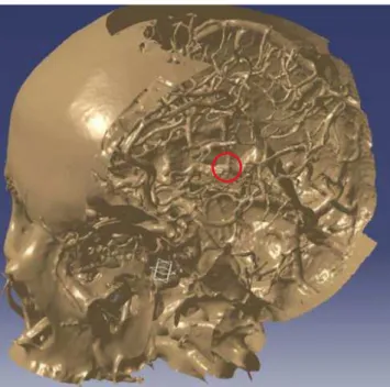
he study consisted of two stages. In the irst, two pa-tients with non-ruptured IAs, who consented to take part in the study, were selected. Both underwent CTA imag-ing. he images were generated in DICOM (Digital Imaging and Communication in Medicine) format and processed by two types of specialized software: InVesalius (Figure 1) and CATIA (Figures 2, 3 and 4). Subsequently the IA biomodels were printed with a speciic 3D printer (Object Conmex 350®). he prototypes were made of a lexible acrylic resin called elastomer (TangoBlackPlus®), which can return to its original shape even after being deformed, compressed or stretched. he prototyping was performed at the Renato Archer Center of Technology and Information, in São Paulo, Brazil.
In the second stage of the study, the malleable prototypes that had been created previously were compared with DSA
Source: Núcleo de Prototipagem e Ferramental (NUFER - UTFPR)
Figure 1. Image from InVesalius Software, highlighting the aneurysm.
images of the same patients, aiming to conirm compatibility of morphology and angioarchitecture. It was imperative that com-plete correlation of the aneurysm morphology was achieved as it is essential information for the biomodel validation.
RESULTS
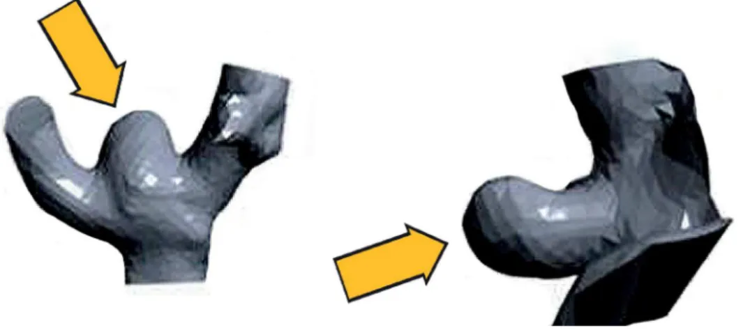
At the end of the irst stage, four 3D elastomer models were made (two models of each aneurysm) to represent the exact shape of the aneurysm in real dimension and size. Model 1 (Figure 2) shows a medium cerebral artery IA, mea-suring 8x5mm. Model 2 (Figure 3) shows a posterior com-municating artery IA, measuring 7x6mm. he four models were produced at the same time (18.5 grams of material), with a cost of U$ 9.39 each. he Center of Technology and Information provided the prototypes without any costs and delivered them in seven days.
In the second stage, the real-size prototypes were validat-ed as perfect copies of the IA morphology when comparvalidat-ed to
their respective DSAs (Figure 5). he aneurysmal necks were precisely recreated in real size.
DISCUSSION
Previous literature has suggested the possibility of produc-ing 3D models for simulation of IA surgery, either microsurgery or endovascular techniques. Rapid prototyping has also been pro-duced in other areas of healthcare, such as in dentistry and vascu-lar surgery, to help in planning the best treatment options7,8,12,13,14,15.
Erbano et al.16 demonstrated the feasibility of making IA
3D models using a non-lexible acrylic resin (FullCure 720®) with the RP method. Four patients were selected with an-eurysms in the most common locations and the biomodels compared to the respective patients’ DSA images, demon-strating that the IA morphology was precisely represented. However, the resin that was used lacked elastic properties to emulate the consistency of the aneurysm.
In the present study, lexible biomodel manufacturing was possible, which could be helpful during surgery planning or pre-operation aneurysm clipping simulation.
he main advantage of IA biomodels is that they allow surgeons to plan ahead and even practice the procedure prior to performing it, as it even gives the opportunity to choose an appropriate metallic clip, thus reducing operating time, vessel manipulation and risk of complications. As the ield of view during an operation is restricted to the surgical access and craniotomy performed, it is not uncommon to ind it dif-icult to see the structures surrounding the aneurysm, such as adjacent vessels. his is yet another feature in which 3D biomodels can help surgery planning11,12,17.
D’Urso et al.10 were the irst to apply the technology of
de-signing IA 3D models using angiotomography. Later, Wurm et al. improved the technique by using 3D rotational CTAs to pro-duce models with higher image resolution12. he technological
Figure 3. Segmented image from patient #2, posterior communicating artery (arrow).
Source: Núcleo de Prototipagem e Ferramental (NUFER - UTFPR)
advances resulted in the design of more accurate printers, with fewer cuts and production of more evolved models.
When it comes to the use of 3D models in endovascu-lar surgery, Chueh et al. and Wetzel et al. demonstrated the possibility of creating prototypes with lumen replication and representation, creating extremely malleable models7,18.
Another important feature of IA prototypes is that they allow a more precise explanation of the procedure proposed to the patient, with a visual and touchable media, facilitat-ing the patient’s and the family’s understandfacilitat-ing of the disease and treatment. Even surgeons feel more conident when they are familiar with the complete, real-size anatomy and mor-phology of the aneurysm to be operated on11,12.
here are still some limitations concerning the future pro-duction of IA prototypes. For example, the models cannot yet
represent the presence of a pre-existing intra-arterial throm-bus or how thick the wall of an aneurysm is. In cases of rup-tured aneurysms, the production of prototypes is not viable due to the long period of time required for the completion of the whole process. It is expected that, in the future, new tech-niques and technologies of 3D printing will make it possible to produce RP models even in urgent situations.
In conclusion, it is possible to produce elastomer IA pro-totypes by means of rapid prototyping. he models have proved to be a faithful reproduction of the CTA. heir lexible material can be used for preoperative planning and simula-tion of surgical strategies, despite not having the elasticity of the actual vessel wall. It is expected that future studies will improve the prototyping technique and apply it to a larger number of patients with non-ruptured IAs.
References
1. Vega C, Kwoon JV, Lavine SD. Intracranial aneurysms: current evidence and clinical practice. Am Fam Physician. 2002;66(4):601-8.
2. Suarez JI, Tarr RW, Selman WR. Aneurysmal subarachnoid hemorrhage. N Engl J Med. 2006;354(4):387-96. doi:10.1056/NEJMra052732
3. Li H, Pan R, Wang H, Rong X, Yin Z, Milgrom DP et al. Clipping versus coiling for ruptured intracranial aneurysms: a systematic review and meta-analysis. Stroke. 2013;44(1):29-37.
doi:10.1161/STROKEAHA.112.663559
4. Guglielmi G, Viñuela F, Duckwiler G, Dion J, Lylyk P, Berenstein A et al. Endovascular treatment of posterior circulation aneurysms by electrothrombosis using electrically detachable coils. J Neurosurg. 1992;77(4):515-24. doi:10.3171/jns.1992.77.4.0515
5. Lawton MT, Spetzler RF. Surgical management of giant intracranial aneurysms: experience with 171 patients. Clin Neurosurg. 1995;42:245-66.
6. Rinne J, Hernesniemi J, Niskanen M, Vapalahti M. Management outcome for multiple intracranial aneurysms. Neurosurgery. 1995;36(1):31-7. doi:10.1227/00006123-199501000-00003
7. Chueh JY, Wakhloo AK, Gounis MJ. Neurovascular modeling: small-batch manufacturing of silicone vascular replicas. AJNR Am J Neuroradiol. 2009;30(6):1159-64. doi:10.3174/ajnr.A1543
8. Müller A, Krishnan KG, Uhl E, Mast G. The application of rapid prototyping techniques in cranial reconstruction and preoperative planning in neurosurgery. J Craniofac Surg. 2003;14(6):899-914. doi:10.1097/00001665-200311000-00014
9. Winder J, Bibb R. Medical rapid prototyping technologies: state of the art and current limitations for application in oral and maxillofacial surgery. J Oral Maxillofac Surg. 2005;63(7):1006-15. doi:10.1016/j.joms.2005.03.016
10. D’Urso PS, Thompson RG, Atkinson RL, Weidmann MJ, Redmond MJ, Hall BI et al. Cerebrovascular biomodelling: a technical note. Surg Neurol. 1999;52(5):490-500. doi:10.1016/S0090-3019(99)00143-3
12. Wurm G, Tomancok B, Pogady P, Holl K, Trenkler J. Cerebrovascular stereolithographic biomodeling for aneurysm surgery. Technical note. J Neurosurg. 2004;100(1):139-45. doi:10.3171/jns.2004.100.1.0139
13. Khan IS, Kelly PD, Singer RJ. Prototyping of cerebral vasculature physical models. Surg Neurol Int. 2014;5(1):11. doi:10.4103/2152-7806.125858
14. Kono K, Shintani A, Okada H, Terada T. Preoperative simulations of endovascular treatment for a cerebral aneurysm using a patient-specific vascular silicone model. Neurol Med Chir (Tokyo). 2013;53(5):347-51. doi:10.2176/nmc.53.347
15. Sugiu K, Martin JB, Jean B, Gailloud P, Mandai S, Rufenacht DA. Artificial cerebral aneurysm model for medical testing, training, and research. Neurol Med Chir (Tokyo). 2003;43(2):69-72. doi:10.2176/nmc.43.69
16. Erbano BO, Opolski AC, Olandoski M, Foggiatto JA, Kubrusly LF, Dietz UA et al. Rapid prototyping of three-dimensional biomodels as an adjuvant in the surgical planning for intracranial aneurysms. Acta Cir Bras. 2013;28(11):756-61. doi:10.1590/S0102-86502013001100002
17. Wurm G, Lehner M, Tomancok B, Kleiser R, Nussbaumer K.
Cerebrovascular biomodeling for aneurysm surgery: simulation-based training by means of rapid prototyping technologies. Surg Innov. 2011;18(3):294-306. doi:10.1177/1553350610395031