DOI: 10.1590/0004-282X20160131
ARTICLE
Effects of crocin on brain oxidative damage
and aversive memory in a 6-OHDA model of
Parkinson’s disease
Efeitos da crocina no dano oxidativo cerebral e na memória aversiva em um modelo
6-OHDA de doença de Parkinson
Rajaei Z1,2,Hosseini M1, Alaei H3
Parkinson’s disease (PD) is a progressive neurodegen-erative disorder that is characterized by the degeneration of dopaminergic nigrostriatal neurons, which leads to mo-tor symptoms of bradykinesia, rigidity, rest tremor, and postural imbalance1. Parkinson’s disease also causes
im-pairments in cognitive performance, and the progression of these deficits can lead to dementia2. The hippocampus,
which is involved in cognitive processes such as learning and memory, is implicated in memory deficits observed in PD since both structural and functional changes of the
hippocampus have been observed in PD patients3,4. Several
MRI studies have also clearly revealed that the reduction of hippocampal volume was accompanied by cognitive defi-cits in PD patients5.
Numerous studies have suggested that oxidative stress plays a major role in the pathogenesis of PD6. Free
radi-cals and other reactive oxygen species (ROS) resulted from dopamine auto-oxidation and metabolism, lipid peroxida-tion, impaired mitochondrial funcperoxida-tion, and deficiencies in endogenous antioxidant systems that may all contribute
1Isfahan University of Medical Sciences, School of Medicine, Department of Physiology, Isfahan, Iran;
2Mashhad University of Medical Sciences, School of Medicine, Neurocognitive Research Center, Mashhad, Iran;
3Isfahan University of Medical Sciences, Alzahra Hospital, Isfahan Neurosciences Research Center, Isfahan, Iran.
Correspondence: Ziba Rajaei; Department of Physiology, School of Medicine, Isfahan University of Medical Sciences, Isfahan, Iran; E-mail: rajaeiz@med.mui.ac.ir
Conflict of interest: There is no conlict of interest to declare.
Support: This study was supported by Isfahan University of Medical Sciences.
Received 16 March 2016; Received in inal form 31 May 2016; Accepted 06 June 2016. ABSTRACT
The purpose of the present study was to investigate the effect of crocin on brain oxidative damage and memory deicits in a 6-hydroxydopamine (6-OHDA) model of Parkinson’s disease. Male Wistar rats were subjected to unilateral injection of 6-OHDA (16 µg) into the medial forebrain bundle and treated with crocin (30 and 60 mg/kg) for six weeks. The rats were tested for memory performance at six weeks after 6-OHDA infusion, and then were killed for the estimation of biochemical parameters. The increase in thiobarbituric acid reactive substances (TBARS) and nitrite levels in the hippocampus were observed in the 6-OHDA lesioned rats, which was accompanied by memory deicits in a passive avoidance test at the end of week 6. Moreover, treatment with crocin decreased TBARS and nitrite levels in the hippocampus, and improved aversive memory. The present study conclusively demonstrated that crocin acts as an antioxidant and anti-inlammatory agent in the hippocampus of parkinsonian rats and could improve aversive memory through its properties.
Keywords: Crocin; 6-hydroxydopamine; oxidative stress; nitric oxide; aversive memory; medial forebrain bundle.
RESUMO
O objetivo do presente estudo foi investigar o efeito da crocina no dano oxidativo cerebral e nos déicits de memória em um modelo 6-OHDA de doença de Parkinson. Ratos Wistar machos foram submetidos à injeção unilateral de 6-OHDA (16 μg) em MFB e tratados com crocina (30 e 60 mg/kg), durante 6 semanas. Os ratos foram testados quanto ao desempenho da memória 6 semanas após a infusão de 6-OHDA, e, em seguida, foram sacriicados para a estimativa dos parâmetros bioquímicos. O aumento nos níveis de TBARS e de nitrito no hipocampo foram observados em ratos 6-OHDA lesionados, acompanhado por déicits de memória em um teste de esquiva passiva no inal da semana 6. Além disso, o tratamento com crocina diminuiu os níveis de nitrito e de TBARS no hipocampo e melhorou a memória aversiva. O presente estudo demonstrou conclusivamente que a crocina age como um antioxidante e um agente anti-inlamatório no hipocampo de ratos parkinsonianos e pode melhorar a memória aversiva através de suas propriedades.
to a progressive loss of dopaminergic neurons7,8. There
is also some evidence showing that the neurotoxicity of 6-hydroxydopamine (6-OHDA), for the modeling of PD, is due to its oxidation and the formation of various oxi-dants and free radicals and the depletion of reduced glu-tathione9. This leads to lipid peroxidation, protein damage
and ultimately degeneration of the nigrostriatal dopami-nergic system. 6-OHDA has also been reported to pro-duce reactive nitrogen species (RNS) such as nitric oxide (NO) by elevated expression of inducible nitric oxide syn-thase (iNOS), especially in neurons10. Reactive oxygen
spe-cies can rapidly interact with NO and subsequently pro-duce more powerful oxidant peroxynitrite. Peroxynitrite is known to structurally and functionally modify critical cellular macromolecules and cause oxidative damages, which finally leads to apoptotic cell death11.
Recently, the major focus of many preclinical studies is
the identiication of drugs or approaches that might pre -vent or inhibit the neurodegenerative process. In this con-text, it has been suggested that antioxidant molecules (ca-rotenoids and polyphenols) and compounds interfering with production of reactive oxygen species and nitric oxide might be protective.
Crocin is a water- soluble carotenoid and an active
con-stituent of safron (Crocus sativus L.). It has been reported
that crocin possesses multiple pharmacological properties, including antioxidative activity12,13, anti-inlammatory14,
pro-tection against cardiovascular diseases, inhibition of tumor cell proliferation, neuroprotection and protection of hepato-cytes12. It has also been shown that the spice safron, which
contains powerful antioxidants such as crocin, protects ni-gral and retinal dopaminergic cells in an acute MPTP mouse model of Parkinson’s disease15. he antioxidant and radical
scavenging activity of crocins have also been shown in sev-eral in vitro models12,16. Based on the role of oxidative and
ni-trosative stress on the pathophysiology of PD and
consider-ing the antioxidant and anti-inlammatory efects of crocin,
the present study was designed to evaluate whether it pro-tects against 6-OHDA-induced oxidative damage and
mem-ory deicits in rats.
METHODS
Animals
Adult male Wistar rats, weighing 250–300g were housed in an air conditioned colony room at 22°C ± 2°C on a standard pellet diet and tap water ad libitum. he Ethics Committee for Animal Experiments at Isfahan University of Medical
Sciences approved the study and all experiments were con-ducted in accordance with the National Institute of Health
Guide for the Care and Use of Laboratory Animals (NIH
Publications Nº 80 23, revised 1996).
Chemicals
Crocin, 6-OHDA, and apomorphine hydrochlo-ride were purchased from Sigma Aldrich Co. 2,2´ Dinitro-5,5´-dithiodibenzoic acid (DTNB), trichloro acetic
acid (TCA), 2-thiobarbituric acid (TBA), Tris-EDTA, chloral hy -drate and hydrochloric acid (HCL) were obtained from Merck.
Experimental design
he animals were randomly divided into four groups,
with eight rats in each group, as follows: normal saline sham-operated group, normal saline-treated lesioned group (6-OHDA), and the crocin-treated lesioned groups (crocin 30 and 60 mg/kg/day). Crocin was dissolved in normal saline and injected at doses of 30 and 60 mg/kg/day
intraperitone-ally, three days before the surgery for six weeks. he surgical
procedures were carried out under general (chloral hydrate,
450 mg/kg, ip) and local anaesthesia (lidocaine 2%). he
rats were placed in a stereotaxic apparatus and the lesion was induced by injection of 6-OHDA (16 µg/4µl 0.2% ascor-bate saline) into the left medial forebrain bundle through
microinjection pump (Kd Scientiic, USA) according to the
coordinates: AP: -4.5 mm; ML: -1.7 mm; DV: -8.2 mm17. he
rats of the sham-operated group also received an identical
volume of the ascorbate saline as the vehicle. he injection
rate was 1 µl/min and the needle was kept in place for an
additional ive minutes before slowly being retracted. At the end of the experiment, the animals were sacriiced and the
hippocampus and cortex were dissected out, washed im-mediately in ice-cold saline, and homogenized in the NaCl
solution by a homogenizer. he hippocampus and cortex
were obtained bilaterally, and the data are measures from combined bilateral tissues.
Lipid peroxidation levels
he lipid peroxidation level of the hippocampus and
cortex was measured as malondialdehyde, which is the
end product of lipid peroxidation. he malondialdehyde re -acts with TBA as a thiobarbituric acid reactive substance (TBARS) and produces a red colored complex that has a peak
absorbance (A) at 535 nm. Briely, a mixture of TCA, TBA, and
HCl were added to 1mL of homogenate, and the mixture was heated for 45 min in a boiling water bath. After cooling, the samples were centrifuged at 1000× g for 10 min and the
ab-sorbance was measured at 535 nm. he level of TBARS was
calculated according to follow equation13. Concentration (Molar) = Absorbance /1.65 x 105.
Total thiol concentration
Total sulfhydryl (SH) groups were measured using DTNB as the reagent. This reagent reacts with the SH groups to produce a yellow colored complex that has a
peak absorbance at 412 nm. Briefly, 1mL Tris-EDTA buf
-fer (pH = 8.6) was added to 50 µL homogenate in 2 mL
against the Tris-EDTA buffer alone (A1). Then, 20 µL of
the DTNB reagent (10 mM in methanol) was added to the mixture and after 15 min, the sample absorbance was read again (A2). The absorbance of the DTNB re-agent was also read as a blank (B). The total thiol con-centration (mM) was calculated by: The total thiol
concentration (mM) = (A2-A1-B) × 1.07/0.05 × 13.613.
Glutathion peroxidase assay
GSH peroxidase concentration was measured with the
GSH peroxidase kit (Randox Labs, Crumlin, UK).
Nitrite levels
he level of nitrite was measured using a colorimetric as
-say kit (Promega Corporation, USA) that involved the Griess reaction. Briely, after adding sulphanilamide solution and
incubation, N-(1-naphthyl) ethylenediamine solution was
added. hen, the sample absorbance was measured by a mi
-croreader in the wavelength of 492 nm. he nitrite concentra -tion of samples was determined by comparing comparison against with the nitrite standard reference curve.
Passive avoidance learning
The passive avoidance test was used in the cur-rent study, because it is a suitable model for evaluating hippocampal-dependent memory deficits in experimental animals. Passive avoidance learning was assessed by shut-tle box at the end of week 6. The apparatus consisted of a light compartment (25×25×20 cm) and a dark compart-ment (50×25×20 cm) with a grid floor and wooden walls. The two compartments were separated by a sliding guillo-tine door. On the day before training, each rat was placed into the apparatus and allowed to move around 5 min for five minutes for habituation. In the training session, ani-mals were placed individually in the light compartment for one minute. After opening the door and entrance of the rat entering into the dark chamber, the door was closed and a 1mA foot electric shock was delivered for 3s through the grid floor. The current intensity for foot shock was se-lected based on previous studies in our laboratory. In the test session, each rat was again placed into the light com-partment. The step-through latency to enter the dark compartment was measured as a positive index of memo-ry performance, with a 300 s cut-off time18.
Histology
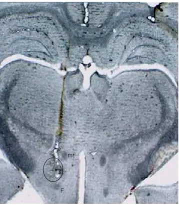
The animals were sacrificed by a high dose of the an-aesthetic. Then, the brains were removed and stored in 10% formalin for 72 h. The brains were sectioned coro-nally at 40 µm by a freezing microtome (Leica, Germany). Sections were mounted on gelatin-coated slides and stud-ied using a light microscope. The track of the needle and injection site of 6-OHDA (Figure 1) was determined by ref-erence to a rat brain atlas17.
Statistical analysis
he data were expressed as mean ± SEM. Statistical
analysis was carried out using one-way ANOVA followed by the LSD post hoc test. A statistical p-value < 0.05 was
con-sidered signiicant.
RESULTS
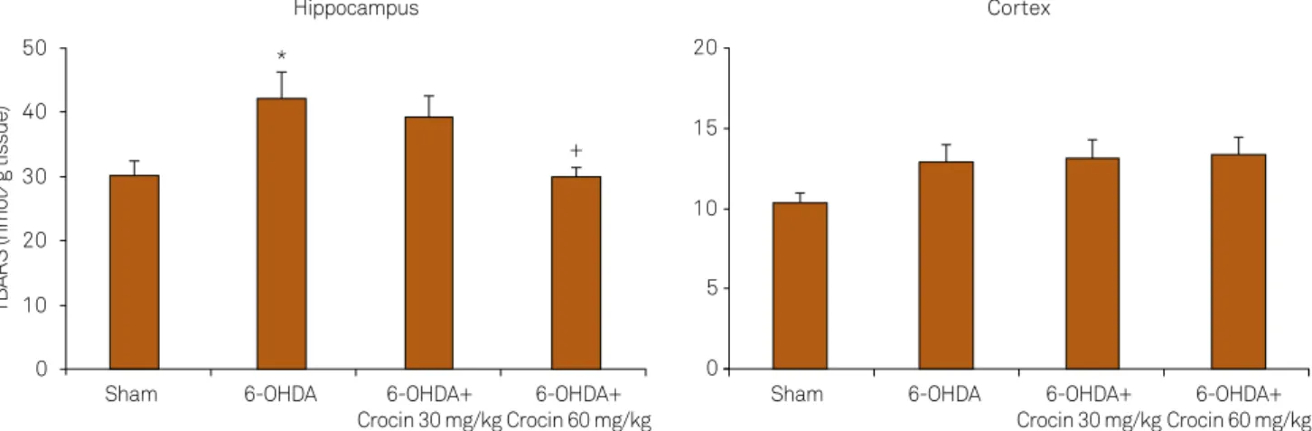
Effects of crocin on lipid peroxidation levels
As shown in Figure 2, a signiicant increase in the levels of
TBARS, an index of lipid peroxidation, was found in the
hip-pocampus of 6-OHDA-lesioned rats (p = 0.01) as compared
with the sham group. Moreover, treatment of lesioned rats with crocin at a dose of 60 mg/kg reduced the TBARS levels in the hippocampus at the end of week 6 (p < 0.05, Figure 2). Meanwhile, 6-OHDA increased the TBARS levels in the cortex
of lesioned rats, although the changes were not signiicant.
Effects of crocin on nitrite levels
Figure 3 shows the nitrite levels in the hippocampus and
cortex of the sham and experimental groups. A signiicant
increase in the nitrite levels in the hippocampus (p < 0.05) of 6-OHDA-lesioned rats was observed as when compared with sham group rats. Treatment of 6-OHDA-lesioned rats
with crocin at a dose of 30 mg/kg signiicantly decreased the
nitrite levels in the hippocampus as when compared to with the lesioned group (p < 0.05, Figure 3). Meanwhile, 6-OHDA
mt: mammilothalamic tract, F: fornix.
Figure 1. Photograph of the coronal section of rat brain
representing injection site of 6-OHDA in the medial forebrain bundle (open circle).
mt
did not change the nitrite levels in the cortex of lesioned rats compared with sham group rats (Figure 3).
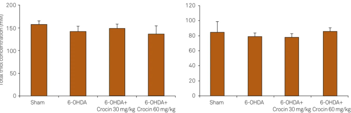
Effects of crocin on total thiol concentration
Figure 4 shows the total thiol concentration in the hip-pocampus and cortex of the sham and experimental groups.
here was no signiicant change in total thiol concentrations
in the hippocampus and cortex of sham and experimental groups (Figure 4).
Effects of crocin on glutathione peroxidase levels
As shown in Figure 5, there was no signiicant diference
in glutathione peroxidase concentration in the hippocam-pus and cortex of sham, 6-OHDA-lesioned rats and lesioned groups treated with crocin at 30 and 60 mg/kg at the end of week 6.
Effects of crocin on passive avoidance learning
As shown in Figure 6, the step-through latency of 6-OHDA-lesioned rats was shorter than the sham group rats
at the end of week 6 (p < 0.05, Figure 6). Moreover, treatment of lesioned rats with crocin at a dose of 30 mg/kg increased the latency as compared with lesioned rats (p < 0.05).
DISCUSSION
In the present study, we evaluated the efect of crocin on
biochemical and behavioral parameters using the 6-OHDA rat model, because this model imitates the pathological and bio-chemical features of PD, such as oxidative stress, mitochondrial dysfunction and apoptosis19. Oxidative stress, imbalance of free
radicals and antioxidants, plays a critical role in the pathogen-esis of PD. Dopaminergic neurons are particularly sensitive to oxidative stress because of their low antioxidant capacity, as evi-denced by low intracellular glutathione20. A number of studies
have also demonstrated that the neurotoxicity of 6-OHDA for the modeling of PD is due to its oxidation, the formation of vari-ous oxidants and free radicals and the depletion of reduced glu-tathione, which leads to lipid peroxidation, protein damage and
Figure 2. Lipid peroxidation levels (thiobarbituric acid reactive substance [TBARS]) in the hippocampus and cortex of the
sham, 6-OHDA-lesioned rats and lesioned rats treated with crocin at doses of 30 and 60 mg/kg at the end of week 6. Data are mean ± SEM for eight animals in each group. *p = 0.01 vs sham group, +p < 0.05 vs 6-OHDA-lesioned group.
0 10 20 30 40 50
Sham 6-OHDA 6-OHDA+
Crocin 30 mg/kg
6-OHDA+ Crocin 60 mg/kg
Sham 6-OHDA 6-OHDA+
Crocin 30 mg/kg
6-OHDA+ Crocin 60 mg/kg
TBARS (nmol⁄g tissue)
Hippocampus
*
+
Cortex
0 5 10 15 20
Figure 3. Nitrite levels in the hippocampus and cortex of the sham, 6-OHDA-lesioned rats and lesioned rats treated with crocin
at doses of 30 and 60 mg/kg at the end of week 6. Data are mean ± SEM for eight animals in each group. *p < 0.05 vs sham group, +p < 0.05 vs 6-OHDA-lesioned group.
*
+
0 1 2 3 4 5 6 7 8
Sham 6-OHDA 6-OHDA+
Crocin 30 mg/kg
6-OHDA+ Crocin 60 mg/kg
Sham 6-OHDA 6-OHDA+
Crocin 30 mg/kg
6-OHDA+ Crocin 60 mg/kg
Nitrite levels (µM/g tissue)
Hippocampus Cortex
ultimately degeneration of the neurons9. In the present study,
we observed an elevated levels of TBARS in the hippocampus of PD rat brains, which was not accompanied by a depleted
gluta-thione peroxidase level. his was, in part, in agreement with pre -vious observations21. Our indings also revealed that long-term
treatment with crocin reduced the TBARS levels in the
hippo-campus of parkinsonian rats. his is in agreement with previ
-ous studies which that report the antioxidant efects of crocin in other models. For example, Rajaei et al.13 reported that
cro-cin attenuated hepatorenal oxidative damage in
streptozoto-cin-induced diabetic rats, as indicated by a signiicant decrease
in TBARS levels and an elevation in total thiol concentrations.
Furthermore, the radical-scavenging and neuroprotective efects
of crocins have also been shown in several in vitro models12,16. For instance, Zhang et al.16 have recently shown that crocin protects
PC12 cells against 1-methyl-4-phenylpyridinium-induced injury. Collectively, it seems that the antioxidant activity of crocin in the hippocampus in a 6-OHDA model of PD is related to its antioxi-dant and radical scavenging activity.
*
0 50 100 150 200 250 300 350
Sham 6-OHDA
+
6-OHDA+ Crocin 30 mg/kg
6-OHDA+ Crocin 60 mg/kg
St
ep
-Thr
ough
La
te
n
cy
(s
)
Figure 6. Step-through latencies in the passive
avoidance test in the sham, 6-OHDA-lesioned rats and lesioned rats treated with crocin at doses of 30 and 60 mg/kg at the end of week 6. Data are mean ± SEM for eight animals in each group. *p < 0.05 vs sham group, +p < 0.05 vs 6-OHDA-lesioned group.
0 160
140
120
100
80
60
40
20
Sham 6-OHDA 6-OHDA+
Crocin 30 mg/kg
6-OHDA+ Crocin 60 mg/kg
Sham 6-OHDA 6-OHDA+
Crocin 30 mg/kg
6-OHDA+ Crocin 60 mg/kg
GPx concentration (u/g tissue)
0 160
140
120
100
80
60
40
20
Hippocampus Cortex
Figure 5. Glutathione peroxidase concentrations in the hippocampus and cortex of the sham, 6-OHDA-lesioned rats and lesioned
rats treated with crocin at doses of 30 and 60 mg/kg at the end of week 6. Data are mean ± SEM for eight animals in each group.
0 50 100 150 200
Sham 6-OHDA 6-OHDA+
Crocin 30 mg/kg
6-OHDA+ Crocin 60 mg/kg
Sham 6-OHDA 6-OHDA+
Crocin 30 mg/kg
6-OHDA+ Crocin 60 mg/kg
Total thiol concentration (mM)
Cortex Hippocampus
0 20 40 60 80 100 120
Figure 4. Total thiol concentrations in the hippocampus and cortex of the sham, 6-OHDA-lesioned rats and lesioned rats treated
A large body of evidence also supports the involvement
of inlammation in the pathogenesis of PD. Microglial acti
-vation is considered as a rapid cellular response to inlam -mation. Activation of microglia induces cytotoxic
media-tors such as NO and inlammatory cytokines, which may
contribute to the PD progression22. NO production
result-ing from induced NOS gene expression and subsequent iNOS enzyme activation is a primary contributor to the
in-lammatory response22,23. Examination of postmortem PD
brains has revealed robust microgliosis and the presence of high levels of iNOS expression in the substantia nigra (SN) compared with control brains24. It has also been
re-ported that unilateral injection of 6-OHDA into the SNc increased NOS expression in the SN and striatum of PD rats25. Along with this, the present indings showed that the
6-OHDA increased the levels of the nitrite, stable NO
me-tabolite, in the hippocampus of parkinsonian rats. he re -sults also showed that treatment with crocin attenuated the nitrite levels in this tissue. To our the best of
knowl-edge, this is the irst study reporting the anti-inlammatory efects of crocin through reducing NO levels in a 6-OHDA
model of Parkinson’s disease, which is in agreement with
its anti-inlammatory efects that have previously been re -ported14. In this context, it has been reported that crocin
in-hibited the lipopolysaccaride-induced NO release from cul-tured rat brain microglial cells14. Moreover, treatment with
crocin decreased NO levels and NOS activity in cortical mi-crovascular homogenates in an ischemic model in rat26.
In the present study, 6-OHDA injections also produced
memory deicit, which acts by increasing oxidative stress with -in the bra-in of rats. Previous studies have also demonstrated that 6-OHDA could produce cognitive impairments in ani-mals, and oxidative stress has been shown to play an impor-tant role in memory impairment27. Reactive oxygen species
induced by 6-OHDA can react with biological target mole-cules and contribute to increased neuronal damage and death through protein oxidation, DNA damage, and peroxidation of membrane lipids. In our study, the passive avoidance test was used to examine whether crocin could improve memory of
parkinsonian rats. his task is based on the motivation of pas -sive avoidance from the fear of foot shock. Crocin at a dose of
References
1. Marsden CD. Parkinson’s disease. Lancet. 1990; 335(8695):948-52.
doi:10.1016/0140-6736(90)91006-V
2. Goetz CG, Emre M, Dubois B. Parkinson’s disease dementia:
deinitions, guidelines, and research perspectives in diagnosis. Ann Neurol. 2008;64(Suppl 2):S81-92. doi:10.1002/ana.21455
3. Joelving FC, Billeskov R, Christensen JR, West M, Pakkenberg
B. Hippocampal neuron and glial cell numbers in Parkinson’s disease: a stereological study. Hippocampus. 2006;16(10):826-33. doi:10.1002/hipo.20212
4. Jokinen P, Brück A, Aalto S, Forsback S, Parkkola R, Rinne
JO. Impaired cognitive performance in Parkinson’s disease is
related to caudate dopaminergic hypofunction and hippocampal atrophy. Parkinsonism Relat Disord. 2009;15(2):88-93. doi:10.1016/j.parkreldis.2008.03.005
5. Laakso MP, Partanen K, Riekkinen P, Lehtovirta M, Helkala EL, Hallikainen
M et al. Hippocampal volumes in Alzheimer’s disease, Parkinson’s disease with and without dementia, and in vascular dementia: an MRI study. Neurology. 1996;46(3):678-81. doi:10.1212/WNL.46.3.678
6. Bhat AH, Dar KB, Anees S, Zargar MA, Masood A, Soi MA et al.
Oxidative stress, mitochondrial dysfunction and neurodegenerative diseases; a mechanistic insight. Biomed Pharmacother.
2015;74:101 -10. doi:10.1016/j.biopha.2015.07.025
30 mg/kg increased the step-through latency during the test-ing session as when compared to parkinsonian rats, in the oth-er words, it produced the amelioration of retention memory in parkinsonian rats. Consistent with this, memory-enhancing
efects of crocin have previously been previously reported in
other models, such as cerebral ischemia.
However, it looks like that 60 mg/kg crocin is more eicient
in decreaseing TBARS in the hippocampus compared to the
30 mg/kg dose, but 60 mg/kg crocin did not signiicantly af
-fect the behavior impairment. his discrepancy could be re -lated to the fact that several other factors, rather other than reactive oxygen species and oxidative stress, are also involved
in the development of cognitive impairments in PD. hese fac -tors include the imbalance in NO production and increased nitrosative stress28, dysfunction of the cholinergic system29, neuroinlammation and apoptosis30. For instance, Kuhad and
Chopra28 have reported a signiicant increase in nitrite levels in
the cortex and hippocampus of diabetic rats having with
cog-nitive deicits. In our study, nitrite levels were also signiicantly increased in the hippocampus of parkinsonian rats. Excessive
production of NO by increased iNOS expression leads to the formation of an extremely potent oxidizing agent, peroxyni-trite (ONOO–), which causes neuronal death10. Peroxynitrite,
which is formed by reaction between superoxide and NO, re-acts with many biological target molecules and damages the neurons by oxidizing or nitrating proteins, lipids, and DNA11. In fact, the cell membrane has no signiicant barrier against difusion of peroxynitrite into cells, thus allowing peroxyni -trite to induce DNA damage and to mediate the activation of apoptosis pathways10. Taken together, the data presented here
suggest that the improvement of memory by 30 mg/kg crocin might be mediated, at least in part, by decreased production of NO and inhibition of nitrosative stress in the hippocampus.
In conclusion, the present study demonstrated that the
crocin acts as an antioxidant and anti-inlammatory agent
7. Cadet JL, Brannock C. Free radicals and the pathobiology of brain dopamine systems. Neurochem Int. 1998;32(2):117 -31. doi:10.1016/S0197-0186(97)00031-4
8. Jenner P. Altered mitochondrial function, iron metabolism and
glutathione levels in Parkinson’s disease. Acta Neurol Scand Suppl. 1993;146:6-13.
9. Kumar R, Agarwal AK, Seth PK. Free radical -generated neurotoxicity
of 6-hydroxydopamine. J Neurochem. 1995;64(4):1703-7. doi:10.1046/j.1471-4159.1995.64041703.x
10. Guo S, Bezard E, Zhao B, Yang X, Bezard E, Zhao B.
Protective effect of green tea polyphenols on the SH SY5Y cells against 6- OHDA induced apoptosis through ROS- NO pathway. Free Radic Biol Med. 2005;39(5):682-95. doi:10.1016/j.freeradbiomed.2005.04.022
11. Liaudet L, Vassalli G, Pacher P. Role of peroxynitrite in the redox
regulation of cell signal transduction pathways. Front Biosci (Landmark Ed). 2009;14(14):4809- 14. doi:10.2741/3569
12. Chen Y, Zhang H, Tian X, Zhao C, Cai L, Liu Y et al.
Antioxidant potential of crocins and ethanol extracts of Gardenia jasminoides Ellis and Crocus sativus L.: a relationship investigation between antioxidant activity and crocin contents. Food Chem. 2008;109(3):484-92. doi:10.1016/j.foodchem.2007.09.080
13. Rajaei Z, Hadjzadeh MA, Nemati H, Hosseini M, Ahmadi M,
Shaiee S. Antihyperglycemic and antioxidant activity of crocin in streptozotocin-induced diabetic rats. J Med Food. 2013;16 206- 10. doi:10.1089/jmf.2012.2407
14. Nam KN, Park YM, Jung HJ, Lee JY, Min BD, Park SU et al.
Anti -inlammatory effects of crocin and crocetin in rat brain microglial cells. Eur J Pharmacol. 2010;648:110-6. doi:10.1016/j.ejphar.2010.09.003
15. Purushothuman S, Nandasena C, Peoples CL, El Massri N,
Johnstone DM, Mitrofanis J et al. Saffron pre-treatment offers neuroprotection to nigral and retinal dopaminergic cells of MPTP-Treated mice. J Parkinsons Dis. 2013;3(1):77-83. doi:10.3233/JPD-130173
16. Zhang GF, Zhang Y, Zhao G. Crocin protects PC12 cells against
MPP+-induced injury through inhibition of mitochondrial
dysfunction and ER stress. Neurochem Int. 2015;89:101-10. doi:10.1016/j.neuint.2015.07.011
17. Paxinos G, Watson C. The rat brain in stereotaxic coordinates. 5th ed.
Amsterdam: Elsevier Academic; 2005.
18. Wang GW, Cai JX. Reversible disconnection of the
hippocampal- prelimbic cortical circuit impairs spatial learning but not passive avoidance learning in rats. Neurobiol Learn Mem. 2008;90(2):365-73. doi:10.1016/j.nlm.2008.05.009
19. Ungerstedt U. 6 -Hydroxy- dopamine induced degeneration of
central monoamine neurons. Eur J Pharmacol. 1968;5(1):107-10. doi:10.1016/0014-2999(68)90164-7
20. Sian J, Dexter DT, Lees AJ, Daniel S, Jenner P, Marsden CD.
Glutathione- related enzymes in brain in Parkinson’s disease. Ann Neurol. 1994;36(3):356- 61. doi:10.1002/ana.410360306
21. Khuwaja G, Khan MM, Ishrat T, Ahmad A, Raza SS, Ashafaq M et al.
Neuroprotective effects of curcumin on 6-hydroxydopamine-induced Parkinsonism in rats: behavioral, neurochemical and immunohistochemical studies. Brain Res. 2011;1368:254 -63. doi:10.1016/j.brainres.2010.10.023
22. Koprich JB, Reske- Nielsen C, Mithal P, Isacson O. Neuroinlammation
mediated by IL -1beta increases susceptibility of dopamine neurons to degeneration in an animal model of Parkinson’s disease. J Neuroinlammation. 2008;5(1):8. doi:10.1186/1742-2094-5-8
23. Mogi M, Togari A, Tanaka K, Ogawa N, Ichinose H, Nagatsu T. Increase
in level of tumor necrosis factor (TNF)-alpha in 6-hydroxydopamine-lesioned striatum in rats without inluence of systemic L- dopa on the TNF- alpha induction. Neurosci Lett. 1999;268(2):101-4. doi:10.1016/S0304-3940(99)00388-2
24. Knott C, Stern G, Wilkin GP. Inlammatory regulators in Parkinson’s
disease: iNOS, lipocortin 1, and cyclooxygenases 1 and 2. Mol Cell Neurosci. 2000;16(6):724 -39. doi:10.1006/mcne.2000.0914
25. Xu R, Zhou Y, Fang X, Lu Y, Li J, Zhang J et al. The possible mechanism
of Parkinson’s disease progressive damage and the preventive effect of GM1 in the rat model induced by 6-hydroxydopamine. Brain Res. 2014;1592:73- 81. doi:10.1016/j.brainres.2014.09.053
26. Zheng YQ, Liu JX, Wang JN, Xu L. Effects of crocin on reperfusion-
induced oxidative/nitrative injury to cerebral microvessels after global cerebral ischemia. Brain Res. 2007;1138:86 -94. doi:10.1016/j.brainres.2006.12.064
27. Hritcu L, Ciobica A, Artenie V. Effects of right- unilateral
6 -hydroxydopamine infusion induced memory impairment and oxidative stress: relevance for Parkinson’s disease. Cent Eur J Biol. 2008;3(3):250- 7. doi:10.2478/s11535-008-0023-8
28. Kuhad A, Chopra K. Curcumin attenuates diabetic encephalopathy
in rats: behavioral and biochemical evidences. Eur J Pharmacol. 2007;576(1-3):34-42. doi:10.1016/j.ejphar.2007.08.001
29. Perry EK, Curtis M, Dick DJ, Candy JM, Atack JR, Bloxham CA et al.
Cholinergic correlates of cognitive impairment in Parkinson’s disease: comparisons with Alzheimer’s disease. J Neurol Neurosurg Psychiatry. 1985;48(5):413-21. doi:10.1136/jnnp.48.5.413
30. Tiwari V, Chopra K. Resveratrol abrogates alcohol-induced cognitive