P
ost-
embryonicdeveloPmentofintramandibularglandsofF
riesellaschrottkyi
(f
riese, 1900) (H
ymenoPtera: a
Pidae)
workersa
ndréH
enriquedeo
liveira¹
l
ucianec
ristinadeo
liveiral
isboa¹
J
osée
duardos
errão²
J
oséc
olaZ
anuncio³
e
dmilsona
maraldes
ouZa¹⁴
ABSTRACT
Exocrine glands play important role in social organization of insects, such as caste and inter-caste differentiation. Due their functional and structural plasticity, morphological studies on such glands contribute to better understanding the biology of social bees. Therefore, the aim of the study was to characterize the sequence of the post-embryonic development of
intraman-dibular glands of Friesella schrottkyi (Friese, 1900) (Hymenoptera: Apidae) workers using
histological and histochemical analyses. The mandibles of pupae at different developmental
stages and newly emerged adults were analyzed. The intramandibular glands of F. schrottkyi
presented two types: class I glands, in the mandible epidermis and class III glands, inside the
mandible cavity that open onto external surface. The intramandibular glands of F. schrottkyi
developed during the transition from the prepupae to the white-eyed pupae, as shown by the
morphological changes. Black-eyed pupae of F. schrottkyi presented fully developed
intraman-dibular glands.
Key-Words: Exocrine glands; Morphology; Development; Stingless bees.
in the colony is related to the worker bee age and morpho-physiology of the exocrine glands. The mor-phology of these glands and the chemical composi-tion of their secrecomposi-tions are parameters to analyze the functional plasticity of these glands (Cruz-Landim & Abdalla, 2002).
INTRODUCTION
Stingless bee workers show plasticity of tasks they perform during their life, which can change ac-cording to the demands of the colony (Van Bethem
et al., 1995). The ability to perform different tasks
www.mz.usp.br/publicacoes www.revistas.usp.br/paz
ISSN impresso: 0031-1049 ISSNon-line:1807-0205
Museu de Zoologia da Universidade de São Paulo
http://dx.doi.org/10.11606/0031-1049.2017.57.25
1. Universidade Federal de Viçosa (UFV), Instituto de Ciências Biológicas e da Saúde, Campus Rio Paranaíba. Rodovia MG-230, km 7, CEP 38810-000, Rio Paranaíba, MG, Brasil.
2. Universidade Federal de Viçosa (UFV), Departamento de Biologia Geral, Campus Viçosa. Avenida Peter Henry Rolfs, s/nº, Campus Universitário, CEP 36570-900, Viçosa, MG, Brasil. 3. Universidade Federal de Viçosa (UFV), Departamento de Entomologia, Campus Viçosa.
Avenida Peter Henry Rolfs, s/nº, Campus Universitário, CEP 36570-900, Viçosa, MG, Brasil. 4. E-mail: edmilson.souza@ufv.br
The main exocrine secretory cells of the insects are class I and III. In class I, the epidermal cells are in close association with the endocuticle and release their secretions to the tegument surface by diffusion through the cuticle, whereas in class III glands, the secretory cells are detached from the epidermis, re-leasing the secretion through a canaliculus connected to a pore in the body surface (Noirot & Quennedey, 1991).
Mandibular glands can be of two types: ecto-mandibular gland (ecto-mandibular gland) and meso-mandibular gland (intrameso-mandibular gland). The ectomandibular glands are better studied than the me-somandibular glands. The term “mandibular glands” is used to distinguish them from the intramandibular glands (Cruz-Landim & Abdalla, 2002). According Nedel (1960), the mandibular glands of bees present secretory activity, spherical shape, and isolated cells with independent canaliculi. Moreover, epidermal hy-pertrophied cells inside the mandibles of some sting-less bees were also described and classified as exocrine glands class I (Costa-Leonardo, 1978).
The function of the intramandibular gland is unknown, but its secretion may contribute to the lubrication of mandibles (Schoeters & Billen, 1994; Amaral & Caetano, 2006) or to release of scent trail pheromones (Martins et al., 2013, 2015) in ants. In the stingless bee Plebeia emerina it is postulate that the intramandibular secretion helps in manipulation of propolis (Santos et al., 2009). The post-embryonic development of the intramandibular glands has been reported in ants (Martins et al., 2013), but is lacking in stingless bees.
Friesella is a monospecific genus of stingless bees
with small workers (3-5 mm). Friesella schrottkyi (Fri-ese, 1900) (Hymenoptera: Apidae) is a highly eusocial bee with labor division, overlapping generations, and food storage (Michener, 1974).
The aim of this study was to characterize the sequence of the post-embryonic development of in-tramandibular glands in the stingless bee F. schrottkyi
using histological and histochemical analyses.
MATERIALS AND METHODS Bee collection
Colonies of F. schrottkyi bees were maintained in Rio Paranaíba (19°11’37”S, 46°14’50”W), state of Minas Gerais, Brazil. Brood combs were removed from colonies and transferred to the Laboratory of Morphology of Arthropods at the Universidade
Fed-eral de Viçosa – Campus de Rio Paranaíba. Worker pupae at different developmental stages character-ized by eye pigmentation (white-, pink-, brown-, and black-eyed pupae) were obtained from brood and newly emerged adults classified by tergite pigmen-tation collected in the nest (Bueno, 1981; Fagundes
et al., 2006).
Histology and histochemistry
The mandibles of F. schrottkyi pupae and adult workers were dissected and transferred to Zamboni fixative solution (Stefanini et al., 1967), dehydrated in a graded ethanol series, embedded in historesin Leica, and sectioned at 3 µm thick. The sections were stained with toluidine blue-borax and analyzed using a light microscope.
Some mandible sections were submitted to Peri-odic acid-Schiff (PAS) staining (Bancroft & Gamble, 2008) for identification of neutral polysaccharides and glycoconjugates and to mercury-bromophenol (Bancroft & Gamble, 2008) for localization of total protein.
RESULTS
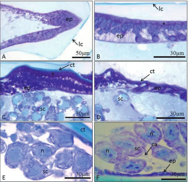
Histological analysis showed variations in the mandible according to the developmental phase of
F. schrottkyi pupae. White- and pink-eyed pupae had
only larval cuticle (Figs. 1A-B). The brown-eyed pu-pae showed both larval (external) and adult (onto the epidermis) cuticles (Figs. 1C-D). Black-eyed pupae had thicker adult cuticle (Fig. 1E).
The epidermis of mandibles in white- and pink-eyed pupae was formed by multiple cell layers with different morphology in the mandible regions. The mandible apex had irregular epidermal cells (Fig. 1A) and the basal region had columnar cells with basophil apex and central spherical nucleus with decondensed chromatin (Fig. 1B). Brown-eyed pupae showed co-lumnar epidermal cells at the base of the mandible (Fig. 1C) and flattened ones in the middle and api-cal mandible regions (Fig. 1D). Black-eyed pupae and newly emerged workers showed flattened cells in the mandible (Figs. 1E-F).
nucleus with decondensed chromatin and cytoplasm with many granules (Fig. 1E). These cells had a cana-liculus that opens on to the mandible surface in black-eyed pupae and newly emerged workers (Fig. 1F).
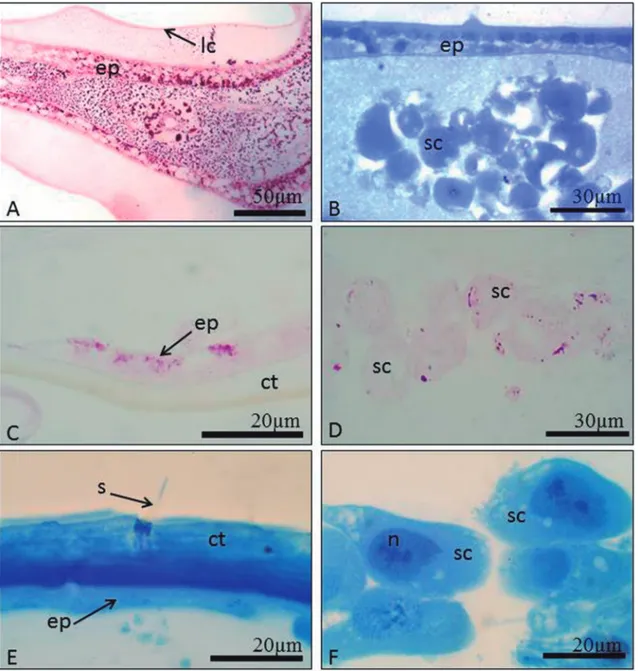
From white to brown-eyed pupae, the epidermal and spherical cells showed a strong positive reaction for PAS (Fig. 2A) and proteins (Fig. 2B). Epidermal cells of black-eyed pupae and newly emerged workers were slightly positive for PAS (Fig. 2C) and the gran-ules of spherical cells were positive (Fig. 2D) and both cell types were positive for protein in black-eyed pu-pae and newly emerged workers (Figs. 2E-F). Sensilla were present on the mandible (Fig. 1D).
DISCUSSION
The morphology of F. schrottkyi intramandibu-lar glands is simiintramandibu-lar to other Hymenoptera species (Cruz-Landim & Abdalla, 2002; Cruz-Landim et al.,
2011; Martins et al., 2013). Epidermal cells of exo-crine glands are class I with secretory features in newly emerged F. schrottkyi workers. However, large nuclei with decondensed chromatin in the spherical class III secretory cells suggest high activity (Silva-Zacarin
et al., 2008).
The new cuticle of brown-eyed F. schrottkyi pu-pae is synthesized in the previous developmental
es because the apex of the epidermal cells was positive for proteins, which may be used in the production of the new cuticle. The epidermal cells of immature in-sects synthesize the cuticle partly constituted by pro-teins, and the cuticular layers are still deposited to a lesser extent in adult insects as reported for Melipona
quadrifasciata (Cruz-Landim et al., 2011).
The multiple layers of epidermal cells in the mandible of white-eyed pupae changes from a single layer of squamous cells to the cubic epidermal cells in
black-eyed pupae, as found for Plebeia emerina (Santos
et al., 2009), Melipona rufiventris and
M. quadrifasci-ata anthioides (Costa-Leonardo, 1978). The presence
such epithelial cells, only in workers, could suggest some exclusive function for workers. Some authors attribute to the intramandibular secretion role in han-dling adhesive resins and propolis in P. emerina
(San-tos et al., 2009; Cruz-Landim et al., 2011).
Precursor cells of the class III glands found in the mandible of white-eyed pupae of F. schrottkyi are
characterized by cytoplasm granules, but with com-plete differentiation in black-eyed pupae. According to Martins et al. (2013), the cells of the class III of the
ant Pachycondyla verenae are also completely
differen-tiated in black-eyed pupae.
The large amounts of glycoconjugates and pro-teins in the mandibular cells from white to brown-eyed pupae of F. schrottkyi suggest a high metabolic rate, perhaps due to the cellular reorganization. The positive staining for polysaccharides and glycocon-jugates in the mandibles of the pupae is probably due to glycogen, which is an energetic fuel for high metabolic activity as shown in the pupae of the ant,
Solenopsis invicta, which use about 75% of their
car-bohydrates during the pupal stage (Wheeler & Buck, 1992). Mandibles of white-eyed pupae of
Pachycon-dyla verenae showed intense cell reorganization with
positive reactions for glycoconjugates and proteins (Martins et al., 2013).
The decrease of polysaccharides and glycoconju-gates in the mandibles of F. schrottkyi during develop-ment of class III cells along the pupae suggest that this gland may be associated with secretion, likely found in the ants Atta sexdens rubropilosa (Amaral & Caetano, 2006) and Pachycondyla verenae (Martins et al., 2013)
The development of intramandibular glands during the transition from prepupae to white-eyed pupae of F. schrottkyi differs from the lack of differ-entiation of the intramandibular glands in P. verenae
prepupae (Martins et al., 2013). The process of dif-ferentiation of intramandibular glands beginning in white-eyed pupae and completed in black-eyed pupae is still little known in stingless bees, but epidermal and non epidermal organs have complete differentia-tion in black-eyed pupae (Neves et al., 2002, 2003; Azevedo et al., 2008).
The post-embryonic development of intraman-dibular glands in F. schrottkyi occurs during the pupal period and completes the development during the black-eyed pupal stage.
RESUMO
As glândulas exócrinas desempenham importantes fun-ções na organização social dos insetos, como a diferen-ciação de castas e inter-castas. Devido à sua plasticidade estrutural e funcional, estudos morfológicos destas glân-dulas contribuem para o melhor entendimento da bio-logia das abelhas sociais. Por isso, o objetivo deste estudo foi acompanhar o desenvolvimento pós-embrionário das glândulas intramandibulares de operárias da abelha sem
ferrão Friesella schrottkyi (Friese, 1900)
(Hymenopte-ra: Apidae) com análises histológicas e histoquímicas. As mandíbulas das pupas em diferentes estágios do desenvol-vimento e operárias recém-emergidas foram analisadas.
As glândulas intramandibulares de F. schrottkyi são
di-vididas em dois tipos: glândulas da classe I na epiderme da mandíbula e da classe III, inseridas na cavidade da mandíbula que se abrem na superfície externa. As
glân-dulas intramandibulares de F. schrottkyi se
desenvol-vem durante a transição de pré-pupa para pupa de olho branco como observado pelas alterações morfológicas nas
células. As pupas de olhos pretos de F. schrottkyi
apre-sentaram glândulas intramandibulares completamente desenvolvidas.
Palavras-Chave: Glândulas exócrinas; Morfologia; Desenvolvimento; Abelhas sem ferrão.
ACKNOWLEDGMENTS
We would like to thank the Coordenação de Aperfeiçoamento de Pessoal de Nível Superior (CAPES), the Conselho Nacional de Desenvolvim-ento Científico e Tecnológico (CNPq), Fundação de Amparo a Pesquisa do Estado de Minas Gerais (FAPEMIG) and Empresa Brasileira de Pesquisa Ag-ropecuária (EMBRAPA) for their financial support.
REFERENCES
Amaral, J.B. & Caetano, F.H. 2006. The intramandibular gland of leaf-cutting ants (Atta sexdens rubropilosa 1908). Micron,
37:154-160.
Azevedo, D.O.; Gus-Matiello, C.P.; Rönnau, M.; Zanuncio, J.C. & Serrão, J.E. 2008. Post-embryonic development of the antennal sensilla in Melipona quadrifasciata anthidioides
(Hymenoptera: Meliponini). Microscopy Research and
Technique, 71:196-200.
Bancroft, J.D. & Gamble, M. 2008. Theory and practice of histological techniques. London, Churchill Livingstone. Bueno, O.C. 1981. Diferenciação dos ovários e determinação
do número de ovaríolos em Apis mellifera (Doctoral Thesis).
Universidade de São Paulo, São Paulo.
Costa-Leonardo, A.M. 1978. Glândulas intramandibulares em abelhas sociais. Ciência & Cultura, 30:835-838.
Cruz-Landim, C. & Abdalla, F.C. 2002. Glândulas exócrinas das
abelhas. Ribeirão Preto, FUNPEC-RP.
Cruz-Landim, C.; Gracioli-Vitti, L.F. & Abdalla, F.C. 2011. Ultrastructure of the intramandibular gland of workers and queens of the stingless bee, Melipona quadrifasciata. Journal of Insect Science, 11:1-9.
Fagundes, I.B.; Campos, L.A.O. & Serrão, J.E. 2006. Tergite pigmentation indicates hypopharyngeal gland developmental degree in Melipona quadrifasciata (Hymenoptera: Apidae: Meliponini). Sociobiology, 48:51-62.
in Pachycondyla verenae (Forel) (Hymenoptera: Formicidae) workers. Sociobiology, 60:154-161.
Martins, L.C.B.; Nascimento, F.S.; Campos, M.C.G.; Lima, E.R.; Zanuncio, J.C. & Serrão, J.E. 2015. Chemical composition of the intramandibular glands of the ant
Neoponera villosa (Fabricius, 1804) (Hymenoptera: Ponerinae).
Chemoecology, 25:25-31.
Michener, C.D. 1974. The social behavior of the bees. Cambridge, Mass., The Belkman Press of Harvard University Press. Nedel, O.J. 1960. Morphologie und Physiologie der
Mandibeldruse einiger Bienen-Arten (Apidae). Zoomorphology,
49:139-183.
Neves, C.A.; Bhering, L.L.; Serrão, J.E. & Gitirana, L.B. 2002. FMRFamide-like midgut endocrine cells during the metamorphosis in Melipona quadrifasciata anthidioides
(Hymenoptera, Apidae). Micron, 33:453-460.
Neves, C.A.; Gitirana, L.B. & Serrão, J.E. 2003. Ultrastructural study of the metamorphosis in the midgut of Melipona
quadrifasciata anthidioides (Apidae, Meliponini) worker.
Sociobiology, 41:443-459.
Noirot, C. & Quennedey, A. 1991. Gland, gland cell, glandular units: some comments on terminology and classification.
Annals of Society Entomologic of France, 27:123-128.
Santos, C.G.; Megiolaro, F.; Serrão, J.E. & Blochtein, B. 2009. Morphology of the head salivary and intramandibular glands of the stingless bee Plebeia emerina (Friese) (Hymenoptera, Meliponini) workers associated with propolis. Annals of the Entomological Society of America, 102:137-143.
Schoeters, E. & Billen, J. 1994. The intramandibular gland, a novel exocrine structure in ants (Insecta, Hymenoptera).
Zoomorphology, 114:125-131.
Silva-Zacarin, E.C.; Taboga, S.R. & Silva de Moraes, R.L. 2008. Nuclear alterations associated to programmed cell death in larval salivary glands of Apis mellifera (Hymenoptera: Apidae). Micron, 39:117-127.
Stefanini, M.; Martino, C.D.E. & Zamboni, L. 1967. Fixation of ejaculated spermatozoa for electron microscopy. Nature,
216:173-174.
Van Bethem, F.D.J.; Imperatriz-Fonseca, V.L. & Velthuis, H.W. 1995. Biology of the stingless bee Plebeia remota (Holmberg): observation and evolutionary implications. Insectes Sociaux,
42:71-87.
Wheeler, D.A. & Buck, N.A. 1992. Protein, lipid and carbohydrate use during metamorphosis in the fire ant, Solenopsis xyloni. Physiological Entomology, 17:397-403.
Aceito em: 22/05/2017 Publicado em: 13/06/2017 Editor Responsável: Kelli dos Santos Ramos
Publicado com o apoio financeiro do Programa de
Apoio às Publicações
Científicas Periódicas da USP
Produzido e diagramado na Seção de Publicações do
Museu de Zoologia da
Universidade de São Paulo
Os periódicos Papéis
A
vulsos de Zoologia
e
Arquivos de Zoologia estão licenciados sob uma Licença CC-BY