online | memorias.ioc.fiocruz.br
Risk factors for vancomycin-resistant enterococci colonisation
in critically ill patients
Deivid William da Fonseca Batistão1/+, Paulo Pinto Gontijo-Filho1,
Natália Conceição2, Adriana Gonçalves de Oliveira2, Rosineide Marques Ribas1
1Laboratório de Microbiologia, Instituto de Ciências Biomédicas, Universidade Federal de Uberlândia, Av. Pará 1720 Bloco 4 C, 38400-902 Uberlândia, MG, Brasil 2Laboratório de Microbiologia, Universidade Federal do Triângulo Mineiro, Uberaba, MG, Brasil
Vancomycin-resistant enterococci (VRE) are important hospital pathogens and have become increasingly com-mon in patients admitted to the intensive care unit (ICU). To determine the incidence and the risk factors associated with VRE colonisation among ICU patients, active surveillance cultures for VRE faecal carriages were carried out in patients admitted to the ICU of the University Hospital of Uberlândia, Minas Gerais, Brazil. Risk factors were assessed using a case-control study. Seventy-seven patients (23.1%) were found to be colonised with vanC VRE and only one patient (0.3%) was colonised with vanA VRE. Independent risk factors for VRE colonisation included nephropathy [odds ratio (OR) = 13.6, p < 0.001], prior antibiotic use (OR = 5.5, p < 0.03) and carbapenem use (OR = 17.3, p < 0.001). Our results showed a higher frequency (23.1%) of Enterococcus gallinarum and Enterococcus casseliflavus, species that are intrinsically resistant to low levels of vancomycin (vanC), without an associated infec-tion, associated with prior antibiotic use, carbapenem use and nephropathy as comorbidity. This study is the first to demonstrate the risk factors associated with vanC VRE colonisation in ICU hospitalised patients. Although vanA and vanB enterococci are of great importance, the epidemiology of vanC VRE needs to be better understood. Even though the clinical relevance of vanC VRE is uncertain, these species are opportunistic pathogens and vanC VRE-colonised patients are a potential epidemiologic reservoir of resistance genes.
Key words: risk factors - vancomycin-resistant enterococci - vanC
Vancomycin-resistant enterococci (VRE) have emerged worldwide as important nosocomial pathogens. The prevalence and incidence of VRE colonisation vary widely among hospitals and studies have suggested that such VRE rates are higher among critically ill patients, particularly those admitted to intensive care units (ICUs), limiting the therapeutic options available (Hendrix et al. 2001, Mutnick et al. 2003, Mazuski, 2008).
Glycopeptide resistance in enterococci is associated with diverse phenotypes (Werner et al. 2008) and their resistance to several antimicrobial agents, whether in-trinsic (low-level resistance to penicillin, cephalosporins and aminoglycosides) or acquired (resistance to glyco-peptides and high concentrations of aminoglycosides), is of great concern (Metallidis et al. 2006).
The most infectious cases are associated with Entero-coccus faecalis and Enterococcus faecium; in these spe-cies, the presence of vanA and vanB genes that encode high-level vancomycin resistance is of great concern due to their spread worldwide (Mazuski 2008).
Enterococcus gallinarum and Enterococcus casse-liflavus are species that are intrinsically resistant to low levels of vancomycin (vanC) and have been found to
colo-Financial support: CNPq
+ Corresponding author: deividwf b@yahoo.com.br Received 8 April 2011
Accepted 10 October 2011
nise the human intestinal tract (Toye et al. 1997, Mondino et al. 2003, Maschieto et al. 2004). Although these spe-cies have been reported to cause infections sporadically, their clinical significance remains uncertain (Mutnick et al. 2003, de Perio et al. 2006, Neves et al. 2009).
Intestinal colonisation by VRE has been associated with a variety of risk factors and asymptomatic coloni-sation by VRE often precedes true infection. Although many studies on the prevalence, incidence, epidemiol-ogy and risk factors of VRE have been performed in Europe and the United States, such data for Brazil are scarce (Warren et al. 2003, Furtado et al. 2005). More-over, despite higher rates of vanC VRE colonisation, the risk factors for colonisation by enterococci of this phe-notype have not been identified (Neves et al. 2009).
The purpose of this study was to determine the inci-dence of VRE colonisation among patients admitted to the ICU of a tertiary teaching hospital and to identify the risk factors that predispose these hospitalised patients to VRE colonisation.
PATIENTS, MATERIALS AND METhODS
Study setting and population - The University Hos-pital of Uberlândia, located in the state of Minas Gerais (MG), southeastern region of Brazil, is a 503-bed tertiary teaching hospital with a 15-bed medical and surgical ICU. We performed a prospective observational study to deter-mine the incidence of VRE colonisation among patients admitted to this ICU between April 2009-January 2010.
remained in the ICU for more than 48 h were acquired once a week until a positive VRE culture was achieved, the patient was discharged from the ICU, or the patient died. Enterococcus was considered to be ICU-acquired if it was detected from a patient who had been on the unit for more than 48 h. Patients who had serious medical conditions that precluded the collection had left the ICU prior to collection, were consenting or who had non-consenting guardians were excluded. The study protocol was approved by the hospital’s ethical committee.
Isolation, identification and susceptibility testing
- The samples obtained were transported to the micro-biology laboratory for selective culturing of VRE. The swabs were inoculated on BBLTM EnterococcoselTM Agar plates (BD, Becton, Dickinson and Company, France), supplemented with vancomycin (6 µg/mL) and incubat-ed aerobically at 35ºC for 48 h. All enterococci samples that were not sensitive to vancomycin were considered VRE. From each plate, some suspected enterococci col-onies were initially identified by Gram staining, growth in 6.5% NaCl broth and bile esculin hydrolysis. All pre-sumed enterococci were further identified as previous-ly described (Teixeira et al. 2007)using the following phenotypic tests: yellow-pigment production, motility,
L-pirrolidonil-β-naftilamida hydrolysis, acid formation
from sorbitol and arabinose and arginine hydrolysis. Antimicrobial susceptibility testing was performed on Müeller-Hinton Agar (BD, Becton, Dickinson and Company) according to recommendations of the Clini-cal and Laboratory Standard Institute (CLSI 2009). The vancomycin minimal inhibitory concentration (MIC) was evaluated by the E-test method (AB Biodisk) and
isolates with a MIC ≥ 32 μg/mL were selected for test -ing by polymerase chain reaction (PCR). The high level resistance to aminoglycosides was carried out by disc-diffusion with gentamicin (120 µg) and streptomycin
(300 μg) (Laborclin Ltda).
The isolates identified as E. gallinarum and E. cas-seliflavus with low-level resistance to vancomycin (MIC = 2-32 µg/mL) were considered as the vanC phenotype.
Epidemiological investigation - A case-control study was then performed. Cases were defined as patients with VRE colonisation, but without other infections, and the controls were defined as patients with neither VRE colo-nisation nor infection from any microorganism. The fol-lowing sociodemographic, clinical and laboratory data were evaluated: age, gender, duration of hospitalisation, prior ICU hospitalisation, underlying diseases, surgery, infections and antibiotic treatment. This information was obtained by a review of medical records and com-puter systems databases.
Prior antibiotic use was defined as the administration of antimicrobials in the 15 days before ICU admission. Prior ICU hospitalisation was defined as an inpatient admission to the ICU for any reason in the 12 months before the current ICU admission.
The defined daily dose (DDD) of antibiotics per 1,000 patients-day was calculated as previously de-scribed (Moreira et al. 2009).
Molecular methods - To assess the presence of the
vanA and vanB genes in enterococci strains with high MIC
for vancomycin (≥ 32 μg/mL), the PCR was performed as
previously described (Woodford et al. 1993). Briefly, ge-nomic DNA was extracted using a QIAamp DNA Mini Kit (Qiagen, Hilden, Germany). The amplification prim-ers for vanA gene were A1 ATGGCAAGTCAGGTGAA-GATGG and A2 TCCACCTCGCCAACAACTAACG, and for vanB gene, B1 TCTGTTTGAATTGTCTGGTAT and B2 GACCTCGTTTAGAACGATG. The cycling con-ditions for the PCR were as follows: 5 min at 94ºC, 30 cycles of 25 s at 94ºC, 40 s at 52ºC and 50 s at 72ºC and a final step of 10 min at 72ºC in a GeneAmp® PCR System 9700 (Applied Biosystems, USA). The PCR products were detected in 1.5% agarose gel by gel electrophoresis and ethidium bromide staining.
Statistical analysis - Statistical analyses were per-formed using GraphPad Prism v.4 (GraphPad Software, San Diego, CA) and Bioestat 5.0 (Belém, PA, Brazil). For determining the risk factors for VRE colonisa-tion, the control and case groups were compared using the Mann-Whitney U test for continuous data and the Fisher exact test or chi-squared test for categorical data, when appropriate. We used a Pearson’s correlation test to calculate the correlation between VRE colonisation per 1,000 patients-day and DDD of antibiotics per 1,000 patients-day. Multiple logistic regression was performed for the significant variables in the univariate analysis. Statistical significance was defined as p≤ 0.05.
RESuLTS
During the study period, 422 patients were admitted to the adult ICU at the University Hospital of Uberlân-dia. Of these patients, 328 stayed there for more than 48 h. Eighty-nine patients were excluded for the reasons listed above. From 333 patients included in the study, 78 (23.4%) were VRE-colonised (18.3/1,000 patients-day) and 35.9% (28/78) of these VRE-colonised patients had acquired the infection in the ICU. We observed that 64.1% (50/78) were already colonised with VRE upon ICU admission, including 46% (23/50) who were trans-ferred from general wards and 54% (27/50) who were from outside of the hospital.
Enterococcus spp were isolated in 90 (27%) of the 333 patients included in this study and two of them (2.2%) presented with more than one species of enterococci in the faecal samples, resulting in a total of 92 samples iso-lated. The enterococci species isolated in our study in-cluded E. casseliflavus (55.4%), E. gallinarum (28.3%),
E. faecium (12%) and E. faecalis (4.3%). We isolated only one E. faecalis strain, which carried the vanA gene.
The distribution of antimicrobial resistance among enterococcal species is presented in Table I. The results showed that only one E. faecalis isolatedwas resistant
peni-cillin resistant and none were ampipeni-cillin resistant. A high-level resistance to aminoglycoside was observed in 16.3% of the strains.
From 78 VRE-colonised patients, 21 of them were included in the case group, because they did not present any infection and the vanA VRE-colonised patient was excluded of the case group because acquired other infec-tions. From 255 VRE non-colonised patients, 143 (56.1%) had no colonisation or infection from any organism and were included in the control group. Twenty-eight VRE-colonised patients were VRE-negative at admission and became colonised during their hospital stay. None of the VRE-colonised patients developed other infections dur-ing the observation time of the study.
The results of the univariate and logistic regression analysis are shown in Tables II, III, respectively. In the univariate analyses, the cases and control groups dif-fered significantly in the following areas of their medical histories: nephropathy (p < 0.001), diabetes mellitus (p = 0.05), prior antibiotic use (p = 0.001), vancomycin use (p = 0.02) and carbapenem use (p< 0.001). In the multivari-ate analysis, significant independent risk factors for VRE colonisation were nephropathy (p< 0.001), prior antibi-otic use (p= 0.03) and carbapenem use (p< 0.001).
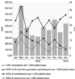
The consumption of vancomycin, carbapenems and third and fourth generation cephalosporins in the ICU represented by the DDD per 1,000 patients-day varied during the study period (Fig. 1). On average, the DDD of cephalosporins, carbapenems and vancomycin were 589.33/1,000, 243.53/1,000 and 152.78/1,000 patients-day, respectively. Despite the high density of cepha-losporin use, only the use of vancomycin (Fig. 2) and carbapenems (Fig. 3) was related to the increased inci-dence of VRE-colonisation per 1,000 patients-day, ac-cording to the Pearson’s correlation (r = 0.61, p = 0.03 and r = 0.53, p = 0.05, respectively).
DISCuSSION
This study of ICU patients found a high prevalence of VRE vanC colonisation among patients admitted to the ICU (23.1%). Prior antibiotic use, including
car-bapenem and vancomycin use in the ICU, as well as diabetes mellitus and nephropathy, were associated with VRE colonisation in ICU patients by univariate analysis. Despite the high rates of colonisation by vanC VRE enterococci among hospitalised patients, no pa-tients infected with bacteria of these phenotypes were detected in the ICU in this study. However, it is impor-tant to report that during the study period, two strains of E. gallinarum-causing bacteraemia were isolated in patients admitted to other hospital units, illustrating their role as potential opportunistic pathogens (Reid et al. 2001, Dargere et al. 2002, Choi et al. 2004).
Patients colonised by vanC VRE are a potential epi-demiologic reservoir of resistance genes. In Brazil, the first case of colonisation by E. gallinarum carrying the
vanA gene was detected in 2002 in a hospital located in the state of Rio Grande do Sul (Camargo et al. 2004) and the first case of a clinical infection was detected in 2002 in the state of Rio de Janeiro (Merquior et al. 2008). Dur-ing routine colonisation surveillance in the same hos-pital in RJ, seven vanA-carrying E. gallinarum isolates were detected (Neves et al. 2009).
Therapy with various antimicrobial agents may con-tribute to increasing colonisation by vanC VRE and oth-ers multiresistant microorganisms (Neves et al. 2009). Several studies have concluded that the consumption of antimicrobial agents is a strong indicator for multiresis-tant microorganism acquisition (Harthug et al. 2002). In our casuistic study, the use of antibiotics was present in approximately 70% of the 333 patients (223 patients) in-cluded in this study; most of these were broad spectrum-cephalosporins (50.4%), carbapenems (34.2%) and van-comycin (26.1%). This overuse of antibiotics, expressed as the DDD per 1,000 patients-day, reflects the high fre-quency of patients with infection in the ICU, although the majority of them were receiving empirical therapy with two drugs. Organisms isolated from patients in ICUs are more likely to be resistant to antibiotics than those isolated from general ward patients or outpatients (Archibald et al. 1997) because there is high antimicro-bial selection pressure in these units (Vincent 2003).
TABLE I
Species distribution of enterococci species according to resistance
Species (number tested)
Antimicrobial n (%)
RIF CLO VAN ERY PEN CIP TET AMP HLGR HLSR
Enterococcus faecium (4) 1 (25) 0 (0) 0 (0) 2 (50) 0 (0) 2 (50) 2 (50) 0 (0) 0 (0) 1 (25)
Enterococcus faecalis (11) 0 (0) 3 (27.3) 1 (9.1) 9 (81.8) 2 (18.2) 6 (54.5) 5 (45.4) 0 (0) 2 (18.2) 1 (9.1)
Enterococcus gallinarum (26) 0 (0) 1 (3.8) 0 (0) 6 (23) 0 (0) 3 (11.5) 8 (30.8) 0 (0) 0 (0) 0 (0)
Enterococcus casseliflavus (51) 1 (2) 6 (11.8) 0 (0) 25 (49) 0 (0) 5 (9.8) 15 (29.4) 0 (0) 1 (2) 10 (19.6)
Total (92) 2 (2.2) 10 (10.9) 1 (1.1) 42 (45.7) 2 (2.2) 16 (17.3) 30 (32.6) 0 (0) 3 (3.3) 12 (13 )
The consumption of third and fourth-generation ce-phalosporins, vancomycin and carbapenems in our ICU is much higher than that in many other countries (Morei-ra et al. 2009). The use of antibiotics, particularly vanco-mycin, has been identified as a risk factor for VRE colo-nisation or infection in some, but not all studies (Fridkin et al. 2001, Furtado et al. 2005, 2006, Sakka et al. 2008). Accordingly, the positive correlation in our study be-tween the incidence of VRE and the consumption of van-comycin and carbapenems in the ICU suggest that the
prevalence of antibiotic use is an important risk factor, particularly for the emergence and spread of VRE.
It has been reported that vancomycin use maintains the intestinal environment in a state that is favourable for VRE growth and promotes the possibility of an indi-vidual VRE carrier becoming a transmitter, additionally increasing the risk of a non-carrier becoming colonised (Furtado et al. 2006). Treatment with vancomycin was more common in VRE-colonised patients in the pres-ent study, but this association was not statistically sig-nificant in multivariate models. Unlike vancomycin use, carbapenem use in the ICU was an independent risk fac-tor for VRE colonisation. Recent studies suggested that some carbapenems are excreted in high concentrations in the bile, resulting in the inhibition of anaerobes and overgrowth of enterococci (Stiefel et al. 2007).
Apart from the use of antimicrobial agents and inpa-tient care in particular units, other risk factors for VRE colonisation and infection that have been identified in other studies with samples of vanA VRE include the fol-lowing: length of hospital stay, solid organ transplants, chronic dialysis, admission to the hospital at least once during the 12 months prior to ICU admission and hos-pitalisation for over three days prior to ICU admission (Ostrowsky et al. 1999, Warren et al. 2003, Furtado et al. 2005). We found that nephropathy was a risk factor for VRE colonisation. Patients with nephropathy are at TABLE II
Risk factors for vancomycin-resistant Enterococcus spp in hospitalized patients
Risk factors
Frequency (%) or mean ± SD Univariate analysis
Cases (n = 21)
Controls (n = 143)
p
(OR; 95% CI)
Age (years) 52.9 (± 17.6) 50.5 (± 18.9) 0.54
Gender
Female 38.1 41.3 0.78 (0.87; 0.34-2.24)
Male 61.9 58.7 0.78 (1.14; 0.44-2.93)
Duration of hospitalization (days) 7.4 (± 4.9) 5.8 (± 3.9) 0.18
Prior ICU hospitalization 71.4 59.4 0.29 (1.70; 0.62-4.66)
Surgery 52.4 53.1 0.94 (0.97; 0.39-2.43)
Underlying diseases
Cancer 14.3 9.8 0.46 (1.54; 0.40-5.87)
Diabetes mellitus 28.6 12.6 0.05 (2.74; 1.01-8.08)
Cardiac disease 42.9 50.3 0.52 (0.74; 0.29-1.86)
Autoimmune disease 4.8 2.8 0.50 (1.74; 0.18-16.34)
Nephropathy 23.8 4.2 < 0.001 (7.13; 1.95-26.06)
Antibiotics treatment
Prior antibiotic use 23.8 4.9 0.001 (6.38; 1.81-22.48)
Vancomycin 19 4.2 0.02 (5.37; 1.38-20.98)
Third or fourth generation cephalosporin 38.1 27.3 0.30 (1.64; 0.63-4.26)
Fluoroquinolones 9.5 5.6 0.62 (1.77; 0.35-8.99)
Carbapenems 28.6 2.8 < 0.001 (13.90; 3.52-54.87)
Overall mortality 14.3 9 0.44 (1.63; 0.43-6.29)
CI: confidence interval; ICU: intensive care unit; OR: odds ratio; SD: standard deviation.
TABLE III
Logistic regression of risk factors for vancomycin-resistant
Enterococcus spp in hospitalized patients
Risk factors p OR (95% CI)
Underlying disease
Diabetes mellitus 0.79 1.22 (0.26-5.82) Nephropathy < 0.001 13.65 (3.24-57.55) Antibiotics
Prior antibiotic use 0.03 5.52 (1.08-28.05)
Vancomycin 0.54 1.70 (0.30-9.67)
Carbapenems < 0.001 17.29 (3.42-87.56)
score) was unavailable and the analysis of comorbid con-ditions was limited by the lack of data on the patient’s underlying disease status.
In conclusion, our results showed a higher frequency of colonisation by VRE vanC, without an associated infection, associated with several risk factors, such as prior antibiotic use, carbapenem use and nephropathy as comorbidity. This study documents the emerging trend that the rates of VRE colonisation and administration of antibiotics (high DDD of antimicrobial) are very com-mon and it may provide an accurate view of the problem in the ICUs of Brazilian university hospitals.
Despite several potential limitations, this study is the first to demonstrate the risk factors associated with vanC VRE colonisation in ICU hospitalised patients. Although vanA and vanB enterococci have the greatest clinical importance, vanC VRE epidemiology should be better understood. Despite its indefinite clinical rel-evance, these species are opportunistic pathogens and vanC VRE-colonised patients are a potential epidemio-logic reservoir of resistance genes.
REFERENCES
Archibald L, Phillips L, Monnet D, McGowan JE Jr, Tenover F, Gaynes R1997. Antimicrobial resistance isolates from inpatients and outpatients in the United States: increasing importance of the intensive care unit. Clin Infect Dis24: 211-215.
Fig. 2: Pearson’s correlation coefficient between vancomycin-resist-ant enterococci (VRE) colonization per 1,000 patients-day and de-fined daily doses (DDD) of vancomycin per 1,000 patients-day.
Fig. 3: Pearson’s correlation coefficient between vancomycin-resistant enterococci (VRE) colonization per 1,000 patients-day and defined daily doses (DDD) of carbapenems per 1,000 patients-day.
an increased risk for acquiring VRE for several reasons, such as extensive contact with the hospitals, presence of multiple comorbid conditions, need for haemodialy-sis, proximity to other VRE-colonised patients, or even infected patients receiving prolonged administration of antibiotics, including vancomycin (Humphreys et al. 2004, Assadian et al. 2007).
The presence of VRE in infections is more com-mon in ICUs in North America and in parts of South America than in European ones (Guzman-Blanco et al. 2000, Mutnick et al. 2003). VRE isolation has been re-ported in some Brazilian hospitals, mainly in the state of São Paulo (Cereda et al. 1997); in our hospital, VRE was described for the first time in 2003 in a post-surgical infection caused by an E. faecalis of vanA phenotype and was the first case to be described in MG (Ribas et al. 2007). However, high level aminoglycoside resis-tance has not been uncommon and frequencies of 18.5% in SP (Maschieto et al. 2004),22% in Brasília (Titze-de-Almeida et al. 2004) and 40.5% in RJ (Mondino et al. 2003) have been observed. In the present study in Uber-lândia, 16.3% of the isolates cultured from colonised individuals showed high levels of aminoglycoside resis-tance. An increase in the frequency of enterococci with high level aminoglycoside resistance has recently been observed due to spread of plasmids and transposons that encode enzymes that inactivate aminoglycosides (Sood et al. 2008, Protonotariou et al. 2010).
Several potential limitations of the present study should be mentioned. First, we had a relatively small number of patients in the case group, thus reducing the statistical power and the ability to study subsets of pa-tients. Another limitation of this study was that informa-tion on the management of the patient’s condiinforma-tion and severity of the underlying illness (e.g., the APACHE II
Assadian O, Askarian M, Stadler M, Shaghaghian S 2007. Preva-lence of vancomycin-resistant enterococci colonization and its risk factors in chronic haemodialysis patients in Shiraz, Iran.
BMC Infect Dis7: 52.
Camargo IL, Barth AL, Pilger K, Seligman BG, Machado AR, Darini AL 2004. Enterococcus gallinarum carrying the vanA gene clus-ter: first report in Brazil. Braz J Med Biol Res 37: 1669-1671. Cereda R, Pignatari AC, Hashimoto A, Sader H 1997. In vitro
an-timicrobial activity against enterococci-isolated in a University Hospital in São Paulo-Brazil. J Infect Dis1: 83-90.
Choi SH, Lee SO, Kim TH, Chung JW, Choo EJ, Kwak YG, Kim MN, Kim YS, Woo JH, Ryu J, Kim NJ 2004. Clinical features and outcomes of bacteraemia caused by Enterococcus casseli-flavus and Enterococcus gallinarum: analysis of 56 cases. Clin Infect Dis38: 53-61.
CLSI - Clinical Laboratory and Standards Institute 2009. Perfor- mance standards for antimicrobial susceptibility testing. �ine- �ine-teenth Informational Supplement M100-S19, CLSI, Wayne, 177 pp. Dargere S, Vergnaud M, Verdon R, Saloux E, Le Page O, Leclercq R,
Bazin C 2002. Enterococcus gallinarum endocarditis occurring on native heart valves. J Clin Microbiol40: 2308-2310.
de Perio MA, Yarnold PR, Warren J, Noskin GA2006. Risk factor and outcomes associated with non-Enterococcus faecalis,
non-Enterococcus faecium enterococcal bacteraemia. Infect Control Hosp Epidemiol27: 28-33.
Fridkin SK, Edwards JR, Courval JM, Hill H, Tenover FC, Lawton R, Gaynes RP, McGowan JE Jr, Intensive Care Antimicrobial Resistance Epidemiology (ICARE) Project, the National Noso-comial Infections Surveillance (NNIS) System Hospitals 2001. The effect of vancomycin and third generation cephalosporins on prevalence of vancomycin-resistant enterococci in 126 USA adult intensive care units. Ann Intern Med135: 175-183.
Furtado GH, Martins ST, Coutinho AP, Wey SB, Medeiros EA 2005. Prevalence and factors associated with rectal vancomycin-resist-ant enterococci colonization in two intensive care units in São Paulo, Brazil. Braz J Infect Dis9: 64-69.
Furtado GH, Mendes RE, Pignatari AC, Wey SB, Medeiros EA 2006. Risk factors for vancomycin-resistant Enterococcus faecalis bac-teremia in hospitalized patients: an analysis of two case-control studies. Am J Infect Control 34: 447-451.
Guzman-Blanco M, Casellas JM, Sader HS 2000. Bacterial resistance to antimicrobial agents in Latin America. The giant is awaken-ing. Infect Dis Clin �orth Am14: 67-81.
Harthug S, Jureen R, Mohn SC, Digranes A, Simonsen GS, Sunds-fjord A, Langeland N, Norwegian Enterococcal Study Group 2002. The prevalence of faecal carriage of ampicillin-resistant and high-level gentamicin-resistant enterococci among inpatients at 10 major Norwegian hospitals. J Hosp Infect50: 145-154. Hendrix CW, Hammond JM, Swoboda SM, Merz WG, Harrington SM,
Perl TM, Dick JD, Borschel DM, Halczenko PW, Pelz RK, Rocco LE, Conway JE, Brower RG, Lipsett PA 2001. Surveillance strate-gies and impact of vancomycin resistant enterococcal colonization and infection in critically ill patients. Ann Surg233: 259-265. Humphreys H, Dolan V, Sexton T, Conlon P, Rajan L, Creamer E,
Walshe J, Donohoe J, Smyth EG 2004. Implications of coloniza-tion of vancomycin-resistant enterococci (VRE) in renal dialysis patients. Learning to live with it? J Hosp Infect58: 28-33. Maschieto A, Martinez R, Palazzo IC, Darini ALC 2004. Antimicro-
Antimicro-bial resistance of Enterococcus sp. isolated from the intestinal tract of patients from a university hospital in Brazil. Mem Inst Oswaldo Cruz99: 763-767.
Mazuski JE 2008. Vancomycin-resistant enterococcus: risk factors, surveillance, infections and treatment. Surg Infect9: 567-571. Merquior VLC, Neves FPG, Ribeiro RL, Duarte RS, Marques EA,
Teixeira LM 2008. Bacteraemia associated with a vancomycin-resistant Enterococcus gallinarum strain harbouring both the
vanA and vanC1 genes. J Med Microbiol57: 244-245.
Metallidis S, Chatzidimitriou M, Tsona A, Bisiklis A, Lazaraki G, Koumentaki E, Gikas A, Alexiou-Daniel S, Nikolaidis P 2006. Vancomycin-resistant enterococci, colonizing the intestinal tract of patients in a university hospital in Greece. Braz J Infect Dis 10: 179-184.
Mondino SS, Castro AC, Mondino PJ, Carvalho M da G, Silva KM, Teixeira LM 2003. Phenotypic and genotypic characterization of clinical and intestinal enterococci isolated from inpatients and out-patients in two Brazilian hospitals. Microb Drug Resist9: 167-174. Moreira MR, Ribas RM, Rodrigues AAA, Gontijo Filho PP 2009.
Consumo de antibióticos e etiologia de pneumonia associada à ventilação em pacientes internados na unidade de terapia inten-siva do Hospital de Clínicas da Universidade Federal de Uberlân-dia. Rev Panam Infectol11: 11-16.
Mutnick AH, Biedenbach DJ, Jones RN 2003. Geographic variations and trends in antimicrobial resistance among Enterococcus faecalis and
Enterococcus faecium in the SENTRY Antimicrobial Surveillance Program (1997-2000). Diag Microbiol Infect Dis46: 63-68. Neves FP, Ribeiro RL, Duarte RS, Teixeira LM, Merquior VL 2009.
Emergence of the vanA genotype among Enterococcus galli-narum isolates colonizing the intestinal tract of patients in a university hospital in Rio de Janeiro, Brazil. Int J Antimicrob Agents33: 211-215.
Ostrowsky BE, Venkataraman L, D’Agata EM, Gold HS, De Giro-lami PC, Samore MH1999. Vancomycin-resistant enterococci in intensive care units: high frequency of stool carriage during a non-outbreak period. Arch Intern Med159: 1467-1472.
Protonotariou E, Dimitroulia E, Pournaras S, Pitiriga V, Sofianou D, Tsakris A 2010. Trends in antimicrobial resistance of clinical isolates of Enterococcus faecalis and Enterococcus faecium in Greece between 2002 and 2007. J Hosp Infect3: 225-227. Reid KC, Cockerill III FR, Patel R 2001. Clinical and epidemiological
features of Enterococcus casseliflavus/flavescens and Entero-coccus gallinarum bacteraemia: a report of 20 cases. Clin Infect Dis32: 1540-1546.
Ribas RM, Darini AL, Moreira TA, Freitas C, Gontijo Filho PP2007. Vancomycin-resistant vanA phenotype Enterococcus faecalis: first case in Minas Gerais state and epidemiological consider-ations. Braz J InfecDis11: 439-440.
Sakka V, Tsiodras S, Galani L, Antoniadou A, Souli M, Galani I, Pan-telaki M, Siafakas N, Zerva L, Giamarellou H 2008. Risk-factors and predictors of mortality in patients colonized with vancomy-cin-resistant enterococci. Clin Microbiol Infect14: 14-21. Sood S, Malhotra M, Das BK, Kapil A 2008. Enterococcal infections
& antimicrobial resistance. Indian J Med Res128: 111-121. Stiefel U, Pultz NJ, Donskey CJ 2007. Effect of carbapenem
admin-istration on establishment of intestinal colonization by vanco-mycin-resistant enterococci and Klebsiella pneumoniae in mice.
Antimicrob Agents Chemother51: 372-375.
Teixeira LM, Siqueira-Carvalho MG, Facklam RR 2007. Enterococ-cus. In PR Murray, EJ Baron, JH Jorgensen, ML Landry, MA Pfaller (eds.), Manual of clinical microbiology, ASM Press, Washington DC, p. 430-442.
Boelens H, Van Belkum A, Felipe MS 2004. Molecular epidemi-ology and antimicrobial susceptibility of enterococci recovered from Brazilian intensive care units. Braz J Infect Dis8: 197-205. Toye B, Shymanski J, Bobrowska M, Woods W, Ramotar K 1997.
Clinical and epidemiologic significance of enterococci intrinsi-cally resistant to vancomycin (possessing the vanC genotype).
J Clin Microbiol35: 3166-3170.
Vincent JL 2003. Nosocomial infections in adult intensive-care units.
Lancet361: 2068-2077.
Warren DK, Kollef MH, Seiler SM, Fridkin SK, Fraser VJ 2003. The epidemiology of vancomycin-resistant Enterococcus
coloniza-tion in a medical intensive care unit. Infect Control Hosp Epide-miol24: 257-263.
Werner G, Coque TM, Hammerum AM, Hope R, Hryniewicz W, Johnson A, Klare I, Kristinsson KG, Leclercq R, Lester CH, Lillie M, Novais C, Olsson-Liljequist B, Peixe LV, Sadowy E, Simonsen GS, Top J, Vuopio-Varkila J, Willems RJ, Witte W, Woodford N 2008. Emergence and spread of vancomycin resist-ance among enterococci in Europe. Euro Surveill 13: 1-11. Woodford N, Morrison D, Johnson AP, George RC 1993.