Xylella fastidiosa
gene expression analysis by DNA microarrays
Regiane F. Travensolo
1,2, Lucia M. Carareto-Alves
1, Maria V.C.G. Costa
1, Tiago J.S. Lopes
1,
Emanuel Carrilho
2and Eliana G.M. Lemos
11
Departamento de Tecnologia, Faculdade de Ciências Agrárias e Veterinárias de Jaboticabal,
Universidade Estadual Paulista “Júlio de Mesquita Filho”, Jaboticabal, SP, Brazil.
2Instituto de Química de São Carlos, Universidade de São Paulo, São Carlos, SP, Brazil.
Abstract
Xylella fastidiosa genome sequencing has generated valuable data by identifying genes acting either on metabolic pathways or in associated pathogenicity and virulence. Based on available information on these genes, new strate-gies for studying their expression patterns, such as microarray technology, were employed. A total of 2,600 primer pairs were synthesized and then used to generate fragments using the PCR technique. The arrays were hybridized against cDNAs labeled during reverse transcription reactions and which were obtained from bacteria grown under two different conditions (liquid XDM2and liquid BCYE). All data were statistically analyzed to verify which genes were differentially expressed. In addition to exploring conditions forX. fastidiosa genome-wide transcriptome analysis, the present work observed the differential expression of several classes of genes (energy, protein, amino acid and nu-cleotide metabolism, transport, degradation of substances, toxins and hypothetical proteins, among others). The un-derstanding of expressed genes in these two different media will be useful in comprehending the metabolic characteristics ofX. fastidiosa, and in evaluating how important certain genes are for the functioning and survival of these bacteria in plants.
Key words: Xylella fastidiosa, DNA microarray, gene expression.
Received: July 21, 2008; Accepted: November 24, 2008.
Introduction
X. fastidiosa (Wells et al., 1987) belongs to the gram-negative group and is restricted to the xylem vessels of host plants. It has been associated with diseases that af-fect diverse plant species, some of which economically im-portant, these including alfalfa, almonds, blackberries, coffee, citrus fruits, grapes, peaches, pears, plums and cer-tain ornamental plants (Hopkins, 1989). Citrus Variegated Chlorosis (CVC) was first detected in Brazil in 1987, and currently constitutes a serious threat to the Brazilian orange juice industry, since it is present in the main cultivation ar-eas, being responsible for significant losses in orange pro-duction (Rossettiet al., 1990). In 2000, a consortium of laboratories in São Paulo State published theX. fastidiosa isolate 9a5c sequenced genome. A main chromosome (2,679,305 base pairs) and two other plasmids (51,158 and 1,285 base pairs) were sequenced, these presenting a total of 2,905 genes, from which half presented similarity with unknown protein functions (Simpsonet al., 2000).
Understanding the complete genome sequence was a substantial advance towards comprehension of metabolic and replicate characteristics, and for starting the first ap-proach in determining pathogenicity mechanisms. Papers published recently have explored the information generated by genomic sequencing, highlighting a series of hypotheses related to the functioning of energy metabolism, nutrient transport, adherence, aggregation, toxicity, the secretion of pathogenicity factors, intercellular interactions, iron ho-meostasis, antioxidant responses and other important pathogenicity mechanisms (Simpsonet al., 2000; Keenet al., 2000; Dow and Daniels, 2000; Lambaiset al., 2000; Silvaet al., 2001; Leiteet al., 2002; Meidaniset al., 2002).
It is known thatX. fastidiosademands a complex me-dium for itsin vitrodevelopment (Holtet al., 1994). With X. fastidiosa genome sequencing, the possible genes in-volved in bacterial metabolism have become known, and as a result, a defined and adequate medium for cultivatingX. fastidiosa, known as XDM2(Xylelladefined medium), was
set up (Lemoset al., 2003). The components of this me-dium have been included based on metabolic pathways found with the help of information obtained from the X. fastidiosagenome. XDM2contains glucose, vitamins
(bio-tin, thiamine, pyridoxine hydrochloride and nicotinic acid) and amino acids (serine, methionine, asparagine and
www.sbg.org.br
Send correspondence to Eliana G.M. Lemos. Departamento de Tecnologia, Faculdade de Ciências Agrárias e Veterinárias, Uni-versidade Estadual Paulista, Via de Acesso Prof. Paulo Donato Castellane s/n, 14884-900 Jaboticabal, SP, Brazil. E-mail: [email protected].
glutamine), as well as iron, phosphate, sulfate and myo-inositol. The XDM2medium has made it possible to
culti-vateX. fastidiosamore successfully than by using the com-plex BCYE modified media (Campanharo et al., 2003), which presents in its formulation only yeast extract and an ACES buffer. Furthermore, differences among X. fastidiosaisolates, obtained from various host plants, have been observed. These are related to their cultivation in me-dia of different compositions, thereby indicating the exis-tence of genetic variability within this group of bacteria (Hopkins, 1989).
The present accumulation of information with the se-quencing of genomes from various organisms has offered an enormous opportunity to understand the biological func-tions of many genes, previously described as unknown (Lashkariet al., 1997). Furthermore, microarray technol-ogy (Shalonet al., 1996) provides a simultaneous way for immediately monitoring the expression of several genes. In practice, it is possible to arrange about 6,000 elements (genes) in an area of less than 1.8 cm2. A nearly complete collection of 4,290Escherichia coliopen reading frames (ORFs) was obtained for analyzing the expression ratio of this bacterium when cultivated in two different media (min-imal and rich). Bacteria cultivated in a rich medium pre-sented accelerated multiplication, the higher number of genes significantly expressed being related to the transla-tion apparatus. On the other hand, bacteria cultivated in the minimal medium showed elevated expression of many of those genes involved in biosynthetic pathways, mainly in the amino acids (Taoet al., 1999).
The aim of this work was to develop a DNA micro-array analysis set, and undertake a transcriptional study of those genes related to the metabolism of theX. fastidiosa 9a5c strain isolated from citrus fruits, when cultivated un-der two distinct conditions, BCYE (complex media) and XDM2(defined media).
Materials and Methods
Cultivation conditions
For genomic DNA extraction,X. fastidiosa isolate 9a5c was cultivated in ‘Petri dishes’ containing BCYE me-dium (Wellset al., 1981) at 28 °C for six days. For RNA ex-traction, the bacteria were cultivated for four days at 30 °C in a 250 mL Erlenmeyer flask containing 30 mL of either liquid XDM2medium (Lemoset al., 2003) or liquid
modi-fied BCYE (Campanharoet al., 2003) under shaking condi-tions (140 rpm).
Isolation of genomic DNA and total RNA
Genomic DNA was extracted according to the de-scribed methodology (Ausubelet al., 1987) with a modifi-cation that includes a step with RNAse treatment as fol-lows: 200mg/mL, 1.5 h at 37 °C. The methodology used for RNA extraction (Chomczynski and Sacchi, 1987) involved
a monophasic solution of phenol and guanidine isothio-cyanate (Trizol - Invitrogen). The RNA samples treated with DNAse I were purified through the NucleoSpin®RNA II BD Bioscience kit (Clontech), resuspended in H2ODEPC
and stored at -80 °C.
Synthesis of fluorescent cDNA from total RNA
The construction of fluorescent cDNA for hybridiza-tion reachybridiza-tions was by means of a CyScribe cDNA Post La-beling kit (Amersham Biosciences) with 30mg of RNA and 15mg of random primers (Amersham Bioscience). The re-verse transcriptase reaction occurred at 37 °C for 3 h in a programmable thermocycler (PTC-100 Programmable Thermal Controller - MJ Research, Inc.). As the control of the transcriptase reaction, 1mL of the synthetic RNA from the Lucidea Universal ScoreCard kit (Amersham Biosci-ences) was used. The reaction was neutralized with 20mL of 2 M of HEPES, and the cDNAs were purified through precipitation with 3 M of sodium acetate and 75 mL of 100% v/v ethanol, and kept at - 20 °C overnight. After cen-trifuging and washing with 70% ethanol, the cDNA was resuspended in 30mL of CyDye diluted in 0.1 M of bicar-bonate of soda, pH 9.0. The sample was kept in the dark at 25 °C for 1 h, and the labeling reactions were stopped by the addition of 15mL of 4 M of hydroxylamine for 15 min at 25 °C. The sample was then resuspended in 400mL of a TE buffer (10 mM of Tris-HCl pH 8.0, 1 mM of EDTA) and concentrated in a Microcon-type column -YM30 (Milli-pore). The efficiency of the reading was monitored by mea-suring absorption at 260 nm (for DNA concentration), 550 nm (for Cy3) and 650 nm (for Cy5).
Amplification ofX. fastidiosagenes
Primers were used to amplify the 2,600 ORFs of the X. fastidiosagenome. These primers were built for both forward and reverse directioning, ranging from 16 to 19 nu-cleotides in length and from 48 °C to 57 °C in Tm (melting temperature). PCR reactions were done in a total volume of 100mL containing a PCR buffer (50 mM of KCl, 200 mM of Tris-HCl pH 8.4), 2 mM of MgCl2, 10 mM of dNTP, 2 U
Microarray construction
The amplified products were suspended in 50% v/v DMSO in a final concentration of 100 to 300 ng/mL and ar-ranged in duplicate at a distance of 250mm in glass slides treated with aminosilane (Corning). Printing of micro-arrays was done by a robot model GMS 417 Arrayer (Affymetrix Inc.). After printing, the DNAs were re-hydrated (42 °C for 10 s), dried (70 °C for 1 min) and fixed in a UV camera cross-link (1300 x 100mJ cm2). The slides were kept at 70 °C for 2 h and then stored under vacuum at room temperature. Genetically distant negative controls were also included in this array, these consisting of human genes (pHUM1 and pHUM7) and plant genes (707050B11 - Rubisco), as well as synthetic controls from various spe-cies (human, mouse, Arabidopsis spp., Archaeabacteria andE. coli) obtained by the Lucidea Universal ScoreCard kit (Amersham Biosciences).
Hybridization and washing
Hybridization and washing were carried out in a GeneTac Hybridization (Genetic MicroSystems) device. Initially, slides containing the microarrays were denatured at 65 °C for 5 min. A solution containing 8mL of blocking liquid (Amersham Biosciences RPN 3601), 19mL of SSC 20x, 5.5mL SDS 2% w/v and 100 pmol of cDNA marked with the fluorescent dyes Cy3 and Cy5, totaling 110mL, was denatured at 95 °C for 2 min, deposited on the slide and kept for 12 h at 42 °C. After hybridization, the slides were washed at 25 °C in the following solutions: 2x SSC/SDS 0.5% w/w, 0.5x SSC and 0.05x SSC. All washing-steps consisted of 10 cycles with 10 s of solution flux and 20 s of incubation. The slides were then dried for 15 min and sub-mitted to fluorescence detection.
Image acquisition and data analyses
The slides were submitted to fluorescence reading in a model GMS 418 Arrayer Scanner (Affymetrix Inc.) under different wavelengths - 550 nm (Cy3) and 650 nm (Cy5). The location and identity of each gene on the slide was de-fined in a text file, created with the aid of the Clone-Tracker2 program (BioDiscovery). The signal was quantified through ImaGene software (v. 4.1, BioDisco-very), in which two images from the Cy3 and Cy5 fluores-cent dyes were overlapped and the spots classified according to morphology and intensity. The computer dis-plays an electronic symbol as a false-color image where a red or green spot corresponds to expression of a gene in sample 1 or 2, respectively, while a yellow-orange spot in-dicates that the gene was expressed at similar levels in both samples. For transformation of data, the background signal was discounted from the signal of each spot using the local background obtained by the GeneSight (BioDiscovery) program. The transformation sequence included back-ground correction, omitted flagged spots, combined
repli-cates and floor, by adding a shifted Log transformation and ratio.
Data obtained from the intensity ratio of the signal measured by the Cy5 (experiment) over the Cy3 (control) were normalized according to the average intensity of the total signal. We used all the genes in our dataset to calculate normalization since this assumes that the majority of the measured genes were not differentially regulated. The nor-malization procedure is a suitable approach for minimizing variations so that a common base for comparison is estab-lished. There are a number of reasons that justify data nor-malization, these including unequal quantities of starting RNA, differences in labeling or detection efficiency among the different fluorescent dyes used, and systematic bias in measured expression levels (Quackenbush, 2002). How-ever, current normalization methods are not applicable to all conditions. Normalization can be carried out in several different ways, such as within the slide in order to adjust dye incorporation efficiency, between two slides for dye swap experiments and across slides for repetition of the same experiments (Yanget al., 2001). In the latter case, ap-plication would be to the entire data set (overall normaliza-tion), instead of to a particular physical data subset or sub grid (local normalization).
Final intensity of hybridization was determined in all the experiments from six replicates per data point, and is representative for three independent determinations (slides) from each media culture. Replicates in duplicate within each slide were combined by the median of their val-ues, whereupon statistical analysis was carried out using the SAM method (Significance Analysis of Microarrays). This method is based ont-test statistics and is employed to calculate the false discovery rate (FDR) and gene error chance (q-value) (Tusheret al., 2001). Significant varia-tions in expression of those genes related toX. fastidiosa metabolism were compared when cultivated in liquid mod-ified BCYE and liquid XDM2median.
Detection of cDNA by micro-chip electrophoresis
The reverse-transcription step for generation of cDNA was performed in a final volume of 20mL using 1mg of total RNA digested with 0.5mM DNAse I. The random primer (1mM) and digested RNA were denatured for 5 min at 70 °C, then immediately cooled on ice for another 5 min and added to a 15mL RT mix containing 2.0 mM of dNTPs, 3 mM of MgCl2, 1x RT buffer and 1mL of ImProm-II RT
(all from Promega, Madison, WI, USA). The mixture was incubated for 5 min at 25 °C, 60 min at 40 °C, and 15 min at 70 °C. PCR reactions were set up in 10mL total reaction volume containing 1x PCR buffer, 2 mM of MgCl2, 10 mM
the following conditions: 94 °C for 2 min, 35 cycles (94 °C for 1 min, 58 °C for 1 min and 72 °C for 1 min and 30 s) and a final step at 72 °C for 5 min. All products were analyzed by using the Bioanalyzer 2100 (Agilent Technologies, Waldbronn, Germany) in conjunction with the LabChip DNA 500 kit, according to manufacturer’s instructions.
Results and Discussion
Synthesis of fluorescent labeled cDNA
In order to verify gene expression differently, we ana-lyzed the growth ofX. fastidiosafrom four days after cul-ture in two different liquid media, modified BCYE and XDM2(Table 2). Based on the genetic analysis of theX.
fastidiosagenome, Lemoset al.(2003) developed certain media with a defined composition, whereby the growth abilities of these bacteria were evaluated in both liquid me-dia and on solid plates.X. fastidiosagrowth was compared in XDM2defined media as well as in BCYE by measuring
cell turbidity and protein content for 14 days at 28 °C under shaking conditions. The authors observed that, after 14 days, the growth rate of bacteria on complex media, such as BCYE was substantially lower than in XDM2, and that, in
the latter, the strains grew equally well both in liquid and on solidified media. However, after four days (96 h) cell tur-bidity and protein content were similar for both.
Preparation of fluorescent labeled cDNAs was car-ried out by total RNA extraction fromX. fastidiosagrowth after four days in liquid modified BCYE and liquid XDM2
media, its concentration being determined by absorbance measurement at 260 nm (A260). RNA integrity was checked
by formaldehyde agarose gel electrophoresis, where the oc-currence of two ribosomal subunit bands (23S and 16S con-taining of 2.9 and 1.5 kb, respectively) was examined (data not shown).
Fluorescent labeled cDNAs were prepared from total X. fastidiosaRNA by reverse transcription. Total RNA was used since most of the mRNAs produced by bacteria do not have a poly (A) tail and are difficult to separate. Labeling efficiency by reverse transcription depends on incorpora-tion efficiency and on the amount of specific nucleotides present in a particular mRNA species. The labeling kit used was developed as a two-step procedure. The first step
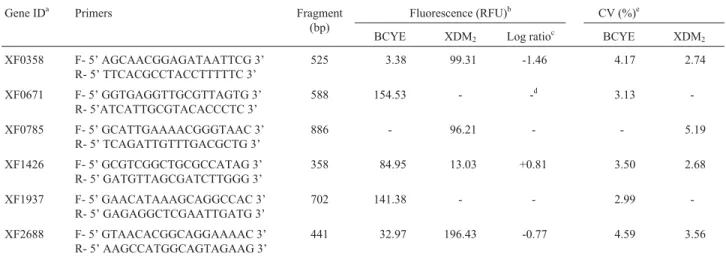
in-Table 1- Nucleotide sequences of the primer used to detect cDNA, fluorescence and the coefficient of variation obtained in microchip electrophoresis.
Gene IDa Primers Fragment
(bp)
Fluorescence (RFU)b CV (%)e
BCYE XDM2 Log ratioc BCYE XDM2
XF0358 F- 5’ AGCAACGGAGATAATTCG 3’ R- 5’ TTCACGCCTACCTTTTTC 3’
525 3.38 99.31 -1.46 4.17 2.74
XF0671 F- 5’ GGTGAGGTTGCGTTAGTG 3’ R- 5’ATCATTGCGTACACCCTC 3’
588 154.53 - -d 3.13
-XF0785 F- 5’ GCATTGAAAACGGGTAAC 3’ R- 5’ TCAGATTGTTTGACGCTG 3’
886 - 96.21 - - 5.19
XF1426 F- 5’ GCGTCGGCTGCGCCATAG 3’ R- 5’ GATGTTAGCGATCTTGGG 3’
358 84.95 13.03 +0.81 3.50 2.68
XF1937 F- 5’ GAACATAAAGCAGGCCAC 3’ R- 5’ GAGAGGCTCGAATTGATG 3’
702 141.38 - - 2.99
-XF2688 F- 5’ GTAACACGGCAGGAAAAC 3’ R- 5’ AAGCCATGGCAGTAGAAG 3’
441 32.97 196.43 -0.77 4.59 3.56
a
Simpsonet al.(2000);bRate of fluorescence (RFU) obtained from replicate media;cLog expression ratios of measured transcript levels determined for the two cultures. The log expression ratio is positive for genes that were more highly expressed in BCYE medium. The log is negative for genes that were more highly expressed in XDM2medium;dGenes that expressed only one condition andeCoefficient of Variation (CV).
Table 2- Components in BCYE and XDM2media, developed forX.
fastidiosa.
Components BCYE XDM2
Glucose (10 g/L) -a +b
K2HPO4(2.1 g/L) - +
KH2PO4(0.8 g/L) - +
MgSO47H2O (0.4 g/L) - +
Ferric pyrophosphate (0.125 g/L)c + +
Aces buffer (10 g/L) +
-Activated charcoal (2 g/L) +
-Yeast extract +
-L-cysteine (0.4 g/L) +
-L-serine (0.4 mg/mL) - +
L-asparagine (1.0 mg/mL) - +
L-methionine (0.4 mg/mL) - +
L-glutamine (4.0 mg/mL) - +
Vitamin stock solution (10 mL/L)d - +
Biotin (0.2 mL/L) - +
Phenol red (0.1%) - +
a
(-) components taken from the media,b(+) components added to the me-dia,cin the BCYE media, the ferric pyrophosphate concentration was
0.25 g/L and thedVitamin stock solution (10 mL/L) contained 0.2 mgD
volves the incorporation of amino allyl-dUTP (AA-dUTP) during cDNA synthesis by using an optimized nucleotide mixture. The second step involves chemically labeled amino allyl-modified cDNA using CyDye NHS-esters. Coupling reactions of amino allyl-modified cDNA were performed separately with Cy3 and Cy5 and both targets were combined in the hybridization solution. The amount of target used for hybridization depends on array format and labeling method. Targets containing 100 pmol of incor-porated fluorescent dye were employed. Such an amount was calculated from the formulas, (OD550x dilution factor x
total volume)/0.15 for Cy3 and (OD650x dilution factor x
total volume)/0.25 for Cy5, where the obtained values are in pmol.
Arraying amplifiedX. fastidiosagenes
DNA arrays were developed through the synthesis of 2,600 amplicons using pairs of primers related to each of theX. fastidiosagenome ORFs. Amplified ORFs were set in a concentration varying from 100 to 300 ng/mL, with fragment-size also varying from 300 to 1,000 bp. The DNA arrays were composed of amplicons that did not need to be purified. The spots were printed in duplicate with a 250mm distance between each, to a total of 5,200 spots, including positive and negative controls. Studies in microarray gene expression analysis, using unpurified amplified products, emphasized non-significant differences between purified and unpurified PCR products, showing a low alteration level in the hybridization signal (6%) in the latter, when compared to the purified version (Diehlet al., 2002).
The comparison of the expression of X. fastidiosa 9a5c genes when isolated from bacteria cultivated in the two different media (liquid modified BCYE and liquid XDM2) was carried out by using SAM software which
de-velops a statistical evaluation of probes’ hybridization pat-terns. The significance of gene expression differences was calculated by the ratio of median fluorescence intensities for each condition, having as parameters a ratio difference of at least 1.5x together with a threshold D of 0.49514. Missing data points were estimated with a K-Nearest-Neighbor imputator equal to 10. The test was undertaken with response format type paired data with a false discov-ery rate (FDR) of less than 0.5%.
Data analysis resulted in a 0.42 false positive rate (FSN - False Significant Number) and 0.31 of false discov-ery genes, thus demonstrating that 99.69% of our experi-ments present positive results and only 0.31% are false. Among the analyzed genes, approximately 5.15% (134) were detected as differentially expressed in both studied conditions. 30 of these (22.4%) showed higher expression in the BCYE medium and 104 (77.6%) in the XDM2
(Table 3). All gene-chip data can be found in the Gene Ex-pression Omnibus (GEO) Repository under accession num-ber GPL7554.
According to the results obtained through microarray analysis, bacteria cultivated in XDM2medium expressed a
higher number of significant genes than those cultivated in BCYE modified medium. This was expected, since the XDM2defined medium offers a smaller variety of nutrients
than the BCYE complex medium. These differences in gene expression patterns were analyzed in detail, as de-scribed below.
Genes involved in energy metabolism
Significantly high expression levels were observed when bacterial cells were cultivated in the XDM2medium
for the following genes: gapA (glyceraldehyde-3-phos-phate dehydrogenase), rfbC (DTDP-4-keto-L-rhamnose reductase), mdh (malate dehydrogenase), odhA (oxoglu-tarate dehydrogenase),pfkA(6-phosphofrutokinase),gcvT (glycine cleavage T protein),fumB(fumarate hydratase), az1(electron transfer protein azurin I), tpiA (triosephos-phate isomerase), yahK (alcohol dehydrogenase), petB (ubiquinol cytochrome C oxidoreductase), atpG (ATP synthase) and acnB (aconitate hydratase 2), whereas, a higher expression was observed for the following genes in those cells cultivated in the BCYE medium: pdhB (dihydrolipoamide acetyltransferase) and pykA (pyruvate kinase type II).
The functionality of the glycolytic pathway in X. fastidiosawas evaluated (Facincaniet al., 2003) by study-ing the enzymes phosphoglucose isomerase, aldolase, gly-ceraldehyde-3-phosphate dehydrogenase and pyruvate kinase from the glycolytic pathway, and glucose-6-phos-phate dehydrogenase from the Entner-Doudoroff, followed by cloning and expression studies of the enolase gene and determination of its activity. These studies showed thatX. fastidiosadoes not use the glycolytic pathway to metabo-lize carbohydrates. As a result, no enzymatic activity was detected for enolase, aldolase and glyceraldehyde-3-phos-phate dehydrogenase, this suggesting thatX. fastidiosamay be using the Entner-Doudoroff pathway to produce pyru-vate as an alternative. Nevertheless, an increase in gene ex-pression of those enzymes related to the glycolytic pathway in the cultivated cells was detected through microarray analysis, this regardless of the supporting XDM2medium.
This set of genes codes for 6-phosphofrutokinase, glyceral-dehyde-3-phosphate dehydrogenase and triosephosphate isomerase (data collected from the cells raised on XDM2),
as well as for pyruvate kinase (data collected from cells raised in a BCYE modified medium).
fastidiosauses cell respiration to obtain energy from glu-cose.
Transport related genes
A total of 140 genes that code for proteins related to the transport of a number of biological molecules were identified in theX. fastidiosa genome, these representing 4.8% of all ORFs (Simpsonet al., 2000). Among these genes, 19 were considered differentially expressed in this comparison, 14 being detected as higher expressed in XDM2medium conditions and five in BCYE. These genes
refer to the transport of anions, cations, carbohydrates, pep-tides, proteins and substances related to secretory path-ways. Genes involved with the secretion of peptides and proteins (xpsHandsecYin XDM2medium,xpsDandsecA
in BCYE) were expressed in both media, these four being related to general secretion and Sec systems. ThexpsHand xpsDgenes expressed code for external membrane proteins that act in the General Secretory Pathway (GSP) Type II. The secreted enzymes in this pathway include polygalac-turonate lyase, endoglucans, a- amylase and proteases (Goughet al., 1988; Huet al., 1992). On the other hand, in gram-negative bacteria such asX. fastidiosa, macromole-cules, which include excreted enzymes, toxins and struc-tures from the surface of the cell, need to pass through both the internal and external membranes before reaching the surface of the cell (Huet al., 1998; Fekkes and Driessen, 1999). The Sec system involves an integral membrane heterotrimer, SecYEG, also known as the translocation complex, which acts together with a homodimeric protein, SecA, which is ATP-dependent. A characteristic of this mechanism is that proteins are translocated in extended conformation, and are frequently bound to SecB or another cytoplasmic chaperonin for proper folding (Berkeset al., 2000).
Most bacteria have other secretory pathways that are distinct from the Sec apparatus (Weineret al., 1998). One of these Sec-independent pathways was named the TAT system (Twin Arginine translocation system) (Sargent et al., 1998), due to precursors activating the pathway through a signal peptide that includes two consecutive arginine resi-dues. The characteristic of the TAT pathway is that it works to transport folded proteins of various sizes through the cytoplasmatic membrane (Berkeset al., 2000). The tatD gene, which is cotranscribed withtatA,tatBandtatC, was expressed under BCYE medium conditions, but apparently does not have any effect on translocation of those proteins containing arginine residues, since it codes for a cyto-plasmatic protein with DNAse activity with no discernible role in tat translocation (Wexleret al., 2000).
Other more expressed genes in the XDM2medium
weremalG,ynhE,yecS,algS,yheSandccmA, which code for proteins belonging to the ABC transport system. This secretory system depends on the mediation of ABC pro-teins, consisting of three cell wall propro-teins, two internal
membrane proteins and an external membrane polypeptide (Binetet al., 1997). ThemalGandalgSgenes are related to the ABC sugar-transportation system. These two genes were significantly expressed in cells cultivated in the XDM2medium, in which glucose was found at a
concentra-tion of 10 g/L, as compared to cells cultivated in BCYE me-dium, with no glucose at all (Lemoset al., 2003). Glucose is the only carbon source found in XDM2which is transported
and used for energy production withinX. fastidiosacells, and whose intermediary compounds are involved in glycol-ysis, the citric acid cycle and the electron-transportation chain, since this microorganism has all the genes related to such energy-associated cycles.
Genes involved in membrane components and surface structures
Six genes that code for proteins related to fimbriae were expressed in both analyzed cultivation media: (XF2542,mrkD,pilU,pilPandpilY-1in the XDM2
me-dium andpilQin the modified BCYE medium). ThepilP, pilY-1andpilQgenes are related to type IV fimbriae in-volved in fimbriae biogenesis, whereaspilUand XF2542 are supposedly responsible for fimbriae retraction and ex-tension, a mechanism known as twitching motility. XF2542 is similar to the subunits ofXanthomonasspp. and Pseudomonasspp. type IV fimbriae. A specific gene ex-pression regulation mechanism of type IV fimbriae was ob-served in different cultivation conditions, suggesting that this is an important factor forX. fastidiosasurvival (Smolka et al., 2003).
In the XDM2medium, transcripts ofmrkD(fimA
fam-ily) genes involved in the adherence ofX. fastidiosa bacte-ria were detected. This protein is similar to others found in a number of bacterial species that infect plants, animals and human beings (Ojanen-Reuhset al., 1997). It is considered to be a key mediator for adhesion and mobility, being an important virulence factor. However, isolated specimens with mutations infimA-andfimF-became pathogenic when inoculated into vine plants (Feil et al., 2003). Hitherto, these two genes have been discarded from involvement in the mechanisms of pathogenicity.
As far as membrane components are concerned, four genes were found expressed in the XDM2medium (dc-14,
XF0881,mreBandmurD) and only one in the BCYE modi-fied medium (mopB). These genes were related to proteins of the internal and external membrane, besides cell wall biogenesis. ThemreB andmurDgenes code for proteins linked to the production of peptidoglycan, which is the main component of bacterial cell walls, and consists of the heteropolymers of N-acetylglucosamine and N-acetyl-muramic acid.
osmo-larity niches (Rawlinget al., 1998). ThemopBgene can be pinpointed as an interesting target, sinceX. fastidiosa sur-vives in a low osmolarity environment when inside xylem vessels.
Genes involved in RNA, DNA and nucleotide metabolism
The analyses revealed higher gene expression in bac-teria cultivated under XDM2conditions for those genes
re-lated to RNA and DNA metabolism (vacC,metG,holA, holB,recGandmutY). On the other hand, for bacteria culti-vated in a modified BCYE medium, onlygltX,ilaIIAand tyrSgenes were considered as showing significant and high expression levels (Table 3). Furthermore, X. fastidiosa showed a higher rate of cell multiplication, when grown in XDM2medium than in, modified BCYE medium (Lemos
et al., 2003). This is in accordance with the levels of expres-sion of those genes related to nucleic acid metabolism,
since a larger number of these genes were expressed under XDM2conditions. Thus a larger number of ribosomes and a
higher speed of protein synthesis were observed for accel-erated cell division cycles (Grunberg-Manago, 1996), as happens in the XDM2medium.
The significant expression difference forX. fastidiosa genes, when cultivated in the XDM media series, was es-sentially related to the production of ribosomal proteins (Nuneset al., 2003). This defined medium presents only glycerol and glutamic acid in its composition, or rather the XDM2precursors used in this paper. The authors suggest
that the majority ofX. fastidiosagenes may be under the control of constitutive promoters, which are induced under nutrient limiting conditions, this representing an important step towards the adaptation of such a bacterium to the ad-verse conditions found within the xylem vessels of infected plants.
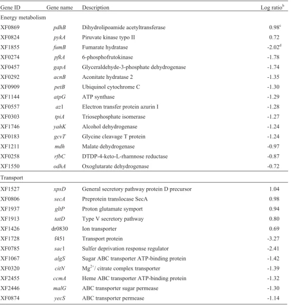
Table 3-Xylella fastidiosagenes induced in BCYE and XDM2media (q-valuea(%) = 0.27 for all genes).
Gene ID Gene name Description Log ratiob
Energy metabolism
XF0869 pdhB Dihydrolipoamide acetyltransferase 0.98c
XF0824 pykA Piruvate kinase typo II 0.72
XF1855 fumB Fumarate hydratase -2.02d
XF0274 pfkA 6-phosphofrutokinase -1.78
XF0457 gapA Glyceraldehyde-3-phosphate dehydrogenase -1.74
XF0292 acnB Aconitate hydratase 2 -1.35
XF0909 petB Ubiquinol cytochrome C -1.30
XF1144 atpG ATP synthase -1.29
XF0557 az1 Electron transfer protein azurin I -1.28
XF0303 tpiA Triosephosphate isomerase -1.27
XF1746 yahK Alcohol dehydrogenase -1.24
XF0183 gcvT Glycine cleavage T protein -1.24
XF1211 mdh Malate dehydrogenase -0.97
XF0258 rfbC DTDP-4-keto-L-rhamnose reductase -0.87
XF1550 odhA Oxoglutarate dehydrogenase -0.72
Transport
XF1527 xpsD General secretory pathway protein D precursor 1.04
XF0806 secA Preprotein translocase SecA 0.98
XF1937 gltP Proton glutamate symport 0.94
XF1913 tatD Type V secretory pathway 0.80
XF1426 dr0830 Ion transporter 0.69
XF1728 f451 Transport protein -3.27
XF0785 sac1 Sulfer deprivation response regulator -2.41
XF1067 algS Sugar ABC transporter ATP-binding protein -1.42
XF0320 citN Mg2+/ citrate complex transporter -1.39
XF2455 ccmA Heme ABC transporter ATP-binding protein -1.32
XF2446 malG ABC transporter sugar permease -1.30
Table 3 (cont.)
Gene ID Gene name Description Log ratiob
XF0933 feoB Ferrous iron transporter protein B -1.13
XF0324 afuA Periplasmic iron-binding protein -0.99
XF2685 sppA Protease IV -0.94
XF1172 secY Preprotein translocase SecY -0.93
XF1476 ynhE ABC tranporter membrane -0.84
XF1520 xpsH General secretory pathway protein H precursor -0.81
XF2133 yheS ABC transporter ATP-binding protein -0.77
Membrane components and surface structure
XF0343 mopB Outer membrane protein 1.56
XF0373 pilQ Fimbrial assembly protein 0.87
XF0103 dc14 Membrane protein -1.22
XF1118 murD UDP-N-acetylmuramoylalanine-D- glutamate ligase -1.08
XF0881 - D-alanil-D-alanina carboxipeptidase -1.04
XF0478 pilY1 Fimbrial assembly protein -1.11
XF0372 pilP Fimbrial assembly protein -1.04
XF1309 mreB Rod shape-determining protein -0.73
XF2542 - Fimbrial protein -1.01
XF0078 mrkD Fimbrial adhesin precursor -0.92
XF1632 pilU Twitching motility protein -0.69
RNA, DNA and nucleotide metabolism
XF0935 ilaIIA Methyltransferase 0.80
XF0822 gltX Glutamyl-tRNA synthetase 0.78
XF0587 purM 5’-phosphoribosyl-5-aminoimidazole synthetase 0.62
XF0169 tyrS Tyrosyl-tRNA synthetase 0.59
XF2178 holA DNA polymerase III, delta subunit -1.85
XF0223 tgt/vacC Queuine tRNA-ribosyltransferase -1.70
XF1909 mutY A/G-specific adenine glycosylase -1.23
XF2672 purE Phosphoribosylaminoimidazole carboxylase, catalytic subunit -1.10
XF0354 recG ATP-dependent DNA helicase -0.89
XF0676 holB DNA polymerase III, delta subunit -0.66
XF0549 metG Methionyl-tRNA synthetase -0.62
XF1297 SCF 11.04 Gluconolactonase precursor -0.60
Biosynthesis of amino acids and proteins
XF2465 metA Homoserine O-acetyltransferase 0.71
XF1427 argM Succinylornithine aminotransferase 0.69
XF1944 dcp Peptidyl-dipeptidase 0.64
XF1189 lon ATP-dependent serine proteinase La 0.62
XF1116 lysA Bifunctional diaminopimelate -2.17
XF0267 pspB Serine protease -1.02
XF2219 hisD Histidinol dehydrogenase -0.81
XF0624 aroE Shikimate 5-dehydrogenase -0.75
XF2324 aroE 3-phosphoshikimate 1-carboxyvinyltransferase -0.71
XF1915 trpG Anthranilate synthase component II -0.64
Biosynthesis of cofactors, prosthetic groups and regulatory functions
Table 3 (cont.)
Gene ID Gene name Description Log ratiob
XF2592 phoR Two-component system, sensor protein 0.83
XF0017 hemF Coproporphyrinogen III oxidase, aerobic 0.82
XF0064 bioB Biotin synthase -1.52
XF0230 panC Pantoate-beta-alanine ligase -1.30
XF0322 tctD Two-component system, regulatory protein -1.20
XF0189 bioA Adenosylmethionine-8-amino-7-oxononanoate aminotransferase -1.05
XF0912 sspB Stringent starvation protein B -0.90
XF1626 algR Two-component system, regulatory protein -0.86
XF0911 sspA Stringent starvation protein A -0.71
XF2336 colR Two-component system, regulatory protein -0.70
XF0950 ribD Riboflavin-specific deaminase -0.69
XF0228 folK 2-amino-4-hydroxy-6-hydroxymethyldihydropteridine pyrophosphokinase -0.66
XF2306 hemB Delta-aminolevulinic acid dehydratase -0.64
XF2545 pilR Two-component system, regulatory protein -0.63
XF0953 ribA GTP cyclohydrolase II/3,4-dihydroxyl-2-butanone 4-phosphate synthase -0.60 Biosynthesis of fatty acids and phospholipids
XF0671 fabG 3-oxoacil-[ACP] reductase 0.71
Degradation of molecules
XF1965 dhaA Haloalkane dehalogenase 0.84
XF1743 est Esterase -1.16
XF1253 lipP Lipase -0.64
Toxins
XF1029 gaa Glutaryl-7-ACA acylase precursor -2.69
XF2759 frpC Haemolysin-type calcium binding protein -0.73
XF1220 cvaB Colicin V secretion ABC transporter ATP-binding protein -0.70 Related to plasmid
XFa0047 taxC Nickase -0.70
Related to phage
XF2478 int Phage-related integrase -0.75
Cell division
XF0796 ftsW Cell division protein -0.81
Others
XF0961 bcp Bacterioferritin comigratory protein -1.32
Hypothetical and conserved proteins
XF0473 - Hypothetical protein 1.16
XF0497 rv2514c Conserved hypothetical protein 0.74
XF1620 - Hypothetical protein 0.69
XF0374 - Hypothetical protein 0.67
XF2041 - Hypothetical protein 0.67
XF1252 b2520 Conserved hypothetical protein 0.66
XF0597 dr1792 Conserved hypothetical protein 0.64
XF2734 - Hypothetical protein 0.59
XF1812 dr0620 Hypothetical protein -2.95
Three ORFs related to nucleotide biosynthesis were expressed in the XDM2 and modified BCYE media:
phosphoribosylaminoimidazole carboxylase (purE), gluco-nolactonase precursor (SCF 11.04) and 5-phosphoribosyl-5-aminoimidazole synthetase (purM). ThepurEandpurM genes are responsible for synthesis of purine ribotides, while SCF11.04 acts on the biosynthesis of nucleo-sides. This reaction is part of purine biosynthesis, starting with the metabolic precursors, ribose-5-phosphate, CO2
and NH3. All the pathways for synthesis of purinic and
pyrimidinic nucleotides have already been described forX. fastidiosa(Simpsonet al., 2000).
Genes involved in the biosynthesis of amino acids and proteins
Through the analysis of those genes related to amino acid biosynthesis, it was possible to observe that X. fastidiosais able to synthesize certain amino acids such as aspartate, cysteine, glutamate, histidine and metionine. Most microorganisms can uptake amino acids from their cultivation medium and oxidize them to sustain energy lev-els, as required by metabolic conditions (Nelson and Cox, 2002). X. fastidiosa presents high biosynthetic capacity, this probably resulting from its success in colonizing the xylem vessels of a number of host plants (Simpsonet al., 2000). However, xylem fluid contains a low concentration
Table 3 (cont.)
Gene ID Gene name Description Log ratiob
XF0172 - Conserved hypothetical protein -2.46
XF1753 - Hypothetical protein -2.32
XF2688 - Conserved hypothetical protein -2.28
XF0358 - Hypothetical protein -2.07
XF2687 - Hypothetical protein -1.70
XFa0028 - Hypothetical protein -1.57
XF0272 - Conserved hypothetical protein -1.39
XF0201 - Conserved hypothetical protein -1.35
XF2428 - Conserved hypothetical protein -1.19
XF1117 - Conserved hypothetical protein -1.16
XF1086 - Conserved hypothetical protein -1.15
XF1798 - Hypothetical protein -1.14
XF2510 - Hypothetical protein -1.13
XF0675 hi0457 Conserved hypothetical protein -1.09
XF2400 - Conserved hypothetical protein -1.05
XF0601 - Conserved hypothetical protein -1.05
XF2449 - Conserved hypothetical protein -1.02
XF2008 tm1181 Conserved hypothetical protein -1.01
XF0638 - Hypothetical protein -1.01
XF2074 - Conserved hypothetical protein -0.68
XFa0018 - Hypothetical protein -0.93
XF2023 - Conserved hypothetical protein -0.67
XF1881 - Hypothetical protein -0.67
XF0357 - Hypothetical protein -0.64
XF1323 - Hypothetical protein -0.62
XF2647 - Conserved hypothetical protein -0.61
XF2363 - Conserved hypothetical protein -0.61
XF2427 - Conserved hypothetical protein -0.60
XF1854 ctp Hypothetical protein -0.59
XF0766 - Hypothetical protein -0.52
aq-value is the lowest False Discovery Rate at which the gene is called significant and measures how significant the gene is: asd
i> 0 increases, the
corre-sponding q-value decreases (Tusheret al., 2001),bLog expression ratios of measured transcript levels determined for the two cultures;cThe log
of organic composts (available energy sources), although it presents a high concentration of amino acids such as gluta-mine and asparagine (Raven, 1984). Glutagluta-mine and arginine are important in the composition of the XDM2
me-dium, as sources of nitrogen and in helpingX. fastidiosa cells to reach the end of their exponential growth phase in less generation-time (Lemoset al., 2003).
Genes related to amino acid biosynthesis were found in both culture media at various expressed levels. Two im-plications arise from this analysis. The first confirms that the TCA cycle is active, since it generates the intermediar-ies for amino acid biosynthesis from the glucose oxidative degradation pathway. The second implication is that the source of amino acids in both media, mainly in XDM2
(which contains arginine, glutamine, metionine and serine), can be used in protein synthesis as well as for supplying the carbon skeleton: a) to replace intermediaries of TCA cycle components in anaplerotic reactions and b) for synthesis of the other amino acids.
The operonsspA-sspBexpression in the XDM2
me-dium was similar to that observed inE. coliduring the sta-tionary phase of the growth curve and under carbon, amino acids and phosphate limiting conditions (Willianset al., 1994). This operon expression level during the four days of X. fastidiosacultivation shows that the active metabolism of the bacterial cells in the XDM2medium results in the
consumption of nutrients up to cells entering the stationary growth phase.
Analyses revealed higher gene expression for the pspBgene which codes serine protease, in bacteria culti-vated in the XDM2medium (Table 3). Serine protease is not
secreted via a type I pathway, but belongs to the auto-transporter family of secreted proteins (Chabeaudet al., 2001). Many proteins belonging to the autotransporter fam-ily are involved in adhesion or auto-aggregation, even though several of them possess the active-site motif of serine protease (Hendersonet al., 1998).
Genes involved in cofactors, prosthetic groups and regulatory functions
Through genomic analysis, it was suggested that vita-mins like thiamin, biotin, nicotinic acid and pyridoxine are synthesized byX. fastidiosacells (Simpsonet al., 2000). The significant expression of genes related to the prosthet-ics groups, cofactors and vitamins, such as those involved with riboflavin (ribA andribD), biotin (bioA andbioB), pantothenate (panC), porphyrin (hemB and hemF), folic acid (folK) and thiamin (thiL), may suggest that the concen-trations used in media composition were insufficient to sus-tain growth, and that the decrease in these levels was responsible for their synthesis. On the other hand, the XDM2medium containing only biotin as a vitamin source
and in a 0.2 mg/L concentration was efficient enough to maintain bacterial cell growth (data not shown). The regu-latory functions category presented six highly expressed
genes in the XDM2medium (sspA,sspB,tctD,algR,colR,
pilR) in comparison to only one gene (phoR) in the BCYE modified medium. Since the functions of most of these sys-tems are still unknown, it is believed that the organisms in which they are expressed may show a higher level of adap-tive answers to certain environmental changes, situations in which the two component systems are induced (Stocket al., 1989).
The phoR gene is induced by phosphate limitation (Hullet, 1996), being significantly expressed only in the BCYE modified medium which does not include ferric pyrophosphate, a possible source of phosphate, in its com-position. The significant expression ofphogenes clearly in-dicates the need to include other sources, for this bacterium to make use of phosphorus for growth.
Genes involved in molecule degradation
Four expressed genes were related to the category of degradation, with emphasis onest(esterase),lipP(lipases) anddhaA (haloalkane dehalogenase). A correlationp be-tween lipase concentration and the production of biofilms might be linked to adhesion and construction of the latter (Smolkaet al., 2003). Lipases hydrolyze ester bonds be-tween the insoluble triacilglycerides interface and the aque-ous phase where the enzyme is dissolved (Anthonsenet al., 1995). In Candida albicans, LIP family lipases are ex-pressed and eliminated during the infection cycle, and it is believed that they contribute to the survival and virulence of this organism in human tissues (Hubeet al., 2000).
Genes related to hypothetical and conserved proteins
Approximately 30% (40 genes) of the 134 differen-tially expressed genes did not present homology with se-quences deposited in GenBank. The involvement of hypo-thetical and conserved proteins, for which functions in other organisms have not yet been described, should be taken as an indication of significant differences in the me-tabolism of this phytopathogen.
Categories of genes expressed only under XDM2
cultivation conditions
The expression of genes related to the toxin catego-ries (frpC, gaa and cvaB), functions related to plasmids (taxC), phago (int), cell division (ftsW) and others (bcp), were only observed in XDM2medium growth conditions.
Categories of genes expressed only in BCYE modified cultivation conditions
The expression of thefadGgene can be assigned to biosynthesis of fatty acids and was observed to be high un-der the BCYE modified medium conditions. TheE. coli fab genes presented higher expression levels in a rich medium, thereby suggesting that the regulation of phospholipid biosynthesis genes might be dependent on the speed of growth, since these genes need a higher number of mem-brane compounds (Taoet al., 1999). However, studies with X. fastidiosademonstrated that growth was higher in the XDM2medium than in the BCYE during the 14-day period
(Lemoset al., 2003). Thus, it is possible thatfabgene ex-pression is mediated by one or more signal molecules found in the modified BCYE medium.
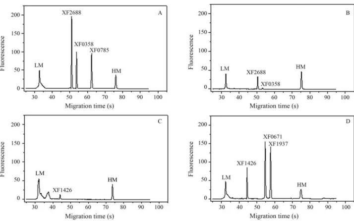
Detection of cDNA by microchip electrophoresis
In order to validate the results from microarrays, six ORFs (XF0358, XF0671, XF0785, XF1426, XF1937 and XF2688) were analyzed for RT-PCR by using microchip electrophoresis. The cDNAs from two different conditions (XDM2and BCYE media) were synthesized and used in
PCR with specific primers (Table 1). The Agilent 2100 Bioanalyzer separated the RT-PCR amplicons and quanti-fied the expression of each gene. As expected, the ratios ob-served in this experiment were similar to those obtained
through the microarray technique. Under XDM2
condi-tions, the ORFs XF0358, XF0785 and XF2688 showed a higher expression in this medium. Similar results were shown in the BCYE medium for ORFs XF1937, XF1426 and XF0671 (Figure 1). It is important to stress that the co-efficient of variance between the replicates for each gene under the conditions studied was seen to be between 2.68 and 5.19%, demonstrating the high level of reliability of the results (Table 1).
Acknowledgments
We thank Alexandre L. Nepomuceno, Jesus A. Ferro, Gonçalo A.G. Pereira and Manoel V. Franco Lemos for their critical review of the manuscript. During this project, R.F.T was supported by FAPESP (00/06289-2).
References
Anthonsen HW, Baptista A, Drablos F and Martel P (1995) Li-pases and esterases: A review of their sequences, structures and evolution. Biotechnol Annu Rev 1:315-371.
Ausubel FM, Brent R, Kingston RE, Moore DD, Seidman JG, Smith JA and Struhl K (1987) Current Protocols in Molecu-lar Biology. 2nd edition. J Wiley & Sons, New York, 637 pp.
Berkes BC, Sargent F and Palmer T (2000) The tat protein export pathway. Mol Microbiol 32:260-274.
Figure 1- Microchip electrophoresis of the reverse transcription-polymerase chain reaction (RT-PCR) products of six ORFs (XF2688, XF0358, XF0785, XF1426, XF0671 and XF1937). A and C: genes expressed in XDM2media; B and D: genes expressed in BCYE media. LM: lower marker. HM: higher
Binet R, Létoffé S, Ghigo JM, Delepelaire P and Wandersman C (1997) Protein secretion by Gram-negative bacterial ABC exporters - A review. Gene 192:07-11.
Campanharo JC, Lemos MVF and Lemos EGM (2003) Growth optimization procedures for the phytopathogen Xylella fastidiosa. Curr Microbiol 46:99-102.
Chabeaud P, Groot A, Bitter W, Tommassen J, Heulin T and Achouak W (2001) Phase-variable expression of an operon encoding extracellular alkaline protease, a serine protease homolog, and lipase in Pseudomonas brassicacearum. J Bacteriol 183:2117-2120.
Chomczynski P and Sacchi N (1987) Single-step method of RNA isolation by acid guanidinium thiocyanate-phenol-chloro-form extraction. Anal Biochem 162:156-159.
Diehl F, Grahlmamm S, Beier M and Hoheisel D (2002) Manufac-turing DNA microarrays from unpurified PCR products. Nucleic Acids Res 30:2-6.
Dow JM and Daniels MJ (2000)Xylellagenomics and bacterial pathogenicity to plant. Yeast 17:263-271.
Facincani AP, Ferro JA, Pizauro JM, Pereira HA, Lemos EGM, Prado AL and Ferro MI (2003) Carbohydrate metabolism of
Xylella fastidiosa: Detection of glycolytic and pentose phos-phate pathway enzymes and cloning and expression of the enolase gene. Genet Mol Biol 26:203-211.
Feil H, Feil WS, Detter JC, Purcell AH and Lindow SE (2003) Sire-directed disruption of thefimA andfimF fimbrial genes ofXylella fastidiosa. J Bacteriol 96:675-682.
Fekkes P and Driessen AJM (1999) Protein targeting to the bacte-rial cytoplasmic membrane. Microbiol Mol Biol Rev 63:161-173.
Gough CL, Dow JM, Barber CE and Daniels MJ (1988) Cloning of two endoglucanase genes ofXanthomonas campestrispv. Campestris: Role of the major enzyme in pathogenesis. Mol Plant Microbe Interact 1:275-281.
Grunberg-Manago M (1996) Regulation of the expression of aminoacyl-tRNA synthetases and translation factor. In: Neidhardt FC, Curtiss R, Ingraham JL, Lin EC, Low KB, Magasanik B, Reznikoff WS, Riley M, Schaechter M and Umbarger HE (eds)Escherichia coliandSalmonella Cellu-lar and MolecuCellu-lar Biology. ASM Press, Washington, pp 1432-1457.
Henderson IR, Navarro-Garcia F and Nataro JP (1998) The great escape: Structure and function of the autotransporter pro-teins. Trends Microbiol 6:370-378.
Holt JG, Krieg NR, Sneath PHA, Staley JT and Williams ST (1994) GenusXylella. In: Holt JG, Krieg NR, Sneath PHA, Staley JT and Williams ST (eds) Bergey’s Manual of Deter-minative Bacteriology. Wilians and Wilkins, New York, pp 1-787.
Hopkins DL (1989)Xylella fastidiosa: Xylem-limited bacterial pathogen of plants. Annu Rev Phytopathol 22:271-290. Hu NT, Hung MN, Chion SJ, Tang F, Chiang DC, Huang HY and
Wu CY (1992) Cloning and characterization of a gene re-quired for the secretion of extracellular enzymes across the
outer membrane by Xanthomonas campestris pv.
Campestris. J Bacteriol 174:2679-2687.
Hu NT, Hung MN, Chen DC and Tsai RT (1998) Insertion muta-genesis of XpsD, an outer membrane protein involved in extracellular protein secretion inXanthomonas campestris
pvcampestris. Annu Rev Microbiol 144:1479-1486.
Hube B, Sterhr F, Bossenz M and Mazur A (2000) Secreted li-pases of Candida albicans: Cloning. Characterization and expression analysis of a new gene family with at least ten members. Arch Microbiol 174:362-374.
Hullet FM (1996) The signal-transduction network for Pho regu-lation inBacillus subtilis. Mol Microbiol 19:933-939. Keen NT, Dumenyo CK, Yang CH and Cooksey DA (2000) From
rags to riches: Insights from the first genomic sequence of a plant pathogenic bacterium. Genome Biol 1:10191-10194. Lambais MR, Goldman MHS, Camargo LEA and Goldman GH
(2000) A genomic approach to the understanding ofXylella fastidiosapathogenicity. Curr Opin Microbiol 3:459-462. Lashkari DA, Derise JL, Mccusker JH, Namath AF, Gentie C,
Hwang SY, Brown PO and Davis RW (1997) Yeast micro-arrays for genome wide parallel genetics and gene expres-sion analysis. Proc Natl Acad Sci USA 94:13057-13062. Leite B, Ishida MI, Alves E, Carrier H, Pascholati SF and Kitajima
EW (2002) Genomic and X-ray microanalysis indicate that Ca2+ and thiols mediate the aggregation and adhesion of
Xylella fastidiosa. Braz J Med Biol Res 35:645-650. Lemos EGM, Carareto-Alves LM and Campanharo JC (2003)
Genomic-based design of defined growth media for the plant pathogen Xylella fastidiosa. FEMS Microbiol Lett 219:39-45.
Meidanis J, Braga MDV and Verjovski-Almeida S (2002) Whole-genome analysis of transporters in the plant patho-genXylella fastidiosa. Microbiol Mol Biol Rev 66:272-299. Nelson DL and Cox MM (2002) Lehninger - Princípios de
Bio-química. 3rd edition. Sarvier, São Paulo, 975 pp.
Nunes LR, Rosato YB, Muto NH, Yanai GM, Silva VS, Leite DB, Gonçalves ER, Souza AA, Coletta-Filho HD, Machado MA,
et al.(2003) Microarray analysis ofXylella fastidiosa pro-vides evidence of coordinated transcription control of later-ally transferred elements. Genome Res 13:570-578. Ojanen-Reuhs T, Kalkkinen N, Westerlund-Wikström B,
van-Door J, Haahtela K, Nurmiaho-Lassila EL, Wengelnik K, Bonas U and Korhonen TK (1997) Characterization of the fimA gene encoding bundle-forming fimbrial of the plant pathogen Xanthomonas campestrispv vesicatoria. J Bacteriol 179:1280-1290.
Quackenbush J (2002) Microarray data normalization and trans-formation. Nat Genet 32:496-501.
Raven JA (1984) Phytophages of xylem and phloem: A compari-son of animal and plant sap-feeders. Adv Ecol Res 13:135-234.
Rawling EG, Brinkman FS and Hancock RE (1998) Roles of the carboxy-terminal half of Pseudomonas aeruginosa major outer membrane proteins OprF in cell shape growth in low osmolarity medium and peptidogycan association. J Bacteriol 180:3556-3562.
Rossetti V, Garnier M and Bove JM (1990) Presénce de bactéries dans le xyléme d’oranger atteints de chorose variegée, une nouvelle maladie des agrumes au Brésil. C R Acad Sci Paris 310:345-349.
Sargent F, Bogsch EG, Satnley NR, Wexler M, Robinson C, Berks BC and Palmer J (1998). Overlapping functions of components of a bacterial Sec-independent protein export pathway. EMBO J 17:3640-3650.
Silva FR, Vettore AL, Kemper EL and Leite A (2001) Fastidiam gum: TheXylella fastidiosaexopolysaccharide possibly in-volved in bacterial pathogenicity. FEMS Microbiol Lett 203:165-171.
Simpson AJG, Reinach FC, Arruda P, Abreu FA, Acencio M, Alvarenga R, Alves LM, Araya JE, Baia GS, Baptista CS,et al. (2000) The genome sequence of the plant pathogen
Xylella fastidiosa. Nature 406:151-159.
Smolka MB, Martins D, Winck FV, Santoro CE, Castellari RR, Ferrari F, Brum IJ, Galembeck E, Coletta-Filho HD, Machado MA,et al(2003) Proteome analysis of the plant pathogen Xylella fastidiosa reveals major cellular and extracellular proteins and a peculiar codon bias distribution. Proteomics 3:224-237.
Stock AM, Robinson VL and Gourdreau PN (1989) Two-com-ponent signal transduction. Annu Rev Biochem 69:183-215.
Tao H, Bausch C, Richmond C, Blattner FR and Conway T (1999) Functional genomics: Expression analysis of Escherichia
coli growing on minimal and rich media. J Bacteriol
181:6425-6440.
Tusher V, Tibshirani R and Chu G (2001) Significance analysis of microarrays applied to transcriptional responses to ionizing radiation. Proc Natl Acad Sci USA 98:5116-5121.
Weiner JH, Bilous PT, Shaw GM, Lubitz SP, Frost L, Thomas GH, Cole JA and Turner RJ (1998) A novel and ubiquitous system for membrane targeting and secretion of cofactor-containing proteins. Cell 93:93-101.
Wells JM, Raju BC, Nyland G and Lowe SK (1981) Medium for isolation and growth of bacteria associated with plum leaf
scald and phony peach disease. Appl Environ Microbiol 42:357-63.
Wells J, Raju BC, Jung HY, Weisburg WG, Mandelco-Paul L and Brenner DJ (1987)Xyllela fastidiosagen nov, sp. Nov. gram negative, xylem limited fastidious plant bacteria related to
Xantomonasspp. Int J Sys Phytopathol 73:136-143. Wexler M, Sargent F, Jack RL, Stanley NR, Bogsch EG, Robison
C, Berks BC and Palmer T (2000) TatD is a cytoplasmic pro-tein with DNAse activity: No requirement for TatD-family protein in Sec-independent protein export. J Biol Chem 275:16717-16722.
Willians MD, Fuchs JA and Flickinger MC (1994) Null mutation in the stringent starvation protein of Escherichia coli dis-rupts lytic development of bacteriophage Pi. Gene 109:21-30.
Yang YH, Buckley MJ and Speed TP (2001) Analysis of cDNA microarrays images. Brief Bioinformatics 2:341-349. Zhong X and Tai PC (1998) When an ATPase is not an ATPase:
At low temperatures the c-terminal domain of the ABC transporter cvaB is a GTPase. J Bacteriol 180:1347-1353.
Internet Resources
Gene Expression Omnibus (GEO) Repository,
http://www.ncbi.nlm.nih.gov/geo.
SAM: Significance Analysis of Microarrays,
http://www.stat.stanford.edu/~tibs/SAM/index.html.
Associate Editor: Carlos F.M. Menck