Article
J. Braz. Chem. Soc., Vol. 26, No. 2, 239-246, 2015. Printed in Brazil - ©2015 Sociedade Brasileira de Química 0103 - 5053 $6.00+0.00
A
*e-mail: [email protected]
Metalloproteomic Profile Determination of Muscle Samples from Nile Tilapia
(
Oreochromis niloticus)
Using AAS and ESI-MS/MS after 2D-PAGE Separation
Bruna Cavecci,a Paula M. de Lima,a João V. de Queiroz,a Camila P. Braga,b Cilene C. F. Padilha,b
Aline L. Leite,c Marília A. R. Buzalaf,c Luiz E. Pezzatoa andPedro M. Padilha*, b
aCollege of Veterinary and Animal Science, bDepartment of Chemistry and Biochemistry,
Institute of Bioscience and cDepartment of Biological Science,
University of São Paulo State University (UNESP), 18618-970 Botucatu-SP, Brazil
This study evaluated the metalloproteomic profile of muscle tissue samples from Nile tilapia
(Oreochromis niloticus). Two-dimensional polyacrylamide gel electrophoresis (2D-PAGE) methods
was used for protein fractionation and identification based on image analysis. Determination of Ca, Cu, Fe, Mn and Zn was performed in the protein spots by flame and/or graphite furnace atomic absorption spectrometry (FAAS, GFAAS), the protein spots were characterized by electrospray ionization-tandem mass spectrometry (ESI-MS/MS). FAAS and GFAAS determinations have indicated the presence of calcium in seven protein spots, iron in only one spot, manganese in six spots, and zinc in two spots. Of the seventeen protein spots where the presence of metal ions was detected, ten were characterized by ESI-MS/MS.
Keywords: metalloproteins, ESI-MS/MS, 2D-PAGE
Introduction
There is consensus in the literature about the importance of metal ions as structural and functional components of fish. From the structural viewpoint, calcium, copper, iron, manganese and zinc are present in metallothioneins. The catalytic role of these ions in enzymatic systems is essential.1
Although metal ions constitute only a small proportion of body tissue (4%), they are essential structural and functional components in many vital processes. One important functional aspect is their role as catalysts in enzymatic systems, by binding to substrates and thus directing the reaction, and as mediators in redox reactions by means of reversible changes in the oxidation state of the metal ion. Regarding the structural aspect, the role of metals as a component of organic compounds is worth mentioning, such as iron in hemoglobin, iodine in thyroxine, cobalt in B12 vitamin, and sulfur in thiamine and biotin. Metal ions stabilize these biomolecules by neutralizing and/or protecting negative charges.1,2
Ion balance is important for bone formation, such as the amounts and ratio of calcium and phosphorus for muscle activity and in the extracellular fluid. Electrolytes, among
which sodium and potassium salts are the most important, represent the main factors for the osmotic control of water metabolism.2,3
The literature estimates that approximately 40% of all proteins and enzymes require the presence of a metal ion, to perform biological activity. Such ions are responsible for many processes, such as energy conversion during photosynthesis and respiration, gene regulation and expression, substrate binding and activation, transportation, and storage, in addition to catalytic processes.4 Metalloproteins are considered distinct from metal-binding proteins. The former are characterized by their high metal-protein interaction affinity. In the latter, the metal-protein interaction has low affinity, and therefore this bond is easily broken.5-7
The study of metalloproteomics in living organisms provides information on how a metal ion is distributed and coordinated to proteins, its essentiality and/or toxicity, and the individual concentration of the metal species, thus contributing to a better understanding of the physiological and functional aspects of these biomolecules.8,9
absorption spectrometry (GFAAS) of calcium, copper, iron, manganese and zinc ions in the protein spots; and the characterization of the protein spots where the studied metal ions were identified by electrospray ionization-tandem mass spectrometry (ESI-MS/MS).
Experimental
Fish allocation, diet preparation and sample collection
The animals were obtained from the Center of Aquaculture of the São Paulo State University - CAUNESP, Jaboticabal, São Paulo State, Brazil, and were brought to the Laboratory of Nutrition of Aquatic Organisms of São Paulo State University - AquaNutri - UNESP, Botucatu, São Paulo State, Brazil. The experimental protocol was approved by the Ethics Committee on the Use of Animals (CEUA), University of São Paulo State (UNESP), under number 04/2011. Five 250 L aquariums were used, and five fish were placed in each aquarium. These aquariums were part of a water recirculation system with mechanical filtering and a biofilter. Aeration was maintained by an air blower. In this system, the water temperature was electronically controlled and adjusted to 27 ± 1 °C. The animals used were adult Nile tilapia with an average individual weight of 250 g, kept in cages and fed with a formulated extruded diet. The animals were manually fed until apparent satiety according to the following protocol: at 8 am, at 11 am, at 3 pm, and at 6 pm. This experiment ran for a period of 45 days, with 15 days of adaptation and 30 days to gain adequate weight for euthanasia.10,11
After this period, the fish from each aquarium were euthanized with a benzocaine solution (100 mg L−1) for the removal of muscle samples. The samples were then transferred to 15 mL polypropylene flasks and stored at –80 °C. Prior to the analytical procedures, a pool of samples was obtained (1 g of the fillet: 1 mL of ultrapure water obtained using PURE LA Elga Ultra Ionic System – www.elgalabwater.com) and macerated using a mortar and pestle. Next, the muscle extracts containing the proteins were centrifuged in a chilled centrifuge (Hettich Zentrifugen, Germany) at 8,000 g for 5 min at 4 °C. The total protein content of the obtained protein extracts was measured, and then the electrophoretic runs were performed.12,13
Sample preparation for electrophoresis
To quantify the protein content in the samples, protein precipitation was performed using ice-cold 80% (v/v) acetone solution at a 1:4 ratio (sample:acetone). The precipitation was performed for 2 h at 10 °C, assuring
quantitative precipitation. Next, the protein precipitate was centrifuged at 8,000 g in a chilled centrifuge for 10 min, and the supernatant was removed. This protein precipitate was washed twice more with the ice-cold acetone solution used for precipitation. Then, the protein precipitate was re-solubilized in buffer containing precast gel IEF (isoelectric focusing stage), 1.5 µg µL−1 solubilized protein in a solution containing 7 mol L−1 urea, 2 mol L−1 thiourea, 2% (m/v) CHAPS, 0.5% (v/v) ampholytes from pH 3 to 10, 0.002% (m/v) bromophenol blue and 2.8 mg of dithiothreitol (DTT). One fraction was used for total protein content determination using the GE Healthcare 2D Quant Kit, and the other fraction was used in the electrophoretic runs. Analytical calibration curves were constructed according to the kit manual’s instructions, and the readings were obtained at a wavelength of 480 nm in the Spectrophotometer Evolution 60 Thermo Fisher Scientific, USA.
Separations by electrophoresis
Prior to the electrophoretic separations, 250 µL aliquots of muscle protein extracts (obtained by re-solubilizing the protein mass precipitated with acetone) were applied in 13 cm strips for IEF, which contained the pre-cast gel with immobilized ampholytes at pH values from 3 to 10. These strips were placed on an apparatus where they remained for 12 h at room temperature to be rehydrated with the protein extract. In addition to the protein extract, approximately 900 µL of mineral oil was added to the strips. After this period, the rehydrated strip was taken to the IEF system for a first-dimensional run of the bi-dimensional electrophoresis, using the following voltage program: Step 1 = 500 V, with accumulation of 500 Vh; Step 2 = 1.000 V, with accumulation of 800 Vh; Step 3 = 10.000 V, with accumulation of 11.300 Vh; Step 4 = 10.000 V, with accumulation of 3.000 Vh.
After the equilibrium step, the second dimension of the electrophoretic process was performed (SDS-PAGE). The strip with muscle protein was applied in a pre-cast 12.5% polyacrylamide gel in a glass plate measuring 180 × 160 × 1.5 mm. The polyacrylamide gels were prepared using the following solutions: acrylamide, N,N’-methylenebisacrylamide,
tris(hydroxymethyl) aminomethane, SDS, N,N’,N,N’
-tetramethylethylenediamine (TEMED), hydrochloric acid, and ammonium persulfate. A piece of filter paper was placed over the polyacrylamide gel next to the strip, over which 10 µL of molecular weight ladder was applied, containing the following proteins: β-phosphorylase (97.0 kDa), albumin (66.0 kDa), ovalbumin (45.0 kDa), carbonic anhydrase (30.0 kDa), trypsin inhibitor (20.1 kDa), and
α-lactalbumin (14.4 kDa). The strip and the filter paper were sealed with hot 0.5% (m/v) agarose solution in adequate buffer to assure their contact with the polyacrylamide gel. Next, the second-dimension electrophoretic run was performed in a 2D-PAGE electrophoresis system, in two steps, using the following program, voltage (V): Step 1 = 90, Step 2 = 250; electric current (mA): Step 1 = 25, Step 2 = 25; power (W): Step 1 = 100, Step 2 = 100; time (h): Step 1 = 0.5, Step 2 = 3. After the running time (ca. 3.5 h) the proteins were fixed for 1 h using a solution containing 10% (v/v) acetic acid and 40% (v/v) ethanol and stained using the colloidal Coomassie dye, which consists of a solution with 8% (m/v) ammonium sulfate, 1.6% (v/v) phosphoric acid, 0.08% (m/v) Coomassie blue G-250, and 25% (v/v) methanol. The dye was in contact with the gel for 72 h, after which it was removed by successive washes with ultrapure water. The gels, obtained in triplicate, were scanned with a GE Healthcare Scanner. The images were analyzed to obtain the correlation between the gel replicates and their spots count, using the ImageMaster platinum 7.0 software.
Determination of Ca, Cu, Fe, Mn and Zn by FAAS and GFAAS
The protein spots obtained in the electrophoretic runs were mineralized with concentrated sulfuric acid (3 mL) and 30% (m/m) hydrogen peroxide (1 mL) in each flask contains 3 spots. The set of digestion flasks was placed in the UNIQUE ultrasonic water bath at a temperature of 40 °C and sonicated at 135 W until the mineralization of the spots was complete (transparent extract). The volume of the acid extract obtained was adjusted to 5 mL with ultrapure water.14 This procedure was performed with spots from three different electrophoretic runs, and gels were obtained in triplicate for each run.
Determination of Ca, Cu, Fe, Mn and Zn by FAAS or GFAAS were performed using SHIMADZU AA-6800 atomic absorption spectrometer (Tokyo, Japan, www.ssi. shimadzu.com), equipped with background absorption correction with deuterium lamp and a self-reverse (SR) system and ASC-6400 automatic sampler. For all determinations it was used SHIMADZU Ca, Cu, Fe, Mn and Zn hollow cathode lamp (using the following wavelengths: 422.7, 327.7, 248.3, 279.5 and 213.0 nm, respectively), operating with a current of 10 mA and a spectral resolution of 0.5 nm. In the GFAAS determinations was used a pyrolytic graphite tube with integrated platform, argon as the inert gas maintaining a constant flow rate of 0.30 L min−1 during the entire heating program except in the atomization step, in which the gas flow was interrupted and the absorbance signals were measured in peak areas. The analytical curves were prepared using Merck Titrisol standard solutions (www.merck-performance-materials. com), and the region of the gel where no protein spots appeared was used for the analytical blank. The accuracy of the methodology metal ions determination was evaluated using the acid extract of the analytical blank that was spiked with 100 mg of reference material (Bovine Muscle Powder, RM 8414 - National Research Council, Canada), containing (mg kg−1): 145 ± 20 of calcium, 2.84 ± 0.45 of copper, 71.2 ± 9.2 of iron, 0.37 ± 0.09 of manganese and 142 ± 14 of zinc. The volume of the acid extract obtained for reference material was adjusted to 5 mL with ultrapure water. The limits of detection (LOD) and quantification (LOQ), were calculated based on the standard deviation of 20 readings of the standard solution blank and on the slope of the analytical curve (LOD = 3σ/slope and LOQ = 10σ/slope).15
Protein spots characterization by ESI-MS/MS
The protein spots were extracted from the gels using a scalpel and cut into segments of approximately 1 mm3, transferred to 2 mL microtubes and prepared for MS according to Shevchenko et al.16 with little modification. Briefly, the gel pieces were destained with 25 mmol L−1 ammonium bicarbonate (Ambic)/acetonitrile (ACN) (50:50 v/v), and after distained the fragments were dehydrated with two ACN bath for 10 min and dried at room temperature (RT). Next, gel pieces were rehydrated with 20 mmol L−1 DTT in 50 mmol L−1 Ambic for 40 min at 56 °C, after this time, the excess of reagent was removed and 55 mmol L−1 iodoacetamide (IAA) in Ambic
50 mmol L−1 was added for 30 min at RT. Next, IAA
After dried samples were incubated overnight at 37 °C with 10 ng µL−1 trypsin in 25 mmol L−1 Ambic for 15 min (Trypsin Gold Mass Spectrometry, Promega, Madison, USA). The peptides were extracted from gel by addition of extraction buffer A (50% ACN with 1% formic acid) to each tube and incubate for 15 min at 40 °C under sonication (40 kHz/30 W, Unique 1600A, Brazil). Supernatant was collected and transferred to new tube. This Step was repeated with extraction buffer B (60% methanol with 1% formic acid) and extraction buffer C (100% ACN). Extracts were dried in a vacuum centrifuge (Eppendorf, Hamburg, Germany) and peptides were dissolved in 10 µL 3% ACN with 0.1% formic acid. The peptides identification was performed on a nanoAcquity UPLC-Xevo QTof MS system (Waters, Manchester, UK). The nanoAcquity UPLC®, was equipped with nanoAcquity HSS T3 (75 µm × 150 mm, 1.8 µm particle size, Waters Manchester, UK) analytical reverse phase column. The column was equilibrated with mobile phase A (0.1% formic acid in water). Then the peptides were separated with a linear gradient of 7-85% mobile phase B (0.1% formic acid in ACN) for 31 min at a flow rate of 0.4 µL min−1. The Xevo® G2 Q-TOF mass spectrometer was operated in positive mode, and data were collected in MSE method in elevated energy (19-45 V). Source conditions were capillary voltage, 2.5 kV; sample cone, 30 V; extraction cone, 5.0 V and source temperature, 80 °C. Data acquisition was obtained during 20 min and the scan range was 50-2000 Da. ProteinLynx Global Server (PLGS) version 3.0 was used to process and search the continuum LC-MSE data, setting carbamidomethylation of cysteines as fixed modification and oxidation of methionines as variable modification, allowing one missing cleavage and maximal error tolerance of 10 ppm.17 Protein identification was obtained with the embedded ion accounting algorithm of the software and searching in Fish database (only reviewed, UniProtKB/Swiss-Prot) downloaded at October 2013 from UniProtKB.18
Results and Discussion
Determination of total protein concentration in samples
The total protein content in the muscle samples was determined to obtain the amount of protein to be applied on each strip contained precast polyacrylamide gels in the isoelectric focusing stage (IEF) of the electrophoretic runs. The results indicated that the extracts of protein pellets from the muscle samples contained 14.28 mg mL−1 of total protein. Based on this result, 250 µL of protein extracts contained 1.5 µg µL−1 of protein was applied to each IEF strip.
Optimization of electrophoretic separations
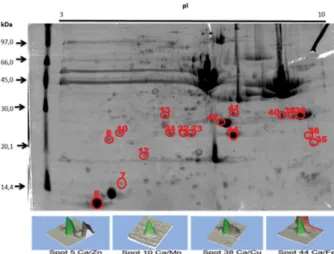
The 2D-PAGE electrophoresis experiments on samples of Nile tilapia muscle tissue were performed with three replicates of runs. Figure 1 shows representative gels obtained from the muscle samples of Nile tilapia. As seen in Figure 1, the gel of the muscle sample extracts exhibited good resolution, demonstrating that protein separation occurred efficiently. The correlation analysis between the obtained gels demonstrated that 73.5% of the protein spots were present in the four gels of the muscle tissue samples. Furthermore, the average number of spots found in the gels was 620, and the exhibited standard deviation was relatively lower than 10%, which is considered very good in protein fractionation by 2D-PAGE.7-9,12,13,18 Thus, the spots were cut, mineralized, and then analyzed by atomic spectrometry (FAAS and/or GFAAS) to detect Ca, Cu, Fe, Mn, and Zn.
Calcium, copper, iron, manganese and zinc determination by FAAS or GFAAS
The accuracy of the method used for calcium, copper, iron, manganese and zinc determined in the protein spots by FAAS and/or GFAAS was assessed using the RM 8414 certified standard. The Table 1 showed the detection limits LOD and quantification LOQ of each element.19,20 It is also observed that the relative standard deviation obtained are less than two percent, which proves that the determination method present great precision (repeatability and reproducibility). The concentrations of calcium, copper, iron, manganese and zinc in the protein spots was calculated based on the protein mass estimated by optical density using the ImageMaster 2D Platinum version 7.0 software program.8,9
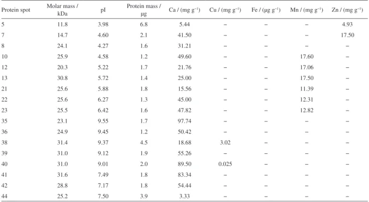
Table 2 lists the concentrations of these metal ions determined in the protein spots highlighted in circles in the Figure 1. Upon examining the results in Table 2, one can see that the calcium, manganese and zinc concentrations in the protein spots range from 3.33 to 97.74 mg kg−1, from 11.39 to 17.60 and 4.93 to 17.50, respectively. It can be observed that the results obtained are within the LOQ (µg kg−1 or mg kg−1) analysis method. In the case of copper only protein spots 38 and 40 showed concentrations of 3.02 and 0.025 mg kg−1, respectively. For iron, just in the protein spot 44 was possible to determine a concentration of 77.50 µg g−1.
Characterization of protein spots by ESI-MS/MS
In the protein spot characterization, more than one protein was found in the database search.21,22 However,
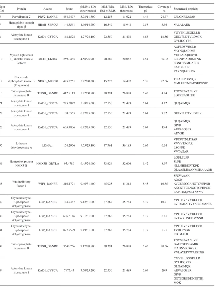
a compromise was made to determine the protein class, considering the highest score and/or coverage and especially the molecular mass data and pI obtained by 2D-PAGE (experimental) in relation to the theoretical molecular mass obtained in the Uniprot database.18 Hence, 17 spots were characterized, and 10 proteins were identified. Table 3 includes the characterization obtained by ESI-MS/MS analysis as the most likely protein identified, based on the conditions described above. Posteriorly, each identified protein was described according to the Uniprot data.
Parvalbumin, found in spot 5, is a protein of low molecular weight (11-12 kDa) that exhibits the isoforms parvalbumin-2, parvalbumin alpha, and parvalbumin beta, all of which normally bind to calcium ions. This protein is directly involved in muscle relaxation processes after contraction occurs and consists mainly of peptide sequences containing serine and alanine: amino acids
Table 1. LOD and LOQ of the each element
Element LOD (m/v) LOQ (m/v) LOD (m/m) LOQ (m/m)
Ca 0.017 mg L−1 0.058 mg L−1 0.85 mg kg−1 2.90 mg kg−1
Cu 0.042 µg L−1 0.14 µg L−1 2.10 µg kg−1 7.00 µg kg−1
Fe 0.084 µg L−1 0.28 µg L−1 4.20 µg kg−1 14.00 µg kg−1
Mn 0.045 µg L−1 0.15 µg L−1 2.25 µg kg−1 7.50 µg kg−1
Zn 0.017 mg L−1 0.056 mg L−1 0.85 mg kg−1 2.80 mg kg−1
Table 2. Molar mass (MM), isoelectric point (pI), protein mass and calcium, copper, iron, manganese and zinc concentrations of the protein spots obtained by 2D-PAGE of muscle tissue samples of Nile Tilapia
Protein spot Molar mass /
kDa pI
Protein mass /
µg Ca / (mg g
−1) Cu / (mg g−1) Fe / (µg g−1) Mn / (mg g−1) Zn / (mg g−1)
5 11.8 3.98 6.8 5.44 − − − 4.93
7 14.7 4.60 2.1 41.50 − − − 17.50
8 24.1 4.27 1.6 31.21 − − − −
10 25.9 4.58 1.2 49.60 − − 17.60 −
12 20.3 5.22 1.7 21.76 − − 17.06 −
13 30.8 5.72 1.4 25.00 − − 17.50 −
21 25.6 5.88 1.8 15.56 − − 11.39 −
22 25.6 6.27 1.3 45.00 − − 12.31 −
23 25.5 6.42 1.6 47.82 − − 12.82 −
35 23.1 9.55 1.7 97.74 − − − −
36 24.9 9.45 1.2 50.42 − − − −
38 31.4 9.37 4.5 18.68 3.02 − − −
39 31.0 9.12 1.9 55.26 − − − −
40 31.0 9.01 2.0 89.50 0.025 − − −
41 31.6 7.49 1.8 83.34 − − − −
42 28.8 7.17 1.8 54.44 − − − −
Table 3. Proteins identified using ESI-MS/MS
Spot
Gel Protein Access Score
pI/MM / kDa experimental
MM / kDa ESI-MS/MS
MM / kDa theoretical
Theoretical pI
Coverage /
% Sequenced peptides 5 Parvalbumin-2 PRV2_DANRE 454.7477 3.98/11.800 12.253 11.622 4.46 24.77 LFLQNFSASAR 7 Hemoglobin subunit
alpha-β HBAB_SERQU 144.5561 4.60/14.700 16.549 15.948 9.58 5.56 VALALAER
8 Adenylate kinase
isoenzyme 1 KAD1_CYPCA 168.1528 4.27/24.100 22.550 21.498 6.88 18.56
YGYTHLSSGDLLR GELVPLDTVLDMIK GYLIDGYPR
10
Myosin light chain 1_ skeletal muscle
isoform
MLE1_LIZRA 2597.485 4.58/25.900 20.582 20.067 4.54 36.02
AGFEDYVEGLR VAYNQIADIMR VEFSADQIEDYR LLGNPSADDMTNK EGNGTVMGAELR EAFGLFDR VAYNQIADIMR
12
Nucleoside diphosphate kinase B
(Fragments)
NDKB_MERBI 425.2751 5.22/20.300 15.225 14.407 5.38 22.66 TFIAIKPDGVQR MMLGETNPADSKPGSIR
13 Triosephosphate
isomerase B TPISB_DANRE 412.9113 5.72/30.800 28.391 26.828 6.45 4.84
TNVSEAVANSVR LDEREAGITEK 21 Adenylate kinase
isoenzyme 1 KAD1_CYPCA 775.5877 5.88/25.600 22.550 21.489 6.64 4.12 QLQAIMQK 22 Adenylate kinase
isoenzyme 1 KAD1_CYPCA 108.0555 6.27/25.600 22.550 21.489 6.64 7.22 GELVPLDTVLDMIK
23 Adenylate kinase
isoenzyme 1 KAD1_CYPCA 605.4806 6.42/25.500 22.550 21.489 6.64 13.4
QLQAIMQK GIVR AEVASGSER ADVSK
35 L-lactate
dehydrogenase A LDHA... 154.2966 9.55/23.100 37.761 36.183 6.67 6.34
VIGSGTNLDSAR VVVVTAGAR LSGFPR VVTAGAR
36 Homeobox protein
HMX3-B HMX3B_ORYLA 95.4789 9.45/24.900 33.624 32.606 6.42 8.97
LGDLSLPR SLPR
NLLNIEDKPTKPK QLAAELEAANMSHAAAQR
38 Wnt inhibitory
factor 1 WIF1_DANRE 216.1721 9.46/31.400 45.925 41.312 8.45 10.85
SPSVAAAK AQQR
AVCEPSCGAHGTCVEPNR ANCSTTCLNGGTCFHPGK EAPETSQPSETNYVV
39
Glyceraldehyde-3-phosphate dehydrogenase
G3P_DANRE 144.2367 9.12/31.000 37.362 35.784 8.19 10.21 VPTPNVSVVDLTVR LVIDGHAITVYSERDPANIK
40
Glyceraldehyde-3-phosphate dehydrogenase
G3P_DANRE 696.6146 9.01/31.000 37.362 35.784 8.19 8.41 VPTPNVSVVDLTVR LVTWYDNEFGYSNR
41
Glyceraldehyde-3-phosphate dehydrogenase
G3P_DANRE 877.7529 7.49/31.600 37.362 35.784 8.19 8.71
VPTPNVSVVDLTVR TVDGPSGK LTGMAFR
42 Triosephosphate
isomerase B TPISB_DANRE 3548.266 7.17/28.800 28.391 26.828 6.45 20.56
TNVSEAVANSVR GAFTGEISPAMIK FIADNVKDWSK VVLAYEPVWAIGTGK
44 Adenylate kinase
isoenzyme 1 KAD1_CYPCA 7975.43 7.50/25.200 22.550 21.489 6.64 29.9
YGYTHLSSGDLLR GYLIDGYPR QLQAIMQK AEVASGSER GIVR
with carboxylic, amino, and hydroxyl groups in their structures, susceptible to complexation with divalent metal ions, especially calcium and magnesium.21 The presence of zinc was also observed, but the literature to date does not show any work that discusses the possibility of zinc binding on parvalbumin or its isoforms. However, for the present protein sites available connections divalent metals may form links with Zn2+.6,21 Thus, it can be inferred that zinc could have been incorporated parvalbumin isoforms by nonspecific bindings forming a protein metal-binding and is not configured as a metal cofactor.6
The metalloprotein hemoglobin subunit alpha β
(spot 7) is involved in oxygen transportation from the gills to the various peripheral tissues in the case of fish.22 This protein displays the “heme” group, responsible for specific complexation with the Fe2+ ion, and is thus considered a metal cofactor. Heme is formed in mature erythrocytes by incorporating a Fe2+ ion to protoporphyrin IX by means of a reaction catalyzed by ferrochelatase. Normally Zinc protoporphyrin (Zn-PP) is formed in small amounts during the synthesis of heme. It binds to the site of the heme-globin in circulating mature cell as a non-functioning metalloporphyrin. If there is enough iron available in the substrate, the zinc ion is replaced in the reaction with protoporphyrin IX. The Zn-PP formed this chelation process is stable and remains in the circulating erythrocyte. The level of intracellular Zn-PP is therefore an indicator of the functional availability of iron during erythroid maturation.23 Besides the globin zinc binding to iron in place, although not functioning, but any other metalloproteinase hemoglobins show peptide sequences in their chains with terminal nitrogen atoms which can form non-specific bindings with other divalent metal ions such as zinc and calcium characterizing this case, protein metal-binding.22
The spots 8, 21, 22, 23, and 44, adenylate kinase isoenzyme 1 was identified, which is a protein of low molecular weight (22 kDa) located in the cellular cytoplasm and has the function of catalyzing the reversible transfer of the terminal phosphate group from ATP to AMP. This protein also exhibits widespread activity as a nucleoside diphosphate kinase. It plays an important role in cell energy homeostasis and in the metabolism of adenine nucleotides. Myosin light chain 1_ skeletal muscle isoform, found in spot 10, is a protein with a molecular weight of 20 kDa and pI of 4.5. Myosin is a hexamer consisting of two heavy chains and four light chains. A motor protein which uses the energy provided by the hydrolysis of ATP to drive movements along actin filaments. Different types of myosin are found in eukaryotic cells. In spot 12 was found Nucleoside diphosphate kinase B (Fragments) is a
metalloprotein whose cofactor is magnesium. This protein has an important role in the synthesis of ATP and other nucleoside triphosphates and is found in cellular cytoplasm and membranes. In these proteins the calcium, iron and the manganese ions were detected by FAAS, the literature does not report the presence these ions in these proteins. However, such as protein contain groups susceptible to complexation with divalent metal ions, so that it can be inferred that those metal ions forms non-specific binding, characterized in this case, as a metal-binding proteins.6
The triosephosphate isomerase B protein identified in spots 13 and 42 has a molecular weight of 28 kDa and a pI of approximately 6.0 and performs catalytic activity in gluconeogenesis, carbohydrate degradation, glycolysis, and making D-glyceraldehyde 3-phosphate from glycerone phosphate. In spot 35, the L-lactate dehydrogenase was identified, which has catalytic activity: (S)-lactate + NAD (+) = pyruvate + NADH, fermentation of pyruvate to lactate and (S)-lactate from pyruvate. It is located in the cellular cytoplasm. In spot 36, we found Homeobox protein HMX3-B, a protein transcription factor involved in the specification of neuronal cell types and necessary for the inner ear and the development of the hypothalamus, most likely related to the lateral line of fish. In the proteins characterized by ESI-MS/MS triosephosphate isomerase B, L-lactate dehydrogenase, and Homeobox protein HMX3-B the calcium and the manganese ions were detected, the literature does not report the presence of these ions in these proteins. But these proteins contain groups susceptible to complexation with divalent metal ions, so that it can be inferred that those metal ions forms non-specific binding, characterized in this case, as a metal-binding proteins.6
In spot 38, the Wnt inhibitory factor 1 was found, which binds to WNT proteins and inhibits their activities. This protein may be involved in the segmentation of the mesodermis. Finally, in spots 39, 40 and 41, the protein glyceraldehyde-3-phosphate dehydrogenase was found, with molecular weight of approximately 31 kDa. All of these spots are bound to calcium and one of them to copper. This protein has both glyceraldehyde 3-phosphate dehydrogenase and nitrosylase activities, thus participating in glycolysis and in nuclear functions, respectively. As in the aforementioned proteins, also occurs here the presence of calcium ions and copper ions, we can say that these proteins are also metal-binding proteins.6
Conclusion
mean correlation obtained in the gel replicates shows that the protein extraction procedures were efficient, preserving the metal-protein structure and thus allowing determination of calcium, copper, iron, manganese, and zinc in the protein spots found by FAAS and/or GFAAS. Analyzing the protein spots in which the presence of the studied metal ions was identified by ESI-MS/MS allowed the characterization of 10 proteins. This metalloproteomics study significantly contributes to the elucidation of the proteome and metallome of Nile tilapia, which is essential for physiological, genetic, and nutritional studies of this fish species.
Acknowledgments
The authors thank Foundation for Research Support of the São Paulo State - FAPESP (process 2011/14902-0 and 2010/51332-5).
References
1. Hilton, J. W.; Aquacult. 1989, 79, 223.
2. As, M. V. C.; Pezzato, L. E.; Barros, M. M.; Padilha, P. M.; Aquacult. 2004, 238, 385.
3. Watanabe, T.; Kiron, V.; Satoh, S.; Aquacult. 1997,151, 185. 4. Haraguchi, H.; J. Anal. At. Spectrom. 2004, 19, 5.
5. Szpunar, J.; Analyst 2005, 130, 442.
6. Garcia, J. S.; Magalhães, C. S.; Arruda, M. A. Z.; Talanta 2006, 69, 1.
7. Mounicou, S.; Szpunar, J.; Lobinski, R.; Chem. Soc. Rev. 2009, 38, 1119.
8. Lima, P. M.; Neves, R. C. F.; Santos, F. A.; Perez, C. A.; Da Silva, M. A. O.; Arruda, M. A. Z.; Castro, G. R.; Padilha, P. M.; Talanta 2010, 82, 1052.
9. Santos, F. A.; Lima, P. M.; Neves, R. C. F.; Moraes, P. M.; Pérez, C. A.; Silva, M. A. O.; Arruda, M. A. Z.; Castro, G. R.; Padilha, P. M.; Microchim. Acta 2011, 173, 43.
10. Miranda, E. C.; Pezzato, A. C.; Pezzato, L. E.; Furuya, W. M.; Acta Sci. 2000, 22, 669.
11. Sa, M. V.; Pezzato, L. E.; Barros, M. M.; Padilha, P. M.; Aquacult. Nutr. 2005, 11, 273.
12. Neves, R. C. F.; Lima, P. M.; Baldassini, W. A.; Santos, F. A.; Moraes, P. M.; Castro, G. R.; Padilha, P. M.; Quim. Nova 2012, 35, 493.
13. Moraes, P. M.; Santos, F. A.; Padilha, C. C. F.; Vieira, J. C. S.; Zara, L. F.; Padilha, P. M.; Biol. Trace Element Res. 2012, 150, 195.
14. Moraes, P. M.; Santos, F. A.; Cavecci, B.; Padilha, C. C. F.; Vieira, J. C. S.; Roldan, P. S.; Padilha, P. M.; Food Chem. 2013, 141, 2614.
15. Neves, R. C. F.; Moraes, P. M.; Saleh, M. A. D.; Loureiro, V. R.; Silva, F. A.; Barros, M. M.; Padilha, C. C. F.; Jorge, S. M. A.; Padilha, P. M.; Food Chem. 2009, 113, 679.
16. Shevchenko, A.; Tomas, H.; Havlis J.; Olsen, J. V.; Mann, M.; Nat. Protoc. 2006, 1, 2856.
17. Li, G. Z.; Vissers, J. P.; Silva, J. C.; Golick, D.; Gorenstein, M. V.; Geromanos, S. J.; Proteomics 2009, 9, 1696.
18. Silva, F. A.; Cavecci, B.; Baldassini, W. A.; Lima, P. M.; Moraes, P. M.; Roldan, P. S.; Padilha, C. C. F.; Padilha, P. M.; J. Food Meas. Charact. 2013, 7, 158.
19. Silva, F. A.; Padilha, C. C. F.; Pezzato, L. E.; Barros, M. M.; Padilha, P. M.; Talanta 2006, 69, 1025.
20. Silva, F. A.; Neves, R. C. F.; Quintero-Pinto, L. G.; Padilha, C. C. F.; Jorge, S. M. A.; Barros, M. M.; Pezzato, L. E.; Padilha, P. M.; Chemosphere 2007, 68, 1542.
21. Berbel, P.; Marco, P.; Cerezo, J. R.; Felipe, J.; Neurosci. Lett. 1996, 204, 65.
22. Edenber, H. J.; Dick, A. M.; Xuei, X.; Tian, H.; Almasy, L.; Bauer, L. O.; Crowe, R. R.; Goate, A.; Hesselbrock, V.; Jones, K.; Kwon, J.; Li, T.-K.; Nurnberger Jr., J. I.; O’Connor, S. J.; Reich, T.; Rice, J.; Schuckit, M. A.; Porjesz, B.; Foroud, T.; Begleiter, H.; Am. J. Hum. Genet. 2004, 74, 705.
23. Kennelly, P. J.; Rodwell, V. W. In Harper’s Illustrated Biochemistry, 27th ed.; Murray, R. K.; Granner, D. K.; Mayes, P. A.; Rodwell, V. W., eds.; McGraw-Hill: New York, USA, 2006, p. 26.
Submitted: July 18, 2014
Published online: November 11, 2014