of
Paramyxovirinae
Michael K. Lo1., Teit Max Søgaard2., David G. Karlin2,3*
1Centers for Disease Control and Prevention, Viral Special Pathogens Branch, Atlanta, Georgia, United States of America,2Division of Structural Biology, Oxford University, Oxford, United Kingdom,3Department of Zoology, University of Oxford, Oxford, United Kingdom
Abstract
The phosphoprotein (P) gene of mostParamyxovirinaeencodes several proteins in overlapping frames: P and V, which share a common N-terminus (PNT), and C, which overlaps PNT. Overlapping genes are of particular interest because they encode proteins originatedde novo, some of which have unknown structural folds, challenging the notion that nature utilizes only a limited, well-mapped area of fold space. The C proteins cluster in three groups, comprising measles, Nipah, and Sendai virus. We predicted that all C proteins have a similar organization: a variable, disordered N-terminus and a conserved,a-helical C-terminus. We confirmed this predicted organization by biophysically characterizing recombinant C proteins from Tupaia paramyxovirus (measles group) and human parainfluenza virus 1 (Sendai group). We also found that the C of the measles and Nipah groups have statistically significant sequence similarity, indicating a common origin. Although the C of the Sendai group lack sequence similarity with them, we speculate that they also have a common origin, given their similar genomic location and structural organization. Since C is dispensable for viral replication, unlike PNT, we hypothesize that C may have originatedde novo by overprinting PNT in the ancestor ofParamyxovirinae. Intriguingly, in measles virus and Nipah virus, PNT encodes STAT1-binding sites that overlap different regions of the C-terminus of C, indicating they have probably originated independently. This arrangement, in which the same genetic region encodes simultaneously a crucial functional motif (a STAT1-binding site) and a highly constrained region (the C-terminus of C), seems paradoxical, since it should severely reduce the ability of the virus to adapt. The fact that it originated twice suggests that it must be balanced by an evolutionary advantage, perhaps from reducing the size of the genetic region vulnerable to mutations.
Citation:Lo MK, Søgaard TM, Karlin DG (2014) Evolution and Structural Organization of the C Proteins ofParamyxovirinae. PLoS ONE 9(2): e90003. doi:10.1371/ journal.pone.0090003
Editor:Darren P. Martin, Institute of Infectious Disease and Molecular Medicine, South Africa
ReceivedDecember 19, 2013;AcceptedJanuary 24, 2014;PublishedFebruary 25, 2014
Funding:This work was supported by the Wellcome Trust grant number 090005 to DK. The funders had no role in study design, data collection and analysis, decision to publish, or preparation of the manuscript.
Competing Interests:The authors have declared that no competing interests exist. * E-mail: [email protected]
.These authors contributed equally to this work.
Introduction
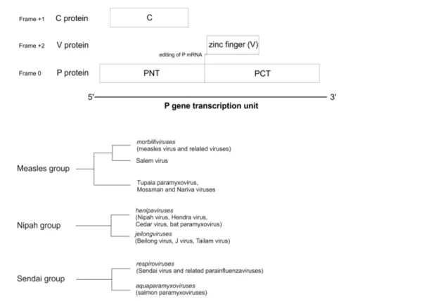
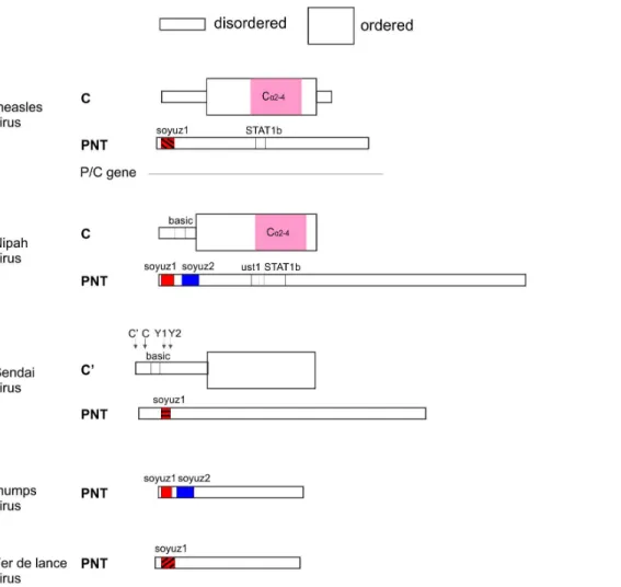
Paramyxovirinaeis a large virus subfamily that contains 9 known human pathogens: measles virus, mumps virus, human parainflu-enza viruses type 1 (hPIV1), 2, 3 and 4, Menangle virus, and the recently emerged, highly pathogenic Nipah and Hendra viruses [1]. Paramyxovirinae encode multiple proteins from the phospho-protein (P) gene transcription unit, including P, V, and C. In almost allParamyxovirinae, the P gene mRNA is edited, resulting in the expression of at least two proteins, P and V, which share an identical N-terminus (PNT), but have a unique C-terminus (Figure 1A) (for a review, see [2]). In addition, several genera, including Morbilliviruses, Henipaviruses, and Respiroviruses, encode a third protein, C, within their P gene, from an overlapping reading frame [2]. The C proteins are expressed by a variety of mechanisms including: leaky scanning [3–5], non-AUG start codons [6,7], ribosomal shunting [8], and proteolytic processing [9]. The region of P that overlaps C, corresponding approximately to PNT (Figure 1A), is disordered [10–13], and contains conserved sequence motifs, such as soyuz1, found in allParamyxovirinae,which binds the viral nucleoprotein, and soyuz2, of unknown function [14].
The two primary functions of the C proteins are their abilities to regulate viral transcription/replication and to antagonize the antiviral responses of the host. These functions are thought to be interconnected, since a decrease in viral transcription/replication often correlates with a decrease in the innate antiviral responses of the host [15–18] (for a review, see [19]). Most paramyxoviral C proteins inhibit viral RNA synthesis, and thereby presumably regulate viral gene expression [20–24]. However, they differ in the degree to which they block host antiviral responses [25]. These responses are composed of two crucial signaling cascades: A) Induction of type I interferon (IFN), following recognition of virus-derived elements by pattern recognition receptors (PRRs) and B) IFN signaling through the JAK/STAT pathway, leading to transcription of antiviral effector genes [26,27].
that this effect is not significant [17,32,33]. Similarly, although the mechanistic details are less clear,HenipavirusC proteins block IFN induction by decreasing viral RNA synthesis, which indirectly inhibits type I IFN induction; but they have minimal effects on IFN signaling [15,34–37]. Like the morbilliviruses, Respirovirus C proteins also counteract IFN induction through two mechanisms: 1) by minimizing production of double-stranded RNA (dsRNA), thereby avoiding PRR activation [16,38]; and 2) by inhibiting IRF3-dependent induction of type I IFN [39]. However, the C proteins of respiroviruses differ from those of Morbilliviruses and Henipavirusesin being also able to inhibit IFN signaling [16,26,38– 53]. Finally, a new role has been reported recently for the C proteins ofrespiroviruses: they regulate the levels of viral genomes and antigenomes produced during infection [54].
Interestingly,henipaviruses andmorbilliviruses can also block IFN signaling, but do so by proteins encoded by the P frame rather than the C frame (i.e. P, V, or a third protein called W), which interfere with the localization or phosphorylation of STAT1 (Signal Transduction Activator of Transcription 1), among other mechanisms [55–62].
Overlapping genes, such as those encoding P and C, are of particular interest because they encode proteins originatedde novo (in contrast to origination by well-characterized processes such as gene duplication or horizontal gene transfer [63,64]). Indeed, overlapping genes are thought to arise by overprinting, a process in which mutations within an existing (‘‘ancestral’’) protein-coding reading frame allow the expression of a second reading frame (the de novo frame), while preserving the expression of the first frame [65–67].De novoproteins have been little studied but are known to play an important role in viral pathogenicity [68,69], for instance by neutralizing the host interferon response [70] or the RNA
interference pathway [71]. In addition, de novo proteins char-acterised so far have previously unknown 3D structural folds [68,71,72] and novel mechanisms of action [71]. Thus, this class of proteins may challenge the notion that nature only utilizes a limited number of different protein folds and that this fold space is well mapped [73,74]. Another particularly interesting feature of overlapping genes is the evolutionary paradox they present, since the overlap imposes sequence constraints which should restrict the ability of the virus to adapt [75–81].
Our study was divided in three strands. First, we predicted the structural organization of the C proteins, and determined whether they had detectable sequence similarity, which could indicate a common origin, guide experimental studies, and facilitate 3D structure determination [82]. Second, we verified our predictions experimentally, by expressing, purifying and characterizing several C proteins in bacteria. Third, we investigated the evolutionary history of the P/C gene overlap, and tried to determine which, of P and C, is the novel frame.
Methods
Sequence Alignment
The accession numbers of the sequences of Paramyxovirinae P used in this study, as well as the abbreviations of species names, are in Table 1. The sequence of the C protein of Pacific salmon paramyxovirus[83,84] was generously made available by Bill Batts and Jim Winton. We used Psi-Coffee [85,86] for multiple sequence alignments (MSAs). All alignments are presented using Jalview [87] with the ClustalX colouring scheme (see Figure 2b and 2d in [88]). The aligned sequences of the C proteins in text format are in File S1.
We used two criteria to estimate the reliability of alignments of the C proteins: 1) the CORE reliability index, which is based on the agreement between the different alignment programs used by Psi-Coffee, and is part of the standard output of Psi-coffee [86]; 2) in the case of the measles and Nipah groups, we also considered the coherence between the alignments of either group separately and the alignment of both groups. We considered as not reliably aligned the positions that either have a low Psi-coffee CORE index, or are not aligned in the same way in these alignments.
Finally, we used TranslatorX [89] to generate a nucleotide alignment of the P/C gene corresponding to an amino acid alignment of the C protein. The alignment of the C proteins (not shown) was created using the MUSCLE program [90] built in TranslatorX, and is thus slightly different from that generated by Psi-coffee, mainly in the region between Ea2and S/Ta4. This has no impact on the results presented.
Sequence Analyses
The secondary structure of individual sequences was predicted using Jpred [91], and was verified in the context of multiple alignments using PROMALS [92]. We predicted disordered regions with MetaPrDOS [93], according to the principles described in [94]. We used HHalign [95] to compare the MSAs of the C proteins of various groups, with a cutoff E-value of 1025
. To identify and cluster homologous C proteins, we performed iterative sequence searches [96] on the C proteins of each taxon, using csi-blast [97] and HHblits [98] with a cutoff E-value of 1023
, as described in [99]. We identified 5 subgroups of homologs (Figure 1), formed by the following taxons: 1) the genusmorbillivirus and Salem virus; 2)Tupaia Paramyxovirus, Mossman virus, andNariva virus; 3) the genus henipavirus; 4) the newly proposed genus
jeilongvirus; and 5) the two generarespirovirusandaquaparamyxovirus (called ‘‘Sendai group’’). Several proteins of subgroups 1 and 3 had a subsignificant (E.1023
) similarity with proteins of subgroups 2 and 4, respectively, indicating that these subgroups may be homologous [99]. We confirmed their homology by using HHalign [95] (E = 5.10211
for the comparison between subgroups 1 and 2, and E = 2.1029
for the comparison between subgroups 3 and 4). We called the combination of subgroups 1 and 2 ‘‘measles group’’ and the combination of subgroups 3 and 4 ‘‘Nipah group’’.
Cloning of the C Genes
To maximize our chances of successfully expressing C proteins, we adopted a high-throughput approach. We cloned full-length synthetic cDNAs (obtained from Genscript) of the C proteins of all 24 species in the measles, Nipah and Sendai groups into the vector pOPIN-F [100] using the InFusion procedure, as described in [100,101]. The resulting fusion proteins have an N-terminal hexahistidine tag followed by a 3C cleavage site immediately upstream of the coding sequence of the C proteins.
Expression of the C Proteins
Proteins were expressed in the bacteria Escherichia coli(E. coli) using the BL21(DE3) Rosetta pLysS strain (Novagen), following the ZYM-5052 auto-induction protocol [102]. Briefly, large scale cultures were inoculated to OD600 of 0.02 and grown for 16 h at 25uC. Cells were harvested and the pellet resuspended 1:3 (w/vol) in lysis buffer (50 mM TrisHCl, 500 mM NaCl, 30 mM Imidazole pH 8.0, 1% vol/vol Protease inhibitor mix (Sigma P8849)) and frozen in liquid nitrogen before storage at280uC.
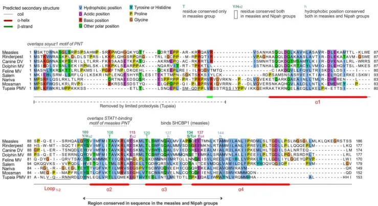
Figure 2. Alignment of the C proteins of the measles group.Numbering corresponds tomeasles virus. The N-terminus of C is highly variable and shown for information only. Only the C-terminal moiety of C (helicesa2 toa4) is reliably aligned; positions that appear conserved but are outside this region are thus not indicated. Residues that have been experimentally substituted (Table 2) are in bold. N-terminal sequences of fragments of Tupaia PMV C obtained after limited proteolysis are underlined. Overlapping motifs of the PNT frame overlapping C are indicated above the alignment.
Purification of the C Proteins
We purified both C proteins in two steps: Nickel Immobilized Affinity Chromatography (IMAC) followed by size-exclusion chromatography (SEC). Pellets were thawed and homogenized (Constant Systems homogenizer) at 25 kpi at 4uC. The lysate was cleared at 50,000 g for 30 minutes before batch incubation of the supernatant (i.e. the soluble fraction of bacteria) on Ni-NTA sepharose FF resin (Qiagen) for 2 hrs at 4uC. The material was collected in an Econo-Pac column (Biorad) and washed in 100 Column Volumes (CV) of lysis buffer. Elution was done in 0.5 CV fractions with lysis buffer containing 500 mM Imidazole. Fractions containing protein were pooled and loaded onto a preparative Superdex 75 (GE Healthcare Life Sciences) size exclusion column pre-equilibrated in 20 mM Tris 150 mM NaCl, 1 mM EDTA, pH 7.5. Peak fractions were pooled and concentrated using 15 ml spin concentrators (Millipore).
Circular Dichroism (CD)
Protein samples were extensively dialyzed into 20 mM NaPho-sphate, 20 mM NaCl pH7.5 and then concentrated to 0.2 mg/ml in spin concentrators (0.5 ml, 3KDa MWCO, Millipore). The Circular dichroism (CD) analysis was done on a JASCO 815 CD spectropolarimeter. Data are averages of 5 independent scans in the 190 nm –250 nm range, and were normalized to the baseline of the dialysis buffer. The data were smoothed using the manufacturer’s software (Jasco SpectraManager) before
interpre-tation. The percentage ofa-helix was calculated according to the formula: percentage ofa-helix = (h208–4000)/(-33000-4000)6100,
whereh208is the ellipticity at 208 nm [103].
Limited Proteolysis
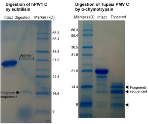
From 1 mg/ml protease stocks, we made 10-fold serial dilutions in 20 mM Hepes, 50 mM NaCl, 10 mM MgSO4, pH 7.5. Proteins were concentrated to 0.6 mg/ml by spin concentrators (0.5 ml, 3 MWCO, Millipore). For limited proteolysis, 10ml of protein was mixed with 3ml of protease and incubated on ice for 30 min, 60 min or 2 hrs. Reactions were stopped by adding 2ml protease inhibitor mix (Sigma P8849). To each reaction, 5ml of 4x SDS PAGE sample buffer was added and samples were heated to 95uC for 2 min before loading on a 1 mm 15% SDS-PAGE gel. A subtilisin digest of hPIV1 C and an a-chymotrypsin digest of Tupaia PMV C gave rise to stable fragments which were blotted to PVDF before submitting the samples for N-terminal sequencing (ALTA bioscience, UK).
Analytical Size Exclusion Chromatography (SEC)
Analytical size exclusion chromatography (SEC) was performed at a flow-rate of 0.5 ml/min using a Superdex 75 10/300 column (GE Healthcare Life Sciences) pre-equilibrated in 20 mM TrisCl, 150 mM NaCl, 1 mM EDTA pH = 7.9. The column was calibrated with a separate run of appropriate globular marker Table 1.Accession numbers ofParamyxovirinaeC proteins.
Virus species Abbreviation Genus Genbank accession number
Atlantic salmon paramyxovirus Atlantic PMV Unclassified ABW38050.1
Beilong virus Beilong Jeilongvirus* YP_512248.1
Bovine parainfluenza virus 3 bPIV3 Respirovirus P06164
Canine distemper virus Canine DV Morbillivirus NP_047203.1
Dolphin Morbillivirus Dolphin MV Morbillivirus NP_945026.1
Bat paramyxovirus/Eid_hel/GH-M74a/GHA/2009 Bat PMV Henipavirus AFH96008.1
Cedar virus Cedar Henipavirus AFP87276.1
Feline morbillivirus Feline MV Morbillivirus AFH55514.1
Hendra virus Hendra Henipavirus O55779
Human parainfluenza virus 1 hPIV1 Respirovirus NP_604434.1
Human parainfluenza virus 3 hPIV3 Respirovirus NP_599251.1
J virus J virus Jeilongvirus* YP_338079.1
Measles virus Measles Morbillivirus NP_056920.1
Mossman virus Mossman Unclassified NP_958051.1
Nariva virus Nariva Unclassified YP_006347585.1
Nipah virus Nipah Henipavirus AAY43913.1
Pacific salmon paramyxovirus Pacific PMV Unclassified AFF60402.1
Peste des petits ruminants virus PPRV Morbillivirus YP_133824.1
Phocine distemper virus Phocine DV Morbillivirus P35940.1
Porcine parainfluenza virus 1 pPIV1 Respirovirus AGR39559.1
Rinderpest virus Rinderpest Morbillivirus ADF32062.1
Salem virus Salem Morbillivirus-like Q9IZB9
Sendai virus Sendai Respirovirus NP_056872.1
Tailam virus Tailam Jeilongvirus* AEU08859.1
Tupaia paramyxovirus Tupaia PMV Unclassified Q9WS38
(*) Proposed genus.
proteins (Gel Filtration LMW Calibration Kit, GE Healthcare Life Sciences).
Results
The C Proteins of Paramyxovirinae Cluster in three Groups: the Measles, Nipah and Sendai Groups
On the basis of sequence analyses (see Methods), the C proteins of Paramyxovirinaecan be divided into three groups: the measles, Nipah and Sendai groups (Figure 1B). The measles group is composed ofmorbilliviruses, of the unclassifiedSalem virus, and of a subgroup comprising the unclassifiedTupaia paramyxovirus, Mossman virusandNariva virus. The Nipah group compriseshenipavirusesand jeilongviruses. Finally, the Sendai group is composed ofrespiroviruses and of the recently described genusaquaparamyxovirus, composed of fish viruses [83,84,104] related to respiroviruses [105,106]. The classification of C into measles and Nipah groups is supported by an examination of the PNT domain of P, which is encoded by the same region as C but in a different frame (Figure 1A). Indeed, the PNT of all species in the Nipah group differ from the PNT of the measles group in having a soyuz2 motif (see Introduction) [14].
We found that otherParamyxovirinaethat do not not express a C frame [2,107] can be classified in two groups based on the phylogeny of their P gene: the mumps group (comprised of the sister generarubulavirusandavulavirus) and the Fer de lance group (formed by the genus ferlavirus [108]). This classification corre-sponds to that of previous analyses [105].
The C Proteins of the Measles and Nipah Groups are Homologous
We separately aligned the C proteins of the measles, Nipah and Sendai groups (Figures 2, 3, and 4 respectively; the aligned sequences in text format are in file S1). In these three groups, we observed a similar organization of the C proteins, composed of a variable N-terminus predicted to be disordered, and of a C-terminus predicted to be ordered and a-helical. We compared these alignments to each other using the profile-profile comparison software HHalign [95] (see Methods). Briefly, a sequence profile is a representation of a multiple alignment that contains information about which amino acids (aas) are ‘‘tolerated’’ at each position of the alignment, and with what probability. Comparing profiles is much more sensitive than comparing single sequences, because the profiles contain information about how the sequences can diverge and thus can identify weak similarities which remain after both sequences have diverged [99,109,110].
HHalign reported that the C proteins of the measles and Nipah groups have statistically significant similarity (E = 461026
) over a region of about 50aa in their C-terminus (shown in Figure 5). This high similarity could in theory result either from convergent evolution or from homologous descent. The fact that the measles and Nipah groups are phylogenetically related [105], and that their C proteins are encoded in the same genomic location makes homologous descent a much more likely explanation. On the other hand, HHalign did not detect any similarity between the C proteins of the Sendai group and those of the measles and Nipah Table 2.Selected experimental substitutions in C and their effect.
Group Virus name C ORF mutation Functional effect(s) of mutation References
MEASLES GROUP Measles virus R44G Ablates nuclear localization [29,156]
S134Y Associated with temperature-sensitive vaccine virus [114] D127–138 (deletion) Ablated interaction with SHCBP1, reduced ability to
inhibit minigenome replication
[115]
NIPAH GROUP Nipah virus No fine mutational data –
SENDAI GROUP Sendai virus CD10–15 (deletion) Inability to interact with and modulate levels of phosphorylated STAT1
[48]
Series of deletions in aa149–157
Loss of nuclear translocation of Y1 by Ran-GTPase pathway [120]
K151A/E153L/R157L (Cm*) Increased IFN-binduction and dsRNA production, induction of antiviral state, increased CPE, apathogenicin vivo
[39,127]
K77A/D80A (Cm2), M139A/ D142A (Cm4),R173A/ E175A/E176A (Cm8)
Inability to bind STAT1, ablated ability to inhibit RNA synthesis, decreased binding to viral polymerase (L protein)
[46,118]
K77R/D80A (Cm2’), D80A Increased cytopathic effect, increased nuclear translocation of IRF3, increased IFN-binduction and production of dsRNA
[39]
K151A/E153A/R154A (Cm5) Attenuated virulencein vivo, inability to block IFN signaling, inability to inhibit replication, inability to skew STAT1/2 phosphorylation and to bind STAT1, decreased binding to L protein
[39,46,118,127]
Human parainfluenza virus 1 (hPIV1)
R84G Increased IFN-bproduction, increased IRF3 nuclear translocation, reduced plaque sizes, non-temperature sensitive mutation contributing to attenuationin vivo
[43,157]
Human parainfluenza virus 3 (hPIV3)
CND25, CND50 (deletions) Increased inhibition of viral RNA synthesis, suppression of viral replication, decreased ability to block Type 1 IFN signaling
[121,122]
K3A, K6A, K12A, E16A, R24A
Increased inhibition of viral RNA synthesis, decreased ability to block Type 1 IFN signaling
[121,122]
aa 90–195 of C Region required for STAT1 binding [119]
These studies used either recombinant viruses, minigenome systems, or eukaryotic expression systems. Substituted residues that are conserved in a group are in bold. For a more comprehensive list of studies onParamyxovirinaeC, please see Table S1.
groups. Thus, either they are not homologous, despite their similar organization, or they are homologous but have diverged in sequence beyond recognition. The latter scenario is possible, in theory, since the relative frame of C compared to P (+1) is the same in the Sendai group and in the measles/Nipah groups (Figure 1A).
Sequence Analysis of the C Proteins of the Measles and Nipah Groups
Figures 2 and 3 present alignments of the C proteins of the measles and Nipah groups, respectively. Above the alignments, we indicated regions of C that overlap conserved motifs of the P frame. The C proteins of the measles and Nipah groups are all composed of a 30–60 amino acid (aa) N-terminus predicted to be
at least partially disordered, and of a 90–120 aa C-terminus comprising a predicteda-helix (a1), a loop of 10–20aa (‘‘loop1–2’’), and three furthera-helices (a2 toa4), followed in some species by C-terminal extensions of at most 20aa (forming helixa5 in some species of the Nipah group).
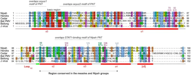
In the C proteins of the measles group, only the region froma2 toa4 is well conserved in sequence; it contains many conserved positions (Figure 2), of which six (boxed) are also conserved in the C proteins of the Nipah group (see below). In contrast, the C proteins of the Nipah group contains two additional, conserved regions (Figure 3): 1) a short N-terminus witha-helical potential (a0, aa 2–19 in Nipah virus), containing a hydrophobic region followed by a basic region (boxed in Figure 3); and 2) a short region at the C-terminus ofa1 (aa 74–83 in Nipah virus) that Figure 3. Alignment of the C proteins of the Nipah group.Conventions are the same as in Figure 2. Numbering corresponds toNipah virus. Several residues that appear conserved have not been indicated, because their alignment is not reliable, or their conservation is probably imposed by the P frame (see text).
doi:10.1371/journal.pone.0090003.g003
Figure 4. Alignment of the C proteins of the Sendai group.Conventions are the same as in Figure 2. Numbering corresponds to the C protein ofSendai virus. Arrows indicate the start of the different isoforms of C. For information, the arrowhead indicates the well-characterized F residue of
respiroviruses(F170 inSendai virus), whose substitution by S reduces innate immune antagonism and attenuation ofin vivopathogenesis by C [39,53,153–155] (see Table S1). The N-terminal sequence of the fragment of hPIV1 C obtained after limited proteolysis is underlined. The variable region between basic region 1 and residue G89 is not reliably aligned and is presented for information only.
contains two conserved acidic positions (E/D). The apparent conservation of other regions of C, which overlap the soyuz1 and soyuz2 motifs of the P frame (Figure 3), should not be over-interpreted, since it may be due to constraints imposed by selection pressures acting in fact on the P frame, which is much more conserved than the C frame in these regions (not shown).
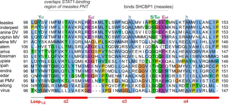
An alignment of the C proteins of both groups (Figure 5) revealed four remarkable positions conserved in nearly all viruses (boxed in Figure 5): a Tyrosine (Y) upstream of helixa2 (Ya2); a Glutamate (Ea2) at the C-terminus of the same helix; a residue with an alcohol group (Serine/Threonine, S/Ta4) at the N-terminus of helix a4; and a Glutamate (Ea4) two residues downstream. Two other positions of hydrophobic nature (indicat-ed by ‘‘h’’) are conserv(indicat-ed in both groups. These conserv(indicat-ed residues are also boxed in Figures 2 and 3, in the separate alignments of the measles and Nipah groups. Other positions that appear conserved in Figure 5 or in Figures 2 and 3 may in fact not be reliably aligned (see Methods) and are therefore not boxed.
Sequence Analysis of the C Proteins of the Sendai Group Figure 4 shows the alignment of the C proteins of the Sendai group. In Sendai virus and human parainfluenza virus 1(hPIV1), as many as four products (C’, C, Y1, Y2) are expressed from the C reading frame by a combination of alternative initiation codons [6–8] and proteolytic processing [9]. Their respective N-termini are indicated by arrows. The C proteins of the Sendai group have a similar organization to that of the measles and Nipah groups. They are composed of a variable, disordered N-terminus of about 80aa, rich in Prolines (P), Serines (S) and Threonines (T), followed by a conserved C-terminus composed of foura-helices (aA toaD). The N-terminus contains a basic region (boxed in Figure 4) within a predicteda-helix (aZ), like the C protein of the Nipah group (Figure 3). In the C protein ofSendai virus, the first half ofaZ was reported to act as a membrane-targeting signal, perhaps by forming an amphipathic a-helix [111]. There are 11 residues
strictly conserved in C across the Sendai group, clustered predominantly in the C-terminus of aC and in aD. aC is particularly rich in K and R (‘‘basic region 2’’ in Figure 4), suggesting it might bind a negatively charged partner.
Obtaining a Reliable Alignment of the Region of PNT Containing STAT1-binding Sites inmeasles virus and Nipah virus
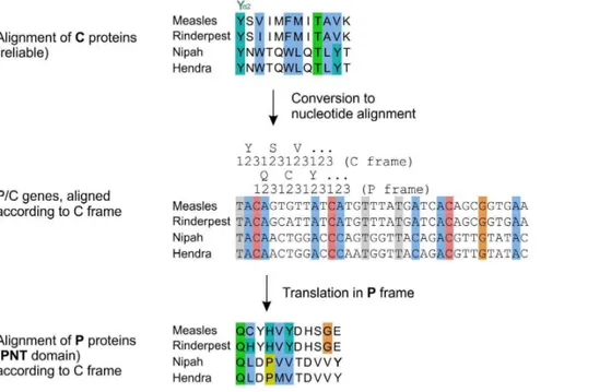
We present in Figure 6 a summary of the structural and functional organization of PNT and C in the different taxa of Paramyxovirinae, to scale, with their functional motifs vis-a`-vis of each other. PNT contains sequences that bind the protein STAT1 in several morbilliviruses (measles virus [55,56], canine distemper virus [57], Rinderpest virus [60]) and henipaviruses (Nipah virus [58] and Hendra virus[59]). The region of PNT that contains these sites is highly variable in sequence (Figure 7), and thus its alignment is not reliable. In contrast, the overlapping region of C is well conserved, and its alignment reliable (Figure 5). Therefore, we used the C frame to construct a reliable alignment of PNT. We proceeded in two steps (see Methods). First, we used the amino acid alignment of the C proteins (Figure 8, top panel) to generate an alignment of the nucleotide sequences of the P/C gene (Figure 8, middle panel and File S2), using TranslatorX [89]. Second, we translated this nucleotide alignment into an amino acid alignment in the P frame (Figure 8, bottom panel). The resulting alignment of PNT of the measles and Nipah groups is presented in Figure 9.
The STAT1-binding Sites of PNT of Nipah Virus and Measles Virus Overlap Different Regions of C and thus Probably Evolved Independently
From the reliable alignment of PNT corrected by using the C frame (Figure 9), we made three observations:
i) The STAT1-binding sites ofmeasles virusandNipah virusPNT are conserved in sequence only in very closely related species
Figure 5. Alignment of the C proteins of the measles and Nipah groups.Conventions are the same as in Figure 2. Several positions appear conserved but have not been indicated, because their alignment is not reliable (see text).
(thick boxes in Figure 9). For instance, in PNT of Feline morbillivirus, which is more distantly related to measles virus than othermorbilliviruses, only 2 aa out of 11 (E110 and I116) correspond to conservative substitutions with respect to the STAT1-binding motif ofmeasles virus(Figure 9). Such a high
number of non-conservative substitutions within a short peptide suggests that it may not bind STAT1.
ii) The STAT1-binding sites ofmeasles virusandNipah virusPNT are not aligned together (Figure 9) (although they overlap Figure 6. Summary of the organization ofParamyxovirinaePNT and C.The figure is to scale, with PNT and C vis-a`-vis of each other. The PNTs are all positioned so that their soyuz1 motifs match. Regions whose homology is proven (by statistically significant similarity) have the same color. Homology of soyuz1 motifs is suspected but not proven [14], thus they have a same color, but different patterns. STAT1b: STAT1-binding site. Ust1: ‘‘upstream of STAT1’’ motif.
doi:10.1371/journal.pone.0090003.g006
slightly, by 4aa), which indicates that they are encoded in different locations of the P/C gene. It is thus highly likely that they have originated independently (see Discussion).
iii) The STAT1-binding sites ofmeasles virusandNipah virusPNT have some limited sequence similarity, as reported earlier [58]: they share a [Y/H]DH[S/G]GE motif, underlined in Figure 8. Procedure to reliably align the central region of PNT of the measles and Nipah groups by using a C frame alignment.
Conventions are the same as in Figure 2. An alignment of C (top panel) is converted in a nucleotide alignment (middle panel) by using TranslatorX (see text), then translated into the P frame (bottom panel), yielding a reliable alignment of the PNT domain of P, which overlaps C. The nucleotide alignment of the P/C genes corresponding to the middle panel is in File S2.
doi:10.1371/journal.pone.0090003.g008
Figure 9. Alignment of the region of PNT of the measles and Nipah groups containing STAT1-binding sites, corrected by using the C frame.Conventions are the same as in Figure 2. This reliable alignment of PNT is based on an alignment of the C frame by the procedure described in Figure 8. The [Y/H]DH[S/G]GE motifs common to the STAT1-binding sites ofmeasles virusandNipah virusPNT are underlined. Note that contrary to the alignment of Figure 7, here they are not aligned together. Y110 ofmeasles virusPNT and Y116 ofNipah virusPNT, which were suggested to be analogous (see text) are in bold.
Figures 7 and 9. However, this similarity is unlikely to be due to homologous descent, since the motifs are not aligned together in the reliable alignment of PNT (Figure 9). Likewise, the tyrosine residues immediately upstream of this motif (Y110 in measles virus PNT, critical for STAT1 inhibition [33,55,112,113], and Y116 in Nipah virus PNT), which were perceived to occur in a similar sequence context [58], are not aligned together either in the reliable alignment of PNT (Figure 9), indicating that they are not homologous either.
Finally, we also noticed an 8aa motif (aa 104–111 inNipah virus) conserved in the PNT of allhenipaviruses(Figure 9, thin box). We called this motif ust1 (for ‘‘upstream of STAT1’’). Its function is unknown, though aa 81–113 ofNipah virusP, which include ust1, are required for the synthesis of viral RNA [58]. We cannot exclude, however, the possibility that the conservation of ust1 is due to constraints imposed by the overlapping C frame.
Functional Organization of the C Proteins in Relation to Their Sequence
We systematically examined mutational studies of Paramyxovir-inaeC and their phenotypic impact. The most relevant studies are in Table 2 and a more extensive list of studies is in Table S1. We found that very few conserved positions identified herein have been subjected to targeted mutagenesis; notable substitutions are indicated in bold in Figures 2 and 4.
In the measles group, experimental substitutions have been performed mostly in the C-terminus of C. In a comparison of a temperature-sensitive strain of measles vaccine, AIK-C, with its parental strain, Edmonston [114], one of several substitutions identified, S134Y, occurs in the S/Ta4position conserved in the measles and Nipah groups (Figures 2 and 5) (Table 2). Although this particular substitution is not responsible for the temperature
sensitive phenotype [114], we note that it is located within a 12aa peptide (aa 127–138) recently shown to inhibit the viral polymerase by interacting with SHCBP1 (Shc Src homology 2 domain-binding protein 1) [115]. This peptide, underlined in Figures 2 and 5, contains two other positions conserved in the measles/Nipah groups (a hydrophobic residue and Ea4). Such conservation suggests that other viruses in the measles/Nipah groups may also bind SHCBP1 to block the viral polymerase. Finally, the role of the disordered N-terminus ofmeasles virusC is poorly known, although it contributes to nuclear localization, which correlates with its ability to block IFN induction [29] (Table 2).
In the Nipah group, there are no fine mutational data published, but it is known that both the N-terminus and the C-terminus of Nipah virus C are required to inhibit minigenome replication [116].
In the Sendai group, experimental substitutions have delineated multiple residues in the C-terminus of C responsible for antagonizing both IFN induction and IFN signaling, and for regulating viral transcription and replication [46,49,117,118] (Table 2 and Table S1). For both Sendai virus and hPIV3, the minimal region required for STAT1-binding corresponds to the structured, well-conserved C-terminus of C [117,119]. Within that domain, aas 149–157 (corresponding roughly to basic region 2, underlined in Figure 4) are critical for nuclear translocation of the Y1 isoform of Sendai virus C, and may also play a role in the inhibition of type-I IFN-stimulated gene expression [120]. This region contains several conserved residues, suggesting that its function may be conserved in the Sendai group. Studies of the N-terminus of C in the Sendai group indicate that it also contributes to antagonizing the innate immune response and to regulate viral transcription and replication [121,122] (Table 2 and Table S1). Taken together, these studies suggest that both the N- and C-terminus of Sendai group C proteins may need to act in
Figure 10. Circular Dichroism (CD) spectra of the C proteins of hPIV1 and Tupaia PMV.
coordinated fashion in order to perform their complete suite of antagonistic and regulatory functions.
Experimental Characterization of one C Protein of the Measles/Nipah Group and of One C Protein of the Sendai Group
In order to check our predictions of structural organization, we attempted to characterize biophysically at least one C protein of the measles/Nipah groups and one of the Sendai group. We systematically tested, in the bacteria E. coli, the expression and solubility of the C proteins of all species in the measles, Nipah and Sendai groups (see Methods). We found that the C proteins of tupaia paramyxovirus (Tupaia PMV) and of hPIV1 were by far the best candidates, for the measles/Nipah groups and Sendai group respectively, in terms of yield and solubility (not shown). We expressed both proteins as hexahistidine-tagged N-terminal fusion proteins in Escherichia coli and purified them from the soluble fraction by immobilized metal affinity chromatography (IMAC) and size exclusion chromatography (SEC) (see Methods). Mass spectrometry confirmed that the C proteins had the exact expected mass. In SDS-PAGE analysis (Figure S1), hPIV1 C migrated at a notably larger size (,31kD) than expected (25.9kD), while Tupaia PMV C migrated at,21kD, only slightly above the expected size (19.7kD). This anomalous migration may be caused by regions that are disordered or have a biased aa composition [123]. Accordingly, the N-terminus of both proteins is predicted disordered, and has a biased composition in the case of hPIV1 C.
We analyzed the secondary structure of the C proteins by Circular Dichroism (CD). The CD spectrum of both proteins (Figure 10) is typical ofa-helical content [124], with two dips in ellipticity at around 208 and 222 nm. The estimated a-helical content was 57% for hPIV1 C and 33% for Tupaia PMV C (see Methods). We also examined the C proteins by analytical SEC (Figure 11). Tupaia PMV C elutes at an apparent molecular mass of 21.4 kDa, close to its theoretical mass of 19.7 KDa. In contrast, hPIV1 C elutes at a much larger MW (38.7 kDa) than expected (25.9 kDa). This discrepancy could correspond to an extended shape, or to self-association in a fast equilibrium between a monomeric and dimeric form (see below).
Limited Proteolysis of hPIV1 C and Tupaia PMV C Confirms That they Have a Flexible N-terminus and a Structured C-terminus
We used limited proteolysis combined with N-terminal sequencing to probe the structural organization of the C proteins of hPIV1 and Tupaia PMV. We tested a range of proteases with different substrate requirements (see Methods), and identified fragments resistant to proteolysis, indicative of folded domains. Digestion of hPIV1 C by subtilisin yielded a stable degradation product of around 14 kD (Figure 12, left panel), whose N-terminal sequence, starting at aa 104, is underlined in Figure 4. The size of this fragment indicates that it comprises the whole C-terminus of C (expected size 14.16 kD), which corresponds well to our sequence predictions (Figure 4). These results are also coherent with cellular experiments that identified a proteolysis-sensitive N-Figure 11. Size Exclusion Chromatography (SEC) on the C proteins of hPIV1 and Tupaia PMV.The curves represent two SEC purifications runs, on the same column and in similar conditions (see text): one for Tupaia PMV C (blue), and one for hPIV1 C (green).
terminus in the C’ proteins ofSendai virus[125]. We note that the presence of a long, disordered region in hPIV1 C is compatible with its high apparent molecular weight observed in SEC (see above) [126].
Digestion of the C protein of Tupaia PMV bya-chymotrypsin yielded a series of bands ranging from 14 kD to 6 kD (Figure 12, right panel); further digestion (not shown) yielded a single 6kD fragment. We obtained N-terminal sequences of the three most abundant fragments, of,14.4, 13, and 6 kD (arrows to the right of Figure 12). They start respectively at aa 30, 43 and 84. This pattern of proteolytic digestion indicates that Tupaia PMV C is composed of a disordered N-terminus and of an ordered C-terminus. This is compatible with our predictions, in which aa 1– 56 are devoid of secondary structure (Figure 2) and aa 1–42 disordered, and in which a predicted loop,a1–2(aa 81–92), could be accessible to proteolysis. The observed fragments of 14.4 and 13kD correspond exactly to C proteins where aa 1–29 and 1–43, respectively, have been digested, whereas the size of the smaller fragment (6kD) corresponds to aa 81–135, indicating that the last 18 C-terminal aa are digested upon extended proteolysis.
In summary, our experiments confirm that in vitro, the C proteins of hPIV1 and Tupaia PMV are predominantlya-helical and contain a disordered N-terminus, whose boundaries are in good agreement with our sequence-based predictions.
Discussion
Substituting the conserved, charged residues we have identified herein should be a powerful way to dissect the function of C. Indeed, charged residues are often on the surface of proteins and thus their conservation is generally the result of functional constraints, rather than constraints imposed by a mere structural
role. The power of this approach has been shown by studies on several regions of respirovirus C [39,46,127], and our thorough sequence analysis of the full-length C proteins of allParamyxovirinae should greatly extend its applicability. In addition, knowing the structural organization of C will allow the design of deletions that have less risk of disrupting its three-dimensional structure.
A Common Origin of the C Proteins?
The C proteins of the Sendai group have no detectable sequence similarity with those of the measles/Nipah groups. However, we consider it unlikely that they have an independent origin, because they are located in the same region of the P gene, in the same frame relative to P, and have a similar structural organization and several similar functions [118,128,129]. Thus we consider that all C proteins most probably have a common origin, as proposed earlier [67,130]. The absence of a C protein in the mumps group is probably due to a loss in the ancestor of that group, since the Sendai group, which has a C protein, is basal in a phylogeny of the P gene [105]. This common origin would imply that inSendai virus, it is the Y1 isoform of C that is the equivalent of C of the measles/Nipah groups, because their start codon have the same location immediately upstream of the soyuz1 motif of the P frame (Figure 6; compare also Figure 4 and Figure 3). Therefore, the C and C’ proteins of Sendai virus would have presumably originated by mutations creating new, alternative start codons upstream of Y1. A common origin ofParamyxovirinae C proteins would also imply that the basic regions in the N-terminus of C have originated independently in the Sendai and Nipah groups, since they occupy different positions with respect to soyuz1 (Figure 6).
Figure 12. Limited proteolysis of the C proteins of hPIV1 and Tupaia PMV.Digestion profiles of hPIV1 C (left) and Tupaia PMV C (right), visualized by SDS-PAGE and Coomassie blue staining. Several fragments (arrowheads) were N-terminally sequenced. Their N-terminal sequences are underlined in Figures 2 and 4.a–chymotrypsin is not visible on the digestion profile of Tupaia PMV C.
Which Frame Originated Earlier, PNT or C?
Overlapping genes typically encode an ancestral frame and a novel frame originated by overprinting it (see Introduction). Our analyses in this work and in an earlier study [14] suggest that the C and PNT frames were probably both present in the ancestor of Paramyxovirinae, making it impossible to conclude which frame is ancestral on the basis of phylogeny. Analysis of codon usage [131] cannot determine which frame is ancestral either, because the codon usages of PNT and C are indistinguishable inParamyxovirinae (Angelo Pavesi, personal communication). However, functional considerations suggest that the PNT frame originated earlier, since it is indispensable to viral replication in vitro [2,132], unlike C [20,133,134]. The ancestry of PNT is supported by a comparison with families related to Paramyxovirinae (Mononegavirales). Most Mononegavirales also encode P proteins with a disordered N-terminus [14,135]; at least inRhabdoviridae, this N-terminus has the same function as Paramyxovirinae PNT, i.e. preventing the nucleoprotein from self-assembling illegitimately [136–139]. Thus, it is reasonable to speculate that the P of the ancestral Mononegavirales already had a disordered N-terminus, which was overprinted by C in the ancestor ofParamyxovirinae.
Convergent Evolution between the STAT1-binding Sites ofmeasles virusandNipah virus?
The STAT1-binding sites ofmeasles virusandNipah virusdo not align together in the reliable alignment of PNT, generated using the C frame (Figure 9). This strongly suggests that they have originated independently. Alternatively, since they overlap by 4aa (Figure 9), these STAT1-binding sites might, in theory, have
originated from a common, short peptide, providing some STAT1-blocking capability, and later have extended respectively upstream and downstream of PNT. However, this scenario is not parsimonious because it would imply several losses in the lineages separating measles virus and Nipah virus. Also, the common 4aa stretch is chemically very different in both viruses (G117EAV in measles virus and V115YHD in Nipah virus, Figure 9). We thus consider it most likely that the STAT1-binding sites ofmeasles virus andNipah virushave originated independently.
Their limited sequence similarity (they share an [Y/H]DH[S/ G]GE motif, underlined in Figure 9) would thus not be the result of homologous descent, but could instead result either from convergent evolution (owing to a common mechanism), or from random chance. Convergent evolution seems a definite possibility, since the mechanisms by which PNT acts are somewhat similar in both viruses (PNT interferes with the phosphorylation of cytoplasmic STAT1) [55,58,112,140], and since the PNT of both viruses bind a similar part of STAT1 [141].
The P/C Gene Exemplifies Three Keys to the Evolutionary Paradox of Overlapping Genes
Overlapping genes are an evolutionary paradox, because they simultaneously encode two proteins whose freedom to mutate is constrained by each other, which should severely reduce the ability of the virus to adapt [75–81].
A first key to the paradox has been suggested earlier [67,77,78,142–146]: overlapping genes frequently encode an ‘‘ancillary’’ frame that can tolerate a higher substitution rate than the other, ‘‘dominant’’ frame; the ancillary frame is often Figure 13. Three patterns of sequence constraints in the overlapping frames P and C.PNT and C are represented vis-a`-vis of each other with same conventions as in Figure 6. Sequence constraints of PNT and C were estimated by their sequence variability.
structurally disordered [68]. Accordingly, a previous sequence analysis ofSendai virusindicated that PNT and C are generally not both under strong constraint [142]; rather, the N-terminus of PNT is markedly more conserved than that of C, whereas the C-terminus of PNT is markedly more conserved than that of C [142]. This is also the case for most of the PNT and C of measles and Nipah virus (Figure 13, evolutionary pattern 1 or 2), with the exception of the region corresponding to the STAT1-binding sites of PNT (see below).
A second key to the paradox of overlapping genes is that it may be beneficial for a virus, under certain conditions, to encode functional motifs simultaneously by using overlapping frames [147]. Initially, we were very surprised to discover that a region of the P/C gene encodes simultaneously, in different frames, two well-conserved regions: the STAT1-binding motif of PNT, and the a2–a4 region of C (Figure 13, evolutionary pattern 3). Intuitively, this arrangement seems to dramatically restrict the capacity of the virus to mutate and to escape host defenses. We were all the more surprised that this arrangement originated twice independently, in measles virusand in Nipah virus(see Figure 6). This seems beyond coincidence, and strongly suggests that the loss of fitness of the virus due to its reduced ability to mutate is compensated by an evolutionary advantage. In fact, this phenomenon had been predicted on the basis of mathematical modeling [147]. Given a high mutation rate, it may be advantageous to encode crucial functional motifs in overlapping frames (provided that they are short), because the superposition of critical amino acids reduce the number of vulnerable positions in the genome. The conditions of application of the model are met here: RNA viruses have one of the highest mutation rate of all organisms [148], and the STAT1-binding sites are short (10–26aa). It will be interesting to investigate whether this evolutionary pattern, in which two reading frames are both under strong constraint, is common in viruses, and whether it does entail a selective advantage. The genome ofHepatitis B virus, for instance, also contains short regions where both the overlapping Polymerase and Glycoprotein frames are under strong constraint [149,150]. A recent innovative methodology that combines experimental and computational approaches [151] could help to tease out the different factors (structural, functional and co-evolutionary) constraining overlap-ping motifs.
Finally, a third key to the paradox of overlapping genes is that they provide a regulatory advantage that may offset the increased constraints they impose on the virus, by encoding two proteins that are co-regulated and have complementary functions [131]. For instance, the expression levels of the C and V proteins of Nipah or measles viruses are co-regulated, since they are transcribed from the same gene transcription unit; in addition, their roles are
complementary, since together they inhibit both viral RNA synthesis and type I IFN induction, enabling an efficient block of the first stage of the host antiviral response [15,17,20,24,152]. In the same vein, the expression of C and P is also co-regulated and they have complementary effects on viral transcription, mediated by binding the same cellular protein, SHCBP1 [115].
Conclusion
In conclusion, we predict that the C proteins of the Sendai group and of the measles/Nipah groups will have the same structural fold, testifying to a common origin, and that this fold will be a previously unobserved one, in keeping with theirde novoorigin [68].
Supporting Information
Figure S1 Purification of the C proteins of hPIV1 and Tupaia PMV. The purifications are visualized by Coomassie blue-stained SDS-PAGE.
(TIF)
Table S1 Effect of experimental substitutions in Para-myxovirinaeC proteins.
(DOCX)
File S1 Multiple sequence alignment of the C proteins of the measles, Nipah, and Sendai groups.
(DOC)
File S2 Multiple sequence alignment of the P/C genes of the measles and Nipah groups, based on an alignment of the C proteins
(DOC)
Acknowledgments
We thank B Bankamp, JM Bourhis, P Devaux, M Jamin, R Neme and A Vianelli for comments on the manuscript. We thank the OPPF-UK for help with expression of the C proteins, and the organizers of the EMBO training ‘‘High-throughput methods for protein production and crystalli-zation’’.
Disclaimer: The findings and conclusions in this report are those of the authors and do not necessarily represent the views of the Centers for Disease Control and Prevention.
Author Contributions
Conceived and designed the experiments: TMS DGK. Performed the experiments: TMS DGK. Analyzed the data: MKL TMS DGK. Contributed reagents/materials/analysis tools: MKL TMS DGK. Wrote the paper: MKL TMS DGK.
References
1. Mayo MA (2002) A summary of taxonomic changes recently approved by ICTV. Arch Virol 147: 1655–1663.
2. Lamb RA, Parks GD (2007) Paramyxoviridae: the viruses and their replication. In: Knipe DM, Howley PM, editors. Fields Virology. Fifth edition ed. Philadelphia: Lippincott Williams & Wilkins. 1449–1496.
3. Bellini WJ, Englund G, Rozenblatt S, Arnheiter H, Richardson CD (1985) Measles virus P gene codes for two proteins. J Virol 53: 908–919.
4. Giorgi C, Blumberg BM, Kolakofsky D (1983) Sendai virus contains overlapping genes expressed from a single mRNA. Cell 35: 829–836. 5. Lo MK, Harcourt BH, Mungall BA, Tamin A, Peeples ME, et al. (2009)
Determination of the henipavirus phosphoprotein gene mRNA editing frequencies and detection of the C, V and W proteins of Nipah virus in virus-infected cells. J Gen Virol 90: 398–404.
6. Curran J, Kolakofsky D (1988) Ribosomal initiation from an ACG codon in the Sendai virus P/C mRNA. EMBO J 7: 245–251.
7. Boeck R, Curran J, Matsuoka Y, Compans R, Kolakofsky D (1992) The parainfluenza virus type 1 P/C gene uses a very efficient GUG codon to start its C’ protein. J Virol 66: 1765–1768.
8. Latorre P, Kolakofsky D, Curran J (1998) Sendai virus Y proteins are initiated by a ribosomal shunt. Molecular and Cellular Biology 18: 5021–5031. 9. de Breyne S, Monney RS, Curran J (2004) Proteolytic processing and
translation initiation: two independent mechanisms for the expression of the Sendai virus Y proteins. J Biol Chem 279: 16571–16580.
10. Karlin D, Longhi S, Receveur V, Canard B (2002) The N-terminal domain of the phosphoprotein of Morbilliviruses belongs to the natively unfolded class of proteins. Virology 296: 251–262.
11. Habchi J, Mamelli L, Darbon H, Longhi S (2010) Structural disorder within Henipavirus nucleoprotein and phosphoprotein: from predictions to experi-mental assessment. PLoS One 5: e11684.
12. Chinchar VG, Portner A (1981) Inhibition of RNA synthesis following proteolytic cleavage of Newcastle disease virus P protein. Virology 115: 192– 202.
14. Karlin D, Belshaw R (2012) Detecting remote sequence homology in disordered proteins: discovery of conserved motifs in the N-termini of Mononegavirales phosphoproteins. PLoS One 7: e31719.
15. Lo MK, Peeples ME, Bellini WJ, Nichol ST, Rota PA, et al. (2012) Distinct and overlapping roles of nipah virus p gene products in modulating the human endothelial cell antiviral response. PLOS ONE 7: e47790.
16. Takeuchi K, Komatsu T, Kitagawa Y, Sada K, Gotoh B (2008) Sendai virus C protein plays a role in restricting PKR activation by limiting the generation of intracellular double-stranded RNA. J Virol 82: 10102–10110.
17. Nakatsu Y, Takeda M, Ohno S, Shirogane Y, Iwasaki M, et al. (2008) Measles virus circumvents the host interferon response by different actions of the C and V proteins. J Virol 82: 8296–8306.
18. Nakatsu Y, Takeda M, Ohno S, Koga R, Yanagi Y (2006) Translational inhibition and increased interferon induction in cells infected with C protein-deficient measles virus. J Virol 80: 11861–11867.
19. Goodbourn S, Randall RE (2009) The regulation of type I interferon production by paramyxoviruses. J Interferon Cytokine Res 29: 539–547. 20. Sleeman K, Bankamp B, Hummel KB, Lo MK, Bellini WJ, et al. (2008) The C,
V and W proteins of Nipah virus inhibit minigenome replication. J Gen Virol 89: 1300–1308.
21. Bankamp B, Wilson J, Bellini WJ, Rota PA (2005) Identification of naturally occurring amino acid variations that affect the ability of the measles virus C protein to regulate genome replication and transcription. Virology 336: 120– 129.
22. Curran J, Marq JB, Kolakofsky D (1992) The Sendai virus nonstructural C proteins specifically inhibit viral mRNA synthesis. Virology 189: 647–656. 23. Cadd T, Garcin D, Tapparel C, Itoh M, Homma M, et al. (1996) The Sendai
paramyxovirus accessory C proteins inhibit viral genome amplification in a promoter-specific fashion. J Virol 70: 5067–5074.
24. Reutter GL, Cortese-Grogan C, Wilson J, Moyer SA (2001) Mutations in the measles virus C protein that up regulate viral RNA synthesis. Virology 285: 100–109.
25. Audsley MD, Moseley GW (2013) Paramyxovirus evasion of innate immunity: Diverse strategies for common targets. World J Virol 2: 57–70.
26. Koyama S, Ishii KJ, Coban C, Akira S (2008) Innate immune response to viral infection. Cytokine 43: 336–341.
27. Chambers R, Takimoto T (2009) Antagonism of innate immunity by paramyxovirus accessory proteins. Viruses 1: 574–593.
28. McAllister CS, Toth AM, Zhang P, Devaux P, Cattaneo R, et al. (2010) Mechanisms of protein kinase PKR-mediated amplification of beta interferon induction by C protein-deficient measles virus. J Virol 84: 380–386. 29. Sparrer KM, Pfaller CK, Conzelmann KK (2012) Measles virus C protein
interferes with Beta interferon transcription in the nucleus. J Virol 86: 796–805. 30. Boxer EL, Nanda SK, Baron MD (2009) The rinderpest virus non-structural C
protein blocks the induction of type 1 interferon. Virology 385: 134–142. 31. Shaffer JA, Bellini WJ, Rota PA (2003) The C protein of measles virus inhibits
the type I interferon response. Virology 315: 389–397.
32. Fontana JM, Bankamp B, Rota PA (2008) Inhibition of interferon induction and signaling by paramyxoviruses. Immunol Rev 225: 46–67.
33. Fontana JM, Bankamp B, Bellini WJ, Rota PA (2008) Regulation of interferon signaling by the C and V proteins from attenuated and wild-type strains of measles virus. Virology 374: 71–81.
34. Mathieu C, Guillaume V, Volchkova VA, Pohl C, Jacquot F, et al. (2012) Nonstructural Nipah virus C protein regulates both the early host proinflammatory response and viral virulence. J Virol 86: 10766–10775. 35. Yoneda M, Guillaume V, Sato H, Fujita K, Georges-Courbot MC, et al. (2010)
The nonstructural proteins of Nipah virus play a key role in pathogenicity in experimentally infected animals. PLOS ONE 5: e12709.
36. Park MS, Shaw ML, Munoz-Jordan J, Cros JF, Nakaya T, et al. (2003) Newcastle disease virus (NDV)-based assay demonstrates interferon-antagonist activity for the NDV V protein and the Nipah virus V, W, and C proteins. J Virol 77: 1501–1511.
37. Lo MK, Rota PA (2008) The emergence of Nipah virus, a highly pathogenic paramyxovirus. J Clin Virol 43: 396–400.
38. Boonyaratanakornkit J, Bartlett E, Schomacker H, Surman S, Akira S, et al. (2011) The C proteins of human parainfluenza virus type 1 limit double-stranded RNA accumulation that would otherwise trigger activation of MDA5 and protein kinase R. J Virol 85: 1495–1506.
39. Irie T, Nagata N, Igarashi T, Okamoto I, Sakaguchi T (2010) Conserved charged amino acids within Sendai virus C protein play multiple roles in the evasion of innate immune responses. PLoS One 5: e10719.
40. Wells G, Addington-Hall M, Malur AG (2012) Mutations within the human parainfluenza virus type 3 (HPIV 3) C protein affect viral replication and host interferon induction. Virus Res 167: 385–390.
41. Schomacker H, Hebner RM, Boonyaratanakornkit J, Surman S, Amaro-Carambot E, et al. (2012) The C proteins of human parainfluenza virus type 1 block IFN signaling by binding and retaining Stat1 in perinuclear aggregates at the late endosome. PLOS ONE 7: e28382.
42. Boonyaratanakornkit JB, Bartlett EJ, Amaro-Carambot E, Collins PL, Murphy BR, et al. (2009) The C proteins of human parainfluenza virus type 1 (HPIV1) control the transcription of a broad array of cellular genes that would otherwise respond to HPIV1 infection. J Virol 83: 1892–1910.
43. Van Cleve W, Amaro-Carambot E, Surman SR, Bekisz J, Collins PL, et al. (2006) Attenuating mutations in the P/C gene of human parainfluenza virus
type 1 (HPIV1) vaccine candidates abrogate the inhibition of both induction and signaling of type I interferon (IFN) by wild-type HPIV1. Virology 352: 61– 73.
44. Malur AG, Chattopadhyay S, Maitra RK, Banerjee AK (2005) Inhibition of STAT 1 phosphorylation by human parainfluenza virus type 3 C protein. J Virol 79: 7877–7882.
45. Komatsu T, Takeuchi K, Yokoo J, Gotoh B (2004) C and V proteins of Sendai virus target signaling pathways leading to IRF-3 activation for the negative regulation of interferon-beta production. Virology 325: 137–148.
46. Kato A, Cortese-Grogan C, Moyer SA, Sugahara F, Sakaguchi T, et al. (2004) Characterization of the amino acid residues of sendai virus C protein that are critically involved in its interferon antagonism and RNA synthesis down-regulation. J Virol 78: 7443–7454.
47. Gotoh B, Takeuchi K, Komatsu T, Yokoo J (2003) The STAT2 activation process is a crucial target of Sendai virus C protein for the blockade of alpha interferon signaling. J Virol 77: 3360–3370.
48. Garcin D, Marq JB, Goodbourn S, Kolakofsky D (2003) The amino-terminal extensions of the longer Sendai virus C proteins modulate pY701-Stat1 and bulk Stat1 levels independently of interferon signaling. J Virol 77: 2321–2329. 49. Garcin D, Marq JB, Strahle L, le Mercier P, Kolakofsky D (2002) All four Sendai Virus C proteins bind Stat1, but only the larger forms also induce its mono-ubiquitination and degradation. Virology 295: 256–265.
50. Garcin D, Curran J, Itoh M, Kolakofsky D (2001) Longer and shorter forms of Sendai virus C proteins play different roles in modulating the cellular antiviral response. J Virol 75: 6800–6807.
51. Gotoh B, Takeuchi K, Komatsu T, Yokoo J, Kimura Y, et al. (1999) Knockout of the Sendai virus C gene eliminates the viral ability to prevent the interferon-alpha/beta-mediated responses. FEBS Lett 459: 205–210.
52. Garcin D, Latorre P, Kolakofsky D (1999) Sendai virus C proteins counteract the interferon-mediated induction of an antiviral state. J Virol 73: 6559–6565. 53. Bartlett EJ, Cruz AM, Esker J, Castano A, Schomacker H, et al. (2008) Human parainfluenza virus type 1 C proteins are nonessential proteins that inhibit the host interferon and apoptotic responses and are required for efficient replication in nonhuman primates. J Virol 82: 8965–8977.
54. Irie T, Okamoto I, Yoshida A, Nagai Y, Sakaguchi T (2014) Sendai virus C proteins regulate viral genome and antigenome synthesis to dictate the negative genome polarity. J Virol 88: 690–698.
55. Caignard G, Guerbois M, Labernardiere JL, Jacob Y, Jones LM, et al. (2007) Measles virus V protein blocks Jak1-mediated phosphorylation of STAT1 to escape IFN-alpha/beta signaling. Virology 368: 351–362.
56. Devaux P, Hudacek AW, Hodge G, Reyes-Del Valle J, McChesney MB, et al. (2011) A recombinant measles virus unable to antagonize STAT1 function cannot control inflammation and is attenuated in rhesus monkeys. J Virol 85: 348–356.
57. Rothlisberger A, Wiener D, Schweizer M, Peterhans E, Zurbriggen A, et al. (2010) Two Domains of the V Protein of Virulent Canine Distemper Virus Selectively Inhibit STAT1 and STAT2 Nuclear Import. Journal of Virology 84: 6328–6343.
58. Ciancanelli MJ, Volchkova VA, Shaw ML, Volchkov VE, Basler CF (2009) Nipah virus sequesters inactive STAT1 in the nucleus via a P gene-encoded mechanism. J Virol 83: 7828–7841.
59. Rodriguez JJ, Wang LF, Horvath CM (2003) Hendra virus V protein inhibits interferon signaling by preventing STAT1 and STAT2 nuclear accumulation. J Virol 77: 11842–11845.
60. Nanda SK, Baron MD (2006) Rinderpest virus blocks type I and type II interferon action: role of structural and nonstructural proteins. J Virol 80: 7555–7568.
61. Chinnakannan SK, Nanda SK, Baron MD (2013) Morbillivirus v proteins exhibit multiple mechanisms to block type 1 and type 2 interferon signalling pathways. PLOS ONE 8: e57063.
62. Chinnakannan SK, Holzer B, Sanz Bernardo B, Nanda SK, Baron MD (2014) Different functions of the common P/V/W and V-specific domains of rinderpest virus V protein in blocking interferon signalling. J Gen Virol 95: 44– 51.
63. Long M, Betran E, Thornton K, Wang W (2003) The origin of new genes: glimpses from the young and old. Nat Rev Genet 4: 865–875.
64. Taylor JS, Raes J (2004) Duplication and divergence: the evolution of new genes and old ideas. Annu Rev Genet 38: 615–643.
65. Keese PK, Gibbs A (1992) Origins of genes: ‘‘big bang’’ or continuous creation? Proc Natl Acad Sci U S A 89: 9489–9493.
66. Carter JJ, Daugherty MD, Qi X, Bheda-Malge A, Wipf GC, et al. (2013) Identification of an overprinting gene in Merkel cell polyomavirus provides evolutionary insight into the birth of viral genes. Proc Natl Acad Sci U S A 110: 12744–12749.
67. Sabath N, Wagner A, Karlin D (2012) Evolution of viral proteins originated de novo by overprinting. Molecular Biology and Evolution 29: 3767–3780. 68. Rancurel C, Khosravi M, Dunker AK, Romero PR, Karlin D (2009)
Overlapping genes produce proteins with unusual sequence properties and offer insight into de novo protein creation. J Virol 83: 10719–10736. 69. Li F, Ding SW (2006) Virus counterdefense: diverse strategies for evading the
RNA-silencing immunity. Annu Rev Microbiol 60: 503–531.
71. Vargason JM, Szittya G, Burgyan J, Hall TM (2003) Size selective recognition of siRNA by an RNA silencing suppressor. Cell 115: 799–811.
72. Meier C, Aricescu AR, Assenberg R, Aplin RT, Gilbert RJ, et al. (2006) The crystal structure of ORF-9b, a lipid binding protein from the SARS coronavirus. Structure 14: 1157–1165.
73. Zhang Y, Hubner IA, Arakaki AK, Shakhnovich E, Skolnick J (2006) On the origin and highly likely completeness of single-domain protein structures. Proc Natl Acad Sci U S A 103: 2605–2610.
74. Skolnick J, Zhou HY, Brylinski M (2012) Further Evidence for the Likely Completeness of the Library of Solved Single Domain Protein Structures. Journal of Physical Chemistry B 116: 6654–6664.
75. Miyata T, Yasunaga T (1978) Evolution of overlapping genes. Nature 272: 532–535.
76. Sander C, Schulz GE (1979) Degeneracy of the information contained in amino acid sequences: evidence from overlaid genes. Journal of Molecular Evolution 13: 245–252.
77. Mizokami M, Orito E, Ohba K, Ikeo K, Lau JY, et al. (1997) Constrained evolution with respect to gene overlap of hepatitis B virus. Journal of Molecular Evolution 44 Suppl 1: S83–90.
78. Hughes AL, Westover K, da Silva J, O’Connor DH, Watkins DI (2001) Simultaneous positive and purifying selection on overlapping reading frames of the tat and vpr genes of simian immunodeficiency virus. J Virol 75: 7966–7972. 79. Maman Y, Blancher A, Benichou J, Yablonka A, Efroni S, et al. (2011) Immune-induced evolutionary selection focused on a single reading frame in overlapping hepatitis B virus proteins. J Virol 85: 4558–4566.
80. Krakauer DC (2000) Stability and evolution of overlapping genes. Evolution 54: 731–739.
81. Simon-Loriere E, Holmes EC, Pagan I (2013) The effect of gene overlapping on the rate of RNA virus evolution. Molecular Biology and Evolution 30: 1916–1928.
82. Jaroszewski L, Li Z, Cai XH, Weber C, Godzik A (2011) FFAS server: novel features and applications. Nucleic Acids Research 39: W38–44.
83. Batts WN, Falk K, Winton JR (2008) Genetic Analysis of Paramyxovirus Isolates from Pacific Salmon Reveals Two Independently Co-circulating Lineages. Journal of Aquatic Animal Health 20: 215–224.
84. Winton JR, Lannan CN, Ranson DP, Fryer JL (1985) Isolation of a new virus from chinook salmon (Oncorhynchus tshawytscha) in Oregon, USA. Fish Pathology 20: 373–380.
85. Di Tommaso P, Moretti S, Xenarios I, Orobitg M, Montanyola A, et al. (2011) T-Coffee: a web server for the multiple sequence alignment of protein and RNA sequences using structural information and homology extension. Nucleic Acids Res 39: W13–17.
86. Taly JF, Magis C, Bussotti G, Chang JM, Di Tommaso P, et al. (2011) Using the T-Coffee package to build multiple sequence alignments of protein, RNA, DNA sequences and 3D structures. Nat Protoc 6: 1669–1682.
87. Waterhouse AM, Procter JB, Martin DM, Clamp M, Barton GJ (2009) Jalview Version 2–a multiple sequence alignment editor and analysis workbench. Bioinformatics 25: 1189–1191.
88. Procter JB, Thompson J, Letunic I, Creevey C, Jossinet F, et al. (2010) Visualization of multiple alignments, phylogenies and gene family evolution. Nat Methods 7: S16–25.
89. Abascal F, Zardoya R, Telford MJ (2010) TranslatorX: multiple alignment of nucleotide sequences guided by amino acid translations. Nucleic Acids Res 38: W7–13.
90. Edgar RC (2004) MUSCLE: a multiple sequence alignment method with reduced time and space complexity. BMC Bioinformatics 5: 113.
91. Cole C, Barber JD, Barton GJ (2008) The Jpred 3 secondary structure prediction server. Nucleic Acids Res 36: W197–201.
92. Pei J, Kim BH, Tang M, Grishin NV (2007) PROMALS web server for accurate multiple protein sequence alignments. Nucleic Acids Res 35: W649– 652.
93. Ishida T, Kinoshita K (2008) Prediction of disordered regions in proteins based on the meta approach. Bioinformatics 24: 1344–1348.
94. Ferron F, Longhi S, Canard B, Karlin D (2006) A practical overview of protein disorder prediction methods. Proteins 65: 1–14.
95. Biegert A, Mayer C, Remmert M, Soding J, Lupas AN (2006) The MPI Bioinformatics Toolkit for protein sequence analysis. Nucleic Acids Res 34: W335–339.
96. Kaushik S, Mutt E, Chellappan A, Sankaran S, Srinivasan N, et al. (2013) Improved Detection of Remote Homologues Using Cascade PSI-BLAST: Influence of Neighbouring Protein Families on Sequence Coverage. PLoS One 8: e56449.
97. Biegert A, Soding J (2009) Sequence context-specific profiles for homology searching. Proc Natl Acad Sci U S A 106: 3770–3775.
98. Remmert M, Biegert A, Hauser A, Soding J (2012) HHblits: lightning-fast iterative protein sequence searching by HMM-HMM alignment. Nature Methods 9: 173–175.
99. Kuchibhatla DB, Sherman WA, Chung BY, Cook S, Schneider G, et al. (2014) Powerful sequence similarity search methods and in-depth manual analyses can identify remote homologs in many apparently ‘‘orphan’’ viral proteins. J Virol 88: 10–20.
100. Berrow NS, Alderton D, Sainsbury S, Nettleship J, Assenberg R, et al. (2007) A versatile ligation-independent cloning method suitable for high-throughput expression screening applications. Nucleic Acids Research 35: e45.
101. Bird LE, Rada H, Flanagan J, Diprose JM, Gilbert RJ, et al. (2014) Application of In-Fusion Cloning for the Parallel Construction of E. coli Expression Vectors. Methods Mol Biol 1116: 209–234.
102. Studier FW (2005) Protein production by auto-induction in high density shaking cultures. Protein Expr Purif 41: 207–234.
103. Greenfield N, Fasman GD (1969) Computed circular dichroism spectra for the evaluation of protein conformation. Biochemistry 8: 4108–4116.
104. Kvellestad A, Dannevig BH, Falk K (2003) Isolation and partial characteriza-tion of a novel paramyxovirus from the gills of diseased seawater-reared Atlantic salmon (Salmo salar L). Journal of General Virology 84: 2179–2189. 105. McCarthy AJ, Goodman SJ (2010) Reassessing conflicting evolutionary
histories of the Paramyxoviridae and the origins of respiroviruses with Bayesian multigene phylogenies. Infect Genet Evol 10: 97–107.
106. Fridell F, Devold M, Nylund A (2004) Phylogenetic position of a paramyxovirus from Atlantic salmon Salmo Salar. Diseases of Aquatic Organisms 59: 11–15.
107. Peeters B, Verbruggen P, Nelissen F, de Leeuw O (2004) The P gene of Newcastle disease virus does not encode an accessory X protein. Journal of General Virology 85: 2375–2378.
108. Kurath G, Batts WN, Ahne W, Winton JR (2004) Complete genome sequence of Fer-de-Lance virus reveals a novel gene in reptilian paramyxoviruses. J Virol 78: 2045–2056.
109. Soding J, Remmert M (2011) Protein sequence comparison and fold recognition: progress and good-practice benchmarking. Curr Opin Struct Biol
21: 404–411.
110. Dunbrack RL (2006) Sequence comparison and protein structure prediction. Current Opinion in Structural Biology 16: 374–384.
111. Marq JB, Brini A, Kolakofsky D, Garcin D (2007) Targeting of the Sendai virus C protein to the plasma membrane via a peptide-only membrane anchor. J Virol 81: 3187–3197.
112. Devaux P, von Messling V, Songsungthong W, Springfeld C, Cattaneo R (2007) Tyrosine 110 in the measles virus phosphoprotein is required to block STAT1 phosphorylation. Virology 360: 72–83.
113. Ohno S, Ono N, Takeda M, Takeuchi K, Yanagi Y (2004) Dissection of measles virus V protein in relation to its ability to block alpha/beta interferon signal transduction. Journal of General Virology 85: 2991–2999.
114. Komase K, Nakayama T, Iijima M, Miki K, Kawanishi R, et al. (2006) The phosphoprotein of attenuated measles AIK-C vaccine strain contributes to its temperature-sensitive phenotype. Vaccine 24: 826–834.
115. Ito M, Iwasaki M, Takeda M, Nakamura T, Yanagi Y, et al. (2013) Measles virus non-structural C protein modulates viral RNA polymerase activity by interacting with a host protein SHCBP1. J Virol 87: 9633–9642.
116. Sleeman K, Bankamp B, Hummel KB, Lo MK, Bellini WJ, et al. (2008) The C, V and W proteins of Nipah virus inhibit minigenome replication. Journal of General Virology 89: 1300–1308.
117. Kato A, Ohnishi Y, Hishiyama M, Kohase M, Saito S, et al. (2002) The amino-terminal half of Sendai virus C protein is not responsible for either counteracting the antiviral action of interferons or down-regulating viral RNA synthesis. J Virol 76: 7114–7124.
118. Grogan CC, Moyer SA (2001) Sendai virus wild-type and mutant C proteins show a direct correlation between L polymerase binding and inhibition of viral RNA synthesis. Virology 288: 96–108.
119. Caignard G, Komarova AV, Bourai M, Mourez T, Jacob Y, et al. (2009) Differential regulation of type I interferon and epidermal growth factor pathways by a human Respirovirus virulence factor. PLoS Pathog 5: e1000587. 120. Irie T, Yoshida A, Sakaguchi T (2013) Clustered Basic Amino Acids of the Small Sendai Virus C Protein Y1 Are Critical to Its Ran GTPase-Mediated Nuclear Localization. PLoS One 8: e73740.
121. Mao H, Chattopadhyay S, Banerjee AK (2010) Domain within the C protein of human parainfluenza virus type 3 that regulates interferon signaling. Gene Expr 15: 43–50.
122. Mao H, Chattopadhyay S, Banerjee AK (2009) N-terminally truncated C protein, CNDelta25, of human parainfluenza virus type 3 is a potent inhibitor of viral replication. Virology 394: 143–148.
123. Iakoucheva LM, Kimzey AL, Masselon CD, Smith RD, Dunker AK, et al. (2001) Aberrant mobility phenomena of the DNA repair protein XPA. Protein Sci 10: 1353–1362.
124. Kelly SM, Jess TJ, Price NC (2005) How to study proteins by circular dichroism. Biochimica Et Biophysica Acta-Proteins and Proteomics 1751: 119– 139.
125. de Breyne S, Stalder R, Curran J (2005) Intracellular processing of the Sendai virus C’ protein leads to the generation of a Y protein module: structure-functional implications. FEBS Lett 579: 5685–5690.
126. Wilkins DK, Grimshaw SB, Receveur V, Dobson CM, Jones JA, et al. (1999) Hydrodynamic radii of native and denatured proteins measured by pulse field gradient NMR techniques. Biochemistry 38: 16424–16431.
127. Kato A, Kiyotani K, Kubota T, Yoshida T, Tashiro M, et al. (2007) Importance of the anti-interferon capacity of Sendai virus C protein for pathogenicity in mice. J Virol 81: 3264–3271.