4518 CRUDE EXTRACT
Lívia Teixeira Duarte, Joyce Batista Tiba, Mariângela Fontes Santiago, Telma Alves Garcia, Maria Teresa Freitas Bara*
Laboratório de Enzimologia, Faculdade de Farmácia, Universidade Federal de Goiás, Goiânia, GO, Brasil.
Submitted: November 03, 2009; Returned to authors for corrections: April 15, 2011; Approved: January 16, 2012.
ABSTRACT
Tyrosinase is an enzyme of industrial interest. The production and characterization of tyrosinase from P.
sanguineus CCT-4518 were investigated. The selection of inductors, luminosity influence, inoculum size
and type of culture medium on the production of tyrosinase and the effect of inhibitors on enzyme activity
were performed. Optimum conditions for intracellular tyrosinase production was observed after 2 days using
0.15% L-tyrosine as inducer, in the presence of light, with inoculum size of 10 mycelium discs, using 2%
malt extract broth medium, incubated at 30°C, and constant agitation of 150 rpm. Tyrosinase activity was
completely inhibited by the addition of 6 mM salicylhydroxamic acid or phenylthiourea, however an
inhibition of 4.15% was recorded by the addition of 0.1 mM sodium azide. No inhibition could be detected
in case of 0.1 mM phenyl methanesulfonyl fluoride addition. Optimal conditions for intracellular tyrosinase
activity using L-dopa as substrate were observed at pH 6.6 and 45°C. Thermal stability studies indicated that
the enzyme is stable at 45°C for 15 minutes. Higher temperatures decreased tyrosinase activity. Enzyme
production was confirmed by non-denaturing polyacrylamide gel electrophoresis and the protein profile was
investigated by denaturing polyacrylamide gel electrophoresis.
Key words:Pycnoporus sanguineus, white-rot fungi, tyrosinase, MBTH, biotechnology.
INTRODUCTION
Tyrosinase (E.C.1.14.18.1) catalyses the o-hydroxylation
of monophenols (monophenolase activity) and the oxidation of
o-diphenols to reactive o-quinones (diphenolase activity), both
reactions using molecular oxygen (22). The o-quinones
undergo non-enzymatic reactions with various nucleophiles,
producing intermediates, which associate spontaneously in
dark brown pigments (8). The monophenolase activity
distinguishes tyrosinase from other phenol-oxidizing enzymes,
such as laccase and peroxidase (26). Accordingly, failure to
oxidize L-tyrosine and ability to oxidize
3,4-dihydroxy-L-phenylalanine (L-DOPA) can be taken as a proof of laccase
activity (22).
In fungi, tyrosinases are associated with melanin
production, which constitute a mechanism of defense to stress
such as UV radiation, free radicals, gamma rays, dehydratation
and extreme temperatures and contribute to the fungal cell-wall
Duarte, L.T. et al. Tyrosinase activity in P. sanguineus
resistance against hydrolytic enzymes, avoiding cellular lysis
(1).
Tyrosinases have many biotechnological and industrial
applications, including treatment of wastewater containing
phenols and dyes (5, 6, 13), synthesis and bioconversion of
o-diphenol drugs, like L-DOPA (7, 17, 18) and protein
cross-linking in food technology (21).
No previous reports about tyrosinase production by P.
sanguineus CCT-4518 (a strain isolated from Amazon forest),
have been published yet, although other strains showed
tyrosinase production (16). In this paper we report the
optimization of in vitro culture conditions of Pycnoporus
sanguineus CCT-4518 for the production of tyrosinase and the
characterization of the tyrosinase produced.
MATERIALS AND METHODS
Organisms and culture conditions
The white-rot fungus Pycnoporus sanguineus CCT-4518
was obtained from Fundação André Tosello, Campinas, São
Paulo, Brazil. Ten 5 mm diameter mycelium discs were
inoculated in 50 mL of malt extract broth (Merck, Darmastadt,
Germany) plus substrate (4, 19, 28). Cultures were grown in
conical flasks (250 ml) with shaking (150 rpm) at 30°C for 7
days (16). Aliquots were withdrawn daily for determination of
enzymatic activity.
Agar spot tests for tyrosinase
Petri dishes containing potato dextrose agar (PDA)
(Acumedia, Maryland, USA) and 100 mg.L-1 p-cresol (Sigma,
St. Louis, USA) were inoculated with one agar plug from the
culture of P. sanguineus and incubated at room temperature for
7 days. Outgrowing mycelial mats received drops of 0.1M
p-cresol in ethanol to their colony margins. Tyrosinase reacts
with p-cresol to form a yellow to red color. Tests were
performed in triplicate and data was obtained by visual
comparison with a control plate without p-cresol (14).
Preparation of cell-free extracts
Cell lysates were prepared daily. Mycelia from three
conical flasks were collected by vacuum filtration in filter
paper, resuspended in 5 mL of sodium phosphate buffer [0.1M
sodium phosphate pH 6.8, 550 mM sorbitol (Sigma, St. Louis,
USA), 0.5 mM PMSF (phenylmethanesulfonyl fluoride) (Acros
Organics, New Jersey, USA), 6.5 L of protease inhibitor
cocktail (Sigma, St. Louis, USA)], lysed in ultrasonic bath.
Cell debris were removed by centrifugation at 4000 rpm for 10
minutes. The resulting supernatant constituted the cell-free
extract and was used immediately for enzymatic activity assays
(16).
Tyrosinase production was optimized by the addition of
0.3% of the following inducers in the culture medium:
L-tyrosine (Acros Organics, New Jersey, USA), L-dopa (L-3,4
dihydroxyphenylalanine) (Acros Organics, New Jersey, USA),
cafeic acid (3,4 dihydroxycinnamic acid) (Acros Organics,
New Jersey, USA), 4-hydroxyphenyl-2 propionic acid and
guaiacol (Sigma, St. Louis, USA). The best inducer was also
tested at 0.15% and 0.075%. Luminosity conditions (presence
and absence of light), quantity of biomass (10 and 20
mycelium discs) and culture media (potato dextrose broth and
malt extract broth) were investigated for the enzymatic
production.
Protein and enzymatic activity assays
Protein concentration was determined by the Bradford’s
method (3), with bovine serum albumin (BSA) (Sigma, St.
Louis, USA) as standard. Laccase activity was determined at
30 °C for 5 minutes using 0.5 mM ABTS (2,2-azino-bis
(3-ethylbezothiazoline-6-sulphonic acid) diammonium salt)
(Sigma, St. Louis, USA) ( 420 = 3.6×10⁴ M.cm-1) (2).
The reaction mixture contained 100 µL of substrate,
sodium acetate buffer (136 mM, pH 5), and 100 µl of the
sample. The oxidation of the substrates was measured after 5
minutes, with a spectrophotometer at 420 nm. Tyrosinse
L-DOPA ( 505 = 2.9×10⁴ M.cm-1) (27). The reaction mixture
contained sodium acetate buffer (50 mM, pH 5), 3.2 mM
L-DOPA, 5 mM MBTH (3-methyl-2-benzothiazolinone
hydrazone) (Sigma, St. Louis, USA), 2%
n-n'-dimethylformamide, 0.1 mM sodium azide and 100 µL of
intracellular crude extract. After incubation at 37 °C for 10
minutes the reaction was stopped by adding 500 µL of 1 M
perchloric acid and absorbance was measured at 505 nm. One
unit of enzyme activity was defined as the amount of enzyme
required to oxidize 1 µM min-1 of the substrate under standard
assay conditions.
Inhibitory effect of different compounds
The effect of inhibitors on tyrosinase and laccase activities
was carried out after incubation at 37 °C for 10 minutes. The
residual activities were measured following the assay
procedure, and the degree of inhibition was expressed as the
percentage of the enzyme activity in the control sample. 0.1
mM sodium azide, 0.1 mM PMSF, 6 mM PTU
(phenylthiourea) (Sigma, St. Louis, USA) and 6 mM SHAM
(salicylhydroxamic acid) (Sigma, St. Louis, USA) were added
to the reaction mixture containing purified laccase (provided by
Dr Telma Garcia, Faculdade de Farmácia – Universidade
Federal de Goiás, Brazil) and commercial tyrosinase from
mushroom (Sigma, St. Louis, USA).
Enzyme characterization
Sodium dodecyl sulfate–polyacrylamide gel
electrophoresis (SDS-PAGE) was carried out according to the
method of Laemmli (20) using a 10% cross-linked
polyacrylamide gel. Coomassie staining was performed to
visualize protein bands from the intracellular crude extract on
the gel and molecular weight was determined by comparison
with protein marker (Fermentas Life Sciences). Tyrosinase
activity was detected in the intracellular crude extract using
L-DOPA as a substrate. After non-denaturing PAGE (10% w/v),
gels were washed and soaked in citrate/phosphate buffer (170
mM, pH 6.6) for 20 minutes at room temperature under mild
agitation, in order to exchange the buffer system. The buffer
was drawn off, and the gels were then incubated in 170 mM
citrate/phosphate buffer (pH 6.6) containing 5 mM L-DOPA, 2
mM MBTH, 3% ethanol and 0.1 mM sodium azide. Protein
bands exhibiting tyrosinase activity stained in red (25).
Enzyme properties
The effect of pH on enzyme activity was determined by
varying the pH of the reaction mixtures using 170 mM sodium
citrate/ sodium phosphate buffer (pH 3.6-7.0) and 170 mM
sodium phosphate buffer (pH 7.6-8.0). The effect of
temperature on enzymatic activity was determined at pH 6.6, in
the range from 25 °C to 70°C. Thermal stability was verified
by incubating the enzyme at 45, 50 and 55 °C during 0, 15, 30,
45, 60 and 90 minutes, and the residual activities were
determined at assay temperature.
Statistical analysis
Statistical analysis was carried out according to Valeriano
et al. (28). Assays were carried out in triplicate, and standard
deviation did not exceed 10% of the average values. Y error
bars indicate the standard error among replicates.
RESULTS
Agar spot tests for tyrosinase
This qualitative test was conducted to verify the tyrosinase
production by P. sanguineus. In the sixth day of observation,
we noticed the appearance of light reddish staining in the
mycelium. Then a few drops of p-cresol 0.1M solution were
added and in the seventh day of incubation the development of
a more intense reddish brown colour in the mycelium was
observed. This colour indicates tyrosinase production.
Tyrosinase production
Duarte, L.T. et al. Tyrosinase activity in P. sanguineus
tyrosinase activities in P. sanguineus extract showed that
tyrosinase is an intracellular enzyme in this fungus. No
extracellular tyrosinase activity was observed during the 7 days
of incubation. The intracellular-tyrosinase activity peaks in the
second day of incubation (18.96 U/L of tyrosinase activity)
(Fig. 1).
0 4 8 12 16 20 24 28
1 2 3 4 5 6 7 8
Incubation tim e (days)
T
y
ro
s
in
a
s
e
a
c
ti
v
it
y
(
U
/L
)
Intracellular Extracellular
Figure 1. Intracellular and extracellular tyrosinase activities in P. sanguineus extract. Assays were done in sodium acetate buffer (50 mM, pH 5), at 37 °C.
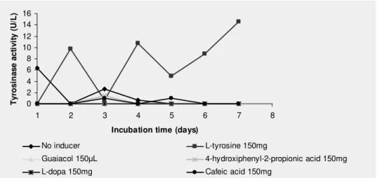
Effect of inducers
L-tyrosine at 0.15% induced intracellular tyrosinase
production at levels considerably higher than the other tested
inducers (L-DOPA, cafeic acid, 4-hydroxiphenyl-2-propionic
acid and guaiacol). The values of tyrosinase activity (U/L) and
the standard deviations (%) were, respectively: 9.77 ± 2.40
(second day); 10.70 ± 3.19 (day 4); 4.98 ± 8.26 (day 5); 8.90 ±
8.91 (day 6) and 14.61 ± 10 (day 7) when L-tyrosine was used
as inducer (Fig.2).The maximum production in the absence of
inducers was 6.34 U/L ± 10%, showing the importance of
inducers for tyrosinase production.
0 2 4 6 8 10 12 14 16
1 2 3 4 5 6 7 8
Incubation time (days)
T
y
ro
s
in
a
s
e
a
c
ti
v
it
y
(
U
/L
)
No inducer L-tyrosine 150mg
Guaiacol 150 L 4-hydroxiphenyl-2-propionic acid 150mg
L-dopa 150mg Cafeic acid 150mg
Figure 2. Inducers selection for tyrosinase production by P. sanguineus.Assays were done in sodium acetate buffer (50 mM, pH
Luminosity influence
When the incubation was performed in the absence of
light, the enzymatic production peaked in the second day
(18.72 U/L of tyrosinase activity). On the other hand, in
presence of light, 3 peaks of production were observed in the
second, fourth and seventh days of incubation (Table 1).
Table 1. Influence of luminosity on tyrosinase production by P. sanguineus
Tyrosinase activity (U/L) Incubation time (days)
Presence of light ± DPR Absence of light ± DPR
1 0.00 ± 0.00 0.00 ± 0.00
2 9.77 ± 2.40 18.72 ± 4.00
3 0.70 ± 10.00 6.54 ± 10.00
4 10.70 ± 3.19 4.47 ± 10.00
5 4.98 ± 8.27 7.31 ± 10.00
6 8.91 ± 8.91 7.98 ± 10.00
7 14.61 ± 10.00 2.05 ± 5.94
Effect of inoculum size
The higher tyrosinase production when incubating 10
mycelium discs occurred in the second day of incubation (18.96
U/L of tyrosinase activity). When using 20 mycelium discs,
optimum tyrosinase production occurred in day 6 (23.22 U/L of
tyrosinase activity) (Table 2). Despite the higher activity found
using 20 mycelium discs, this activity occurred in the day 6, four
days later than the peak using 10 mycelium discs.
Table 2. Correlation between biomass production and tyrosinase activity
Tyrosinase activity (U/L) Incubation time (days)
10 mycelium discs ± DPR 20 mycelium discs ± DPR
1 0.00 ± 0.00 0.00 ± 0.00
2 18.97 ± 5.02 9.24 ± 2.33
3 0.00 ± 0.00 10.83 ± 6.31
4 5.90 ± 10.00 14.39 ± 4.74
5 3.77 ± 10.00 10.79 ± 6.37
6 4.80 ± 10.00 23.22 ± 7.54
7 5.86 ± 8.71 8.45 ± 4.32
Effect of culture medium
Optimal tyrosinase production was observed when potato
dextrose broth was used in the second, third and sixth days. When
malt extract broth 2% was used, the optimum tyrosinase
production was in the second day of incubation (Table 3). For this
reason malt extract broth 2% was chosen for tyrosinase
production, since this peak occurred in a shorter incubation time (2
days) and the enzymatic activity value was higher than the others.
Table 3. Influence of culture media on tyrosinase production by P. sanguineus
Tyrosinase activity (U/L) Incubation time
(days) Malt extract broth 2% ± DPR Potato dextrose broth ± DPR
1 0.00 ± 0.00 0.00 ± 0.00
2 18.97 ± 5.02 12.53 ± 5.53
3 0.00 ± 0.00 14.71 ± 5.14
4 5.90 ± 10.00 3.93 ± 10.00
5 3.77 ± 10.00 5.44 ± 7. 35
6 4.80 ± 10.00 14.22 ± 7 .04
Duarte, L.T. et al. Tyrosinase activity in P. sanguineus
Inhibitory effect of different compounds
Laccase and tyrosinase activities were investigated after 10
minutes of incubation of the enzyme with several inhibitory
compounds (Table 4).
In tests performed with purified laccase, an activity of 103.83
U/ L was found in the absence of sodium azide and an activity of
2.78 U/ L was found in the presence of sodium azide. Soon, there
was a 97.32% inhibition of laccase sample by sodium azide which
is a classical inhibitor of metal containing oxidases.
On testing the effect of SHAM and PTU addition (6 mM) on
laccase activity, it was noticed that there is a pronounce decrease
in the relative activities estimated as 3.62 % (3.98 U/ L) and 4.62
% (5.074 U/ L) respectively as compared with the non-treated
sample (100 %; 109.82 U/ L). Results obtained suggest that
laccase is strongly inhibited by SHAM and PTU.
Sodium azide weakly affected the activity of tyrosinase, with
the maintenance of 95.85% of its activity after incubation with 0.1
mM sodium azide. Despite this drop in tyrosinase activity, the use
of sodium azide was chosen since for the accurate tyrosinase
quantification in samples also containing laccase is essential that it
be inhibited.
The tyrosinase activity in the absence of PMSF (21.72 U/ L)
was not lower than the activity in the presence of the inhibitor
(23.82 U/ L). Results suggest that PMSF does not inhibit
tyrosinase. In addition, the tyrosinase activity was completely
inhibited by 6 mM SHAM and PTU.
Table 4. Effect of several inhibitory compounds on laccase and tyrosinase activities (incubation at 37 ºC for 10 minutes)
Compound Concentration (mM) Laccase relative activity (%) Tyrosinase relative activity (%)
None - 100 100
Sodium azide 0.1 2.68 95.85
PMSF 0.1 - 100
PTU 6 4.62 0
SHAM 6 3.62 0
Electrophoresis and enzymatic activities in gels
SDS-PAGE of the intracellular crude extract of P.
sanguineus showed four bands of low intensity (estimated sizes
of 95 kDa, 78 kDa, 64 kDa and 54 kDa) (Fig. 3).
Tyrosinase activity gel staining revealed one red zone of
tyrosinase (Fig. 4).
The substrate L-dopa used in the activity staining of
tyrosinase is also a substrate for laccase. As the laccase
inhibitor (sodium azide) was not used in this test, the bands
shown laccase activity were also stained. Commercial
tyrosinase and crude extract of P. sanguineus were previously
quantified for tyrosinase and laccase activities (Data not
shown). Laccase activity was null in commercial tyrosinase
while crude extract presented laccase and tyrosinase activities,
confirming the identity of the bands shown in Fig. 4.
Figure 3. SDS-PAGE pattern of the intracellular crude extract of P. sanguineus Coomassie stained. Electrophoresis was
carried out using a 10% cross-linked polyacrilamide gel. Lane
1: molecular mass marker proteins. Lane 2: intracellular crude
Figure 4. Activity staining of tyrosinase. Lane 1: protein band of commercial tyrosinase exhibiting tyrosinase activity is
marked. Lane 2: protein band of P. sanguineus intracellular
crude extract exhibiting tyrosinase activity is marked.
Thermal stability behavior
pH profile of P. sanguineus CCT-4518 tyrosinase showed
optimum activity at pH 6.6. Tyrosinase was more active at
45°C, though it showed considerable activity over the range of
40-60°C. The crude extract presented a fast loss of activity
when pre-incubated for relatively short periods at 50 and 55°C.
Pre-incubation of tyrosinase at 50°C for 15 minutes resulted to
the reduction of activity by about 53.87 %, however a complete
loss of activity was recorded with samples pre-incubated at
50°C and 55°C for 45 and 15 minutes respectively. The
enzyme kept 50% of relative activity after 45 minutes of
incubation (Data not shown).
DISCUSSION
The practical application of tyrosinases in biotechnology
requires large quantities of enzymes; hence, the identification
of new Pycnoporus strains as potential producers of tyrosinases
is of biotechnological importance (16).
Initially, to investigate the potential of this strain to
produce tyrosinase the qualitative test was performed.
According to Gramss et al. (14), the appearance of yellow to
red color at the ends of fungal colony margins, after adding
p-cresol, is an indication of the presence of intracellular
tyrosinase. This is in accordance with Mayer and Harel (23),
who stated that tyrosinase was an intracellular enzyme in most
fungi they investigated. The tyrosinase (cresolase) test was not
influenced by laccase or peroxidase. The indicator, p-cresol, is
stained reddish brown by tyrosinase only, but remains
colourless by laccase and peroxidase (14).
Once detected the ability of the amazonic strain P.
sanguineus CCT-4518 to produce the enzyme of interest, the
work was then directed towards optimization of its in vitro-
production. The ideal conditions of P. sanguineus cultivation
for tyrosinase production were determined and 0.15%
L-tyrosine was the best inducer, in the presence of light, with 10
mycelium discs as inoculum, medium malt extract broth 2%,
incubation at 30°C, 150 rpm agitation, during 2 days. To our
knowledge this is the first report of tyrosinase production
optimization in P.sanguineus. In this connection Halaouli et al.
(16) reported that optimum incubation temperature for P
sanguineus CBS 614.73 is 30°C in agreement with our results.
Despite the higher enzimatic activity observed during the
incubation in the absence of light, the preferred condition for
the tyrosinase production was in the presence of light, in an
attempt to inhibit the laccase production, since it has increased
production in the dark (10).
Aiming to investigate the effect of compounds inhibiting
tyrosinase activity of P. sanguineus (CCT-4518) and having
the parallel laccase production in the cultivation conditions
(0.15% L-tyrosine, in the presence of light, at 30°C and 150
rpm agitation) been noticed, the inhibition of this interfering
was initially searched. Laccase was almost completely
inhibited by sodium azide, which is a classical inhibitor of
metal containing oxidases (11, 27). Laccase was found to be
inhibited by 6 mM SHAM (96.38% ) or PTU (95.38% ),
suggesting that laccase is also inhibited by SHAM and PTU,
Duarte, L.T. et al. Tyrosinase activity in P. sanguineus
tyrosinase inhibitors (15, 24, 25, 30).
P. sanguineus (CCT-4518) tyrosinase barely affected in
reaction mixtures containing 0.1 mM of sodium azide or
PMSF. There is no previous report of tyrosinase inhibition by
sodium azide or PMSF. P. sanguineus (CCT 4518) tyrosinase
activity was completely inhibited by PTU and SHAM at 6 mM
final concentration. According to Gunther et al. (15), Miranda
et al. (24), Rescigno et al. (25) and Winder (30), the tyrosinase
is strongly inhibited by PTU and SHAM.
The protein profile in electrophoresis allows monitoring
the conditions of production and the purity degree of the
purified enzyme. A recent article by Fan and Flurkey (9),
reports two major stained bands of protein along with a less
stained protein, in a sample containing purified tyrosinase of
Portabella mushrooms. The estimated sizes of the bands were
43 kDa, 48 kDa and 60 kDa. It is supposed that the bands in the
43-48 kDa are the active proteolyzed forms of tyrosinase that
many other investigators have observed (12, 29, 31).
According to Halaouli et al. (16), the P. sanguineus (CBS 251
614.73) purified tyrosinase has 45 kDa and is a monomeric
enzyme. To identify which of the four bands detected shows
tyrosinase activity in this study it is necessary to implement the
purification of the enzyme.
The activity determination is a tool to confirm an assumed
activity of the found protein and evaluate the number of
isoforms produced by microorganisms. Under partially
denaturing SDS-PAGE, with samples not boiled or treated with
reducing agents, several isoforms of tyrosinase were detected
in crude extracts of Portabella mushroom (9), contrasting the
observed in this work, where only one tyrosinase isoform was
verified in the non denaturing PAGE.
When the commercial tyrosinase was stained for laccase
and tyrosinase activities by Rescigno et al. (25), protein band
exhibiting laccase activity appeared bellow the protein band
exhibiting tyrosinase activity, as well as the bands observed in
this study.
Due to the still limited number of reports about tyrosinase
and its biotechnological applications, it is necessary to have
knowledge about the enzyme under study. In the biochemical
characterization, the optimum pH of intracellular tyrosinase
production by P. sanguineus (CCT 4518) was found to be 6.6
which coincides with the corresponding value found in case of
the purified tyrosinase from P. sanguineus (CBS 614.73) (pH
6.5-7.0) (16). The optimum temperature for P. sanguineus
(CCT-4518) was 45°C. The P. sanguineus (CBS 614.73)
purified tyrosinase has 60-65°C of the optimum temperature
and remained active in a wide range of temperature (25-70°C)
(16). The P. sanguineus (CCT-4518) tyrosinase showed to be
less thermostable, maintaining only about 50% activity after 15
minutes incubation at 50°C. Differently, the P. sanguineus
(CBS 614.73) purified tyrosinase showed to be more
thermostable, presenting considerable stablility below 60°C. At
50 °C, the enzyme half-life was longer than 120 minutes, while
at 60°C it was almost completely inactivated after 20 minutes
of incubation (16).
In conclusion, P. sanguineus CCT-4518 produces one
intracellular protein with tyrosinase activity when induced with
L-tyrosine. Further study will be required to investigate the
application of crude extract in compounds biotransformation,
bioremediation and biosensors.
ACKNOWLEDGEMENTS
This work was supported by grants from Secretaria de
Ciência e Tecnologia do Estado de Goiás (SECTEG-GO). We
thank Debora Teixeira Duarte for reviewing the manuscript.
REFERENCES
1. Beel, A.A.; Wheeler, M.H. (1986). Biosynthesis and functions of fungal melanins. Annu. Rev. Phytopathol. 24, 411-451.
2. Bourbonnais, R.; Paice, M.G. (1990). Oxidation of non-phenolic substrates. An expanded role for laccase in lignin biodegradation. FEBS Lett. 267, 99-102.
of microgram quantities of proteins utilizing the principle of protein-dye binding. Anal. Biochem. 72, 248-254.
4. Cavallazzi, J.R.P.; Kasuya, C.M.; Soares, M.A. (2005). Screening of inducers for laccase production by Lentinula edodes in liquid medium. Braz. J. Microbiol. 36, 383-387.
5. Chiacchierini, E.; Restuccia, D.; Vinci, G. (2004). Bioremediation of food industry effluents: recent applications of free and immobilised polyphenoloxidases. Food. Sci. Technol. Int. 10 (6), 373-382.
6. Duran, N.; Esposito, E. (2000). Potential applications of oxidative enzymes and phenoloxidase-like compounds in wastewater and soil treatment: a review. Appl. Catal. B. 28, 83-99.
7. Duran, N.; Rosa, M.A.; D’annibale, A.; Gianfreda, L. (2002). Applications of laccases and tyrosinases (phenoloxidases) immobilized on different supports: a review. Enzyme Microb. Technol. 31, 907-931. 8. Espin, J.C.; Morales, M.; Var.n, R.; Tudela, J.; Garcia-Canovas, F.
(1995). A continuous spectrophotometric method for determining the monophenolase and diphenolase activities of apple polyphenol oxidase. Anal. Biochem. 231, 237-246.
9. Fan, Y.; Flurkey, W.H. (2004). Purification and characterization of tyrosinase from gill tissue of Portabella mushrooms. Phytochemistry. 65, 671-678.
10. Garcia, T.A.; Santiago, M.F.; Ulhoa, C.J. (2006). Properties of laccases produced by Pycnoporus sanguineus induced by 2,5-xylidine. Biotechnol. Lett. 28, 633-636.
11. Garcia, T.A.; Santiago, M.F.; Ulhoa, C.J. (2007). Studies on the Pycnoporus sanguineus laccase purified by hydrophobic interaction chromatography. Appl. Microbiol. Biotechnol. 75, 311-318.
12. Gerritsen, Y.A.M.; Chapelon, C.G.J.; Wichers, H.J. (1994). The low isoeletric point tyrosinase of Agaricus bisporus may be a glycoprotein. Phytochemistry. 35, 573-577.
13. Girelli, A.M.; Mattei, E.; Messina, A. (2006). Phenols removal by immobilized tyrosinase reactor in on-line high performance liquid chromatography. Anal. Chim. Acta. 580, 271-277.
14. Gramss, G.; Gunther, T.H.; Fritsche, W. (1998). Spot tests for oxidative enzymes in ectomycorrhizal, wood-, and litter decaying fungi. Mycol. Res. 102 (1), 67-72.
15. Gunther, T.H.; Perner, B.; Gramss, G. (1998). Activities of phenol oxidizing enzymes of ectomycorrhizal fungi in axenic culture and in symbiosis with Scots pine (Pinus sylvestris L.). J. Basic Microbiol. 38, 197-206.
16. Halaouli, S.; Asther, Mi.; Kruus, K.; Guo, L.; Hamdi, M.; Sigoillot, J.C.; Asther, M.; Lomascolo, A. (2005). Characterization of a new tyrosinase from Pycnoporus species with high potential for food technological applications. J. Appl. Microbiol. 98, 332-343.
17. Halaouli, S.; Asther, M. Sigoillot, J.C.; Hamdi, M.; Lomascolo, A. (2006). Fungal yrosinases: new prospects in molecular characteristics, bioengineering and biotechnological applications. J. Appl. Microbiol. 100, 219-232.
18. Haq, I.; Ali, S.; Qadeer, M.A. (2002). Biosynthesis of L-DOPA by Aspergillus oryzae. Bioresour. Technol. 85, 25-29.
19. Herpoel, I.; Moukha, S.; Lesage-Meessen, L.; Sigoillot, J.C.; Asther, M. (2000). Selection of Pycnoporus cinnabarinus strains for laccase production. FEMS Microbiol. Lett. 183, 301-306.
20. Laemmli, U.K. (1970). Cleavage of structural proteins during the assembly of the head of bacteriophage T4. Nature. 227, 680-685. 21. Lantto, R. (2007). Protein cross-linking with oxidative enzymes and
transglutaminase. Effects in meat protein systems. Espoo 2007. VTT Publications 642. 114 p.
22. Mayer, A.M. (1987). Polyphenol oxidases in plants – recent progress. Phytochemistry. 26, 11-20.
23. Mayer, A.M.; Harel, E. (1979). Review: Polyphenol oxidases in plants. Phytochemistry. 18, 193-215.
24. Miranda, M.; Bonfigli, A.; Zarivi, O.; Ragnelle, A.M.; Pacioni, G.; Botti, D. (1992). Truffle tyrosinase: Properties and activity. Plant. Sci. 81, 175-182.
25. Rescigno, A.; Zucca, P.; Fluekey, A.; Inlow, J.; Flurkey, W.H. (2007). Identification and discriminaton between some contaminant enzyme activities in commercial preparations of mushroom tyrosinase. Enzyme Microb. Technol. 41, 620-627.
26. Sanchez-Ferrer, A.; Rodriguez-Lopez, J.N.; Canovas, F.; Garcia-Carmona, F. (1995). Tyrosinase: a comprehensive review of its mechanism. Biochim. Biophys. Acta. 1247, 1-11.
27. Sugumaran, M. (1995). A caution about azide inhibition of enzymes associated with electrophilic metabolites. Biochem. Biophys. Res. Commun. 212, 834-839.
28. Valeriano, V.S.; Silva, A.M.F.; Santiago, M.F.; Bara, M.T.F.; Garcia, T.A. (2009). Production of laccase by Pycnoporus sanguineus using 2,5-xylidine and ethanol. Braz. J. Microbiol. 40, 790-794.
29. Wichers, H.J.; Gerritsen, Y.A.M.; Chapelon, C.G.J. (1996). Tyrosinase isoforms from the fruitbodies of Agaricus bisporus. Phytochemistry. 43 (2), 333-337.
30. Winder, A.J. (1994). A stopped spectrophotometric assay for the dopa oxidase activity of tyrosinase. J. Biochem. Biophys. Methods. 28, 173-183.
31. Zhang, X.; Leeuwen, J.; Wichers, H.J.; Flurkey, W.H. (1999). Characterization of tyrosinase from the cap flesh of Portabella mushrooms. J. Agric. Food Chem. 47, 374-378.