Biological X-ray irradiator characterization for use
with small animals and cells
A. Colello Bruno
1, S.J. Mazaro
2, L.L. Amaral
1, E.M. Rego
1, H.F. Oliveira
1and J.F. Pavoni
21Servic
¸o de Radioterapia, Hospital das Clínicas, Faculdade de Medicina de Ribeirão Preto, Universidade de São Paulo, Ribeirão Preto, SP, Brasil 2Departamento de Física, Faculdade de Filoso
fia, Ciências e Letras de Ribeirão Preto, Universidade de São Paulo, Ribeirão Preto, SP, Brasil
Abstract
This study presents the characterization of an X-ray irradiator through dosimetric tests, which confirms the actual dose rate that small animals and cells will be exposed to during radiobiological experiments. We evaluated the linearity, consistency, repeatability, and dose distribution in the positions in which the animals or cells are placed during irradiation. In addition, we evaluated the performance of the X-ray tube (voltage and tube operating current), the radiometric survey (leakage radiation) and safety devices. The irradiator default setting was established as 160 kV and 25 mA. Tests showed that the dose rate was linear overtime (R2=1) and remained stable for long (constant) and short (repeatability) intervals between readings. The mean dose rate inside the animal cages was 1.27±0.06 Gy/min with a uniform beam of 95.40% (above the minimum threshold guaranteed by the manufacturer). The mean dose rate inside the cell plates was 0.92±0.19 Gy/min. The dose rate dependence with tube voltage and current presented a quadratic and linear relationship, respectively. There was no observed mechanical failure during evaluation of the irradiator safety devices and the radiometric survey obtained a maximum ambient equivalent dose rate of 0.26 mSv/h, which exempts it from the radiological protection requirements of the International Atomic Energy Agency. The irradiator characterization enables us to perform radiobiological experiments, and assists or even replaces traditional therapy equipment (e.g., linear accelerators) for cells and small animal irradiation, especially in early research stages.
Key words: X-ray irradiator; Dosimetric characterization; Radiotherapy; Dosimetry; Radiochromicfilm
Introduction
Studies using small animals and cell experiments have become indispensable for cancer research before clinical implementation of a new therapy (1,2). They assist with the understanding of ionizing radiation interactions with tissues and cells, which is crucial for translational research of new effective radiotherapy techniques.
There are specific small animal irradiators designed
especially for preclinical studies, used to evaluate and optimize new treatment modalities (3,4). Delineating set-up protocols with the equipment (linear accelerators) used clinically for patient treatments is a slow process, and using these irradiators for cells and small animal experiments in the preliminary stages of research would save time.
The most common irradiators used are the gamma-ray irradiators that employ radioactive isotopes such as cobalt-60 or cesium-137. However, recently, it has become
increasingly difficult to purchase such irradiators because
their manufacturing was interrupted. Additionally, the inter-national transportation of isotopes involves radiation pro-tection issues that complicate the process (5).
Thus, X-ray irradiators are an alternative for the gamma-ray irradiator and are being increasingly used due to their low cost and absence of a radioactive source (6,7). Other factors such as no facility-licensing requirements, and less rigorous and easier maintenance also add to the advantages of an X-ray unit (2,8).
For all ionizing radiation machines, certain quality assurance (QA) procedures are required to ensure basic operating conditions. However, there is no international QA recommendation for X-ray irradiators. One of the main goals of the QA procedures is to minimize errors related to dose delivery, which can be prevented using radiation
detectors, such as ionization chambers, dosimetricfilms,
or semiconductor detectors.
This study presents QA tests for X-ray irradiator characterization including dosimetric and safety tests, and a radiometric survey. Irradiator characterization is important for determining the dose distribution pattern and for evaluating the operating parameters in order to guarantee the dose deposition during irradiation. Both
Correspondence: A. Colello Bruno:<[email protected]>
characteristics are essential for the quality of the transla-tional research being developed.
Material and Methods
This study was developed at the Radiotherapy Depart-ment of Ribeirão Preto Hospital and Clinics.
The X-ray irradiator (RS 2000 Biological System irradiator, Rad Source, USA) (Figure 1A) was character-ized in order to establish the reference values for a QA program implementation in this machine. There is no inter-national recommendation describing what tests should be applied or their frequency. We selected some tests to characterize this machine, evaluating its linearity, con-stancy, repeatability, dose distribution in the irradiation chamber, X-ray tube performance, in addition to safety test and radiometric survey.
The evaluated irradiator has six height levels available in its exposure chamber. A mobile tray with samples can be positioned at these levels and irradiated; therefore,
six different dose rates can be achieved. On this tray, there are six circles that correspond to the size of the
radiationfield at a corresponding height (Figure 1B).
We chose the default position in the ionization
cham-ber for film measurements, corresponding to the region
inside circle 6 with the mobile tray at level 1 (Figure 1B). The default irradiation parameters for this irradiator were established at 160 kV (operating voltage) and 25 mA (operating current).
For the dosimetric characterization tests, we used an electrometer (Model Accu-Dose/2086, Radcal Corpora-tion, USA), an ionization chamber (model 10X6-06-3,
Radcal Corporation) and radiochromic films (Gafchromic
EBT2, Ashland Advanced Materials, USA). A holder was used for positioning the ionizing camera on the region of interest. We also used a Thyac III Survey Meter (model 490, Victoreen Instrument Company, USA) for the radio-metric leakage test.
Linearity
Linearity is an important characteristic of the instrument that guarantees the equipment output. This is achieved
when a specific change in the selected irradiation time
generates a proportional change in the radiation gener-ated. A linear relation between the irradiation time and the measured doses is expected.
To test the irradiator linearity, we measured the radiation doses produced for irradiation times varying from 0.5 to 10 min, using the ionization chamber located at the default position.
Constancy
Constancy is another important characteristic as it is desirable that the equipment maintains the same output overtime. To evaluate the irradiator constancy, its dose rate or radiation output was measured at least once per month, for one year, with the ionization chamber in the default position. The acceptable variation of different
read-ings was ±3% of the value obtained for the irradiator
dosimetry. This follows the same criteria established for a medical linear accelerator by the American Association of Physicists in Medicine and Biology (9).
Repeatability
Repeatability refers to the variation in measurements taken by a single person with the same instrument param-eters, under the same conditions and within a short period of time; the agreement between the measurements guar-antees that the instrument presents a precise output.
To evaluate the repeatability of dose measurements in the irradiator, seven dose measurements using a beam with a 1-min exposure were repeated with the ionization chamber in the default position.
Dose distribution
The radiation dose distribution of any device is an important parameter to determine the absorbed dose
distributions along the irradiated volume. The dose eval-uation at any point on the irradiated volume considers the equipment dosimetry, usually performed at a reference
point in the center of the radiation field, and by the
radiation dose profiles, acquired in two orthogonal
direc-tions in the plane perpendicular to the radiation beam and
in the vertical direction of the beam’s central axis.
Radiation dose profiles inside the exposure chamber
from front to back (frontal) and left to right (lateral) were calculated. Radiation dose measurements were performed for 1 min using the ionization chamber; the chamber was moved by sliding the frontal and lateral axes in increments of 1 cm in the central region and 2 cm in peripheral regions. The tray was positioned at level 1 for all the
meas-urements in order to characterize the largest field size
available for irradiation.
Another profile was measured in the vertical direction
inside the exposure chamber, and dose measurements were performed for 1 min in the beam central axis and varying the tray position from levels 1 to 5.
Small animals (e.g., mice and rats) are usually enclosed in a standard, acrylic animal cage during irradiation (Figure 2A and B). During this procedure, the tray is
removed and the cage is placed on thefloor of the
expo-sure chamber, centered on a shielded base.
The animal cagefits perfectly inside the shielded base,
which reduces stress in the animals and ensures that beam uniformity is greater than 95% on any horizontal plane. To evaluate the dose distribution inside the cage,
we measured the average dose rate atfive different points
in the cage (at the four corners and at the center). Measurements were repeated with and without the cage cover (setup 1 and 2, respectively). Setup 1 was also repeated without the shielded base (setup 3).
The cell culture plate irradiation test was performed with samples positioned inside the circle corresponding to its respective tray level, as recommended by the man-ufacturer. To evaluate the dose distribution along the irradiation circle and inside cell culture plates, we used
radiochromicfilms positioned in each of the six wells of the
four culture plates that covered the entire irradiation circle. The cell culture plates were positioned in circle 1 with the
tray on thefirst level (default setting for experiments with
cells) and they were irradiated for 1 min.
X-ray tube performance
The X-ray tube operating range varies from 30 to 160 kV, and its current varies from 5 to 25 mA; both are adjustable. The default irradiation parameters for this irradiator were established as 160 kV and 25 mA. It is
known that these parameters directly influence the
radia-tion output. In order to evaluate this influence, the dose
rate per current was measured at all operating voltages, with the ionization chamber in the default position.
Safety test and radiometric surveying
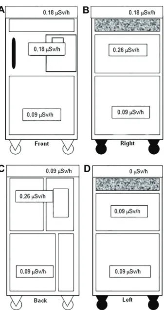
All safety devices were tested, including door locks, warning lights, emergency buttons, irradiation inter-ruption, and apparatus intrinsic stability (warm-up). They were performed 72 times during an 18-month period. A radiometric survey was performed around the irradiator to evaluate the radiation levels in these areas. Equivalent dose rate measurements were monitored at 10 cm from each side of the irradiator (front, rear, right and left), using a Thyac III Survey Meter. At each side, three positions were measured: top, middle, and bottom.
Results and Discussion
Linearity
The measured absorbed dose values for the respec-tive irradiation time followed a linear pattern, indicated by
the correlation coefficient (R2) equal to one (Figure 3).
This result confirmed the expected linearity for irradiation
times up to 10 min.
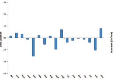
Constancy
The dose rate variation during 1 year is presented in Figure 4. The maximum variation was 1.54%, which is
below the±3% acceptable limit.
Repeatability
The ionization chamber’s readings for the repeatability
study are shown in Table 1, including the mean value and the standard deviation. The values were very similar, with
a standard deviation of 0.001 and a maximum deviation of
0.2%, confirming the equipment’s output precision.
Dose distribution
In exposure chamber. Frontal and lateral dose profiles were achieved (Figure 5). Based on these data, it was
possible to calculate the irradiation field symmetry, a
parameter that describes the maximum percentage devia-tion between the doses at opposite sides of the central
axis inside theflattened area (10). The symmetry for the
frontal and lateral profiles was 3.47 and 0.35%,
respec-tively. The asymmetry in the front axis was due to the anode heel effect, since the X-ray tube is positioned on this axial direction. The dose distribution along the beam central axis decreased following the inverse-square law, as the dose measurement point moves away from the radiation source (Figure 6).
In animal cages. The mean dose rate inside the stand-ard animal cage measured for setups 1, 2, and 3 were
Table 1. Repeatability of dose measurements using the ionization chamber (during 1 min).
Measurement Dose (Gy)
1 1.183
2 1.185
3 1.182
4 1.183
5 1.186
6 1.183
7 1.183
Mean 1.184
Standard deviation 0.001
Figure 3.Irradiator linearity in the 0.5 to 10 min range.
Figure 4.Variation of dose rates during 1 year. The data were obtained by the constancy evaluation in this period.
Figure 5.Dose distribution along the front and lateral axes of the exposure chamber, with the tray positioned at level 1.
1.272±0.058, 1.343±0.041, and 0.989±0.07 Gy/min, respectively. For setups 1 and 2, the beam uniformity was higher than 95% (95.4 and 96.9%, respectively). However,
for setup 3 this value was 92.7%. These values confirm
the manufacturer information that the shielded base homog-enizes the beam on the horizontal plane within the animal cage. It was also possible to determine that the cage
cover attenuates 5.3% of the beam, a significant
attenua-tion that should be considered during animal irradiaattenua-tion procedures.
In cell plates. The mean dose rate measured along circle 1, at the default setting for experiments with cells,
was 0.917±0.189 Gy/min, with standard deviation
corre-sponding to 20.6% of the dose rate value. However, with a repetition of this measurement covering irradiation circle 3, a smaller standard deviation was found: the mean dose
rate was 1.117±0.062 Gy/min, with the standard
devia-tion corresponding to 5.5% of the dose rate value. The main reason for these high variations can be attrib-uted to the cell plate materials, especially the cover, which attenuates the beam. This attenuation can be enhanced by increasing the distance from the irradiation circle center,
increasing in the beam’s incidence angle on the plates.
X-ray tube performance
The dependence of the dose rate on the tube voltage was evaluated at the default position. As expected, it showed a good quadratic relationship (Figure 7), whereas dose rate was linearly dependent on the tube current (Figure 8).
Figure 7. Relationship between dose rate and tube voltage (default position).
Figure 8. Relationship between dose rate and tube current (default position).
Safety test and radiometric surveying
There was no observed mechanical failure in the eval-uation of the irradiator safety devices.
Figure 9 presents the radiometric survey results with the equivalent dose rate values at the various measured points. All the values were below the minimum limit set by
the International Atomic Energy Agency, which is 1mSv/h
at 10 cm around the equipment (11).
The reference values for a quality control program to be implemented in the X-ray irradiator were achieved by performing linearity, constancy, repeatability, dose distri-bution, and X-ray tube performance and safety tests. Special measurements were applied to evaluate dose
distributions and their deviations for cells and small ani-mal settings. The characterization of this biological X-ray irradiator enables us to perform radiobiological experi-ments, in order to assist, or even replace traditional therapy equipment (e.g., linear accelerators) for cells and small animal irradiation, especially in the early research stages.
Acknowledgments
The acquisition of the irradiator used in this study was supported by grant #2009/54218-1, São Paulo Research Foundation (FAPESP).
References
1. Tillner F, Thute P, Bütof R, Krause M, Enghardt W. Pre-clinical research in small animals using radiotherapy technology -a bidirection-al tr-ansl-ation-al -appro-ach.Z Für Med Phys2014; 24: 335–351, doi: 10.1016/j.zemedi.2014.07.004 .
2. McCurdy B, Mueller L, Backman E, Venkataraman S, Fleming E, Asuni G, et al. SU-GG-T-150: Commissioning and validation of a novel measurement based IMRT QA method, incorporating dose recalculation on patient CT data. Med Phys2008; 35: 2760, doi: 10.1118/1.2961901. 3. Pidikiti R, Stojadinovic S, Speiser M, Song KH, Hager F, Saha
D, et al. Dosimetric characterization of an image-guided stereo-tactic small animal irradiator.Phys Med Biol2011; 56: 2585. 4. Pedersen KH, Kunugi KA, Hammer CG, Culberson WS,
DeWerd LA. Radiation biology irradiator dose verification survey. Radiat Res 2016; 185: 163–168, doi: 10.1667/ RR14155.1.
5. Mehta K, Parker A. Characterization and dosimetry of a practical X-ray alternative to self-shielded gamma irradia-tors.Radiat Phys Chem2011; 80: 107–113, doi: 10.1016/ j.radphyschem.2010.08.011.
6. Janatpour K, Denning L, Nelson K, Betlach B, MacKenzie M, Holland P. Comparison of X-ray vs. gamma irradiation
of CPDA-1 red cells. Vox Sang 2005; 89: 215–219, doi: 10.1111/j.1423-0410.2005.00699.x.
7. Gibson BW, Boles NC, Souroullas GP, Herron AJ, Fraley JK, Schwiebert RS, et al. Comparison of Cesium-137 and X-ray irradiators by using bone marrow transplant reconstitution in C57BL/6J mice. Comp Med 2016; 65: 165–172.
8. Kuess P, Bozsaky E, Hopfgartner J, Seifritz G, Dörr W, Georg D. Dosimetric challenges of small animal irradiation with a commercial X-ray unit. Z Für Med Phys2014; 24: 363–372, doi: 10.1016/j.zemedi.2014.08.005.
9. Kutcher GJ, Coia L, Gillin M, Hanson WF, Leibel S, Morton RJ, et al. Comprehensive QA for radiation oncol-ogy: report of AAPM Radiation Therapy Committee Task Group 40. Med Phys 1994; 21: 581–618, doi: 10.1118/ 1.597316.
10. IAEA/TECDOC 1151.Aspectos fı´sicos da garantia da quali-dade em radioterapia - protocolo de controle de qualiquali-dade. Rio de Janeiro: INCA; 2000.