RESEARCH ARTICLE / ARTIGO
A destructive new disease of
Syzygium samarangense
in Thailand caused by the new species
Pestalotiopsis
samarangensis
Sajeewa S.N. Maharachchikumbura1,2,3, Liang-Dong Guo3, Ekachai Chukeatirote1,2, Eric H.C. McKenzie4
& Kevin D.Hyde1,2
1Institute of Excellence in Fungal Research, Mae Fah Luang University, Chiang Rai 57100, Thailand; 2School of Science,
Mae Fah Luang University, Chiang Rai 57100, Thailand; 3Key Laboratory of Systematic Mycology & Lichenology, Institute
of Microbiology, Chinese Academy of Sciences, Beijing 100190, P.R. China; 4Landcare Research, Private Bag 92170,
Auckland, New Zealand
Authors for correspondence: Sajeewa S.N. Maharachchikumbura, e-mail: sajeewa83@yahoo.com; Kevin D. Hyde, e-mail: kdhyde3@gmail.com
ABSTRACT
A new fungal species, Pestalotiopsis samarangensis, was isolated from fruit rot in Syzygium samarangensefrom markets in
Chiang Mai and Chiang Rai provinces, Thailand. Initially small, circular, black, slightly sunken spots developed on fruits. Later, the spots enlarged rapidly, became sunken, and resulted in a soft decay of the fruit flesh. Molecular analysis of the ITS, β-tubulin, and tef1 combined gene sequences distinguish P. samarangensis from other species in the genus. Pathogenicity testing proved that wounding triggers the
disease symptoms and thus careful handling of fruits during transport and storage helps to prevent the disease. Key words: β-tubulin, fruit rot, ITS, neighbor-joining, phylogenetic, tef1.
INTRODUCTION
Syzygium samarangense Merr. (Myrtaceae) is
widely grown for its fruits throughout Cambodia, Laos, India, Philippines, Samoa, Sri Lanka, Taiwan, Thailand, and Vietnam (Srisaard, 2003; Vara-Ubol et al., 2006). In Thailand, the wax apple fruit is commonly known as chomphu and harvested the year round with a peak during January to March from the cultivars ‘Dang Indo’, ‘Phet Ban Plew’, ‘Phet Jin Da’, ‘Phet Nam Pueng’, ‘Phet Sai Rung’, ‘Phet Sam Phran’, ‘Thub Thim Chan’, and ‘Thun Klao’. In 2004, Thailand earned US$ 26.5 million from 69,608 tons of wax apple planted over 10,240 ha (Shü et al., 2008). The fruits have a thin, delicate skin and are thus easily susceptible to pest and disease attack. Diseases of wax apple include
Pestalotiopsis fruit rot (Pestalotiopsis eugeniae (Thüm.)
S. Kaneko), shoot dieback (Fusarium sp.), anthracnose
(Colletotrichum gloeosporioides (Penz.) Penz. & Sacc.), Phytophthora fruit rot (Phytophthora palmivora (E.J.
Butler) E.J. Butler), Dothiorella fruit rot (Dothiorella sp.), and Pseudocercospora fruit rot (Pseudocercospora sp.)
(Janick & Paull, 2008; Lan, 2001), although these species need to be confirmed by molecular data (Phoulivong et al., 2010; Ko Ko et al., 2011).
Pestalotiopsis spp. are important plant-pathogenic
species known mostly from the tropics, where they cause leaf blights (Guba, 1961) in many plant species (Hyde
& Fröhlich, 1995; Xu et al., 1999; Das et al., 2010; Maharachchikumbura et al., 2011). Species may also cause rots of fruit and other post-harvest diseases (Ullasa & Rawal, 1989; Korsten et al., 1995; Xu et al., 1999). Several post-harvest diseases are caused by species of Pestalotiopsis, e.g.,
postharvest decay of mangos by P. glandicola (Castagne)
Steyaert (Ullasa & Rawal, 1989), fruit rot of grapevine by P. menezesiana (Bres. & Torrend) Bissett as well as P. uvicola (Speg.) Bissett (Xu et al., 1999), and fruit rot of rambutan by Pestalotiopsis sp. (Sangchote et al., 1998).
Scabby fruit canker of guava is caused by P. psidii (Pat.)
Mordue (Kaushik et al., 1972).
We surveyed market fruit disease of wax apple in Chiang Mai and Chiang Rai provinces in Thailand in 2010 and 2011, and constantly observed a distinctive fruit rot disease. In this study, we introduce this new Pestalotiopsis fruit rot
disease of wax apple, with a description of morphological and molecular characteristics of the fruit rot agent.
MATERIALS AND METHODS
Symptoms and sample collection
Samples were sorted by symptoms and the presence of wounds and incubated in a clean polythene bag to promote fungal development. The type herbarium specimen (BPI
406804) of P. eugeniae, a pathogen previously recorded
from wax apple was borrowed and studied.
Isolation and identification of the causal agent of the
disease
A single-conidium culture technique was applied to obtain pure colonies of the fungi following a modified method as outlined by Chomnunti et al. (2011). In short, a small piece of diseased fruit bearing acervuli was immersed in 300 µL of sterile distilled water and left a few minutes so that conidia liberate from the conidiomata. A suspension was made and small drops of this suspension were dispersed on water agar (WA) in Petri dishes and kept at room temperature in the laboratory. Conidia were left for 8-12 hours to germinate and single germinating conidia were transferred to fresh PDA plates. The plates were incubated at 25ºC for 7 to 10 days. Colonies were grown on PDA plates, later transferred to PDA slants, and stored at 4 ºC for further study. Sporulation was induced using sterilized (autoclaved) carnation leaves, which were aseptically placed on the surface of the PDA medium with growing mycelium. The morphology of fungal colonies was recorded following the method of Hu et al. (2007). Fungal mycelium and spores were observed under the light microscope and photographed. All microscopic measurements were made with Tarosoft image framework (v. 0.9.0.7).
Pathogenicity tests
Ten-days-old sporulating cultures of Pestalotiopsis
strains on PDA (25ºC, day light) were flooded with Czapek-Dox nutrient solution (Lingappa & Lingappa, 1965), and the mycelia were scraped using a spatula. The conidiomata were crushed using a glass rod. The mycelial suspension was passed through glass wool to remove hyphal fragments and the filtrate containing conidia was collected. The filtrate was centrifuged at 3000 rpm for 3 min and a suspension of 103 mL-1 conidia was prepared. In vitro pathogenicity
testing was carried out on 15 healthy fruits of wax apple bought from the market. The inoculations were carried out in April 2010 as well as September and November 2011. The surface of the fruits was wiped with 70% ethanol and allowed to air-dry. Fruits were separated into three groups of five fruits each. One group was wounded by pin-pricking. A conidial suspension (20 µL) was placed on the surface of the five unwounded fruits and on five wounded fruits. The remaining five fruits were maintained as controls and inoculated with drops of sterile deionized water. The fruits were placed in polythene bags, labeled, and the moisture was maintained for 10 days by the addition of wet sterile cotton wool. Daily observations were made on the development of disease symptoms. Fruits showing symptoms were removed and the pathogen in the diseased tissue was re-isolated on PDA as described above.
DNA extraction and PCR conditions
Total genomic DNA was extracted by the modified protocol of Guo et al. (2000). Fresh fungal mycelium (500 mg) was scraped from the margin of a PDA plate incubated at 25ºC for 7 to 10 days and transferred into a 1.5 mL centrifuge tube with 100 µL of preheated (60ºC) 2× CTAB extraction buffer (2% (w/v) CTAB, 100 mM Tris-HCl, 1.4 M NaCl, 20 mM EDTA, pH 8.0) and 200 mg sterilized quartz sand. Mycelium was ground using a glass pestle for 5 min, and an extra 500 µL 2× CTAB preheated (60ºC) was added and incubated in a 65ºC water bath for 30 min with occasional shaking. 500 µL of phenol:chloroform (1:1) was added to each tube and shaken thoroughly to form an emulsion. The mixture was spun at 11900 gfor 15
min at 25ºC in a microcentrifuge and the supernatant phase decanted into a fresh 1.5 mL tube. Supernatant containing DNA was re-extracted with phenol:chloroform (1:1) at 4ºC until no interface was visible. 50 µL of 5 M KOAc (KO(C2H3O)) was added into the supernatant followed
by 400 µL of isopropanol and inverted gently to mix. The genomic DNA was precipitated at 9200 gfor 2 min at 4ºC
in a microcentrifuge. The DNA pellet was washed twice with 70% ethanol and dried using SpeedVac® (AES 1010; Savant, Holbrook, NY, USA). The DNA pellet was then resuspended in 100 µL TE buffer (10 mM Tris-HCl, 1 mM EDTA).
PCR amplification
The ITS and 5.8S region of the rDNA molecule was amplified using primer pairs ITS4 (5’-TCC TCC GCT TAT TGA TAT GC-3’) and ITS5 (5’-GGA AGT AAA AGT CGT AAC AAG G-3’) (White et al., 1990), the β-tubulin gene region was amplified with primer pairs BT2A (5’-GGT AAC CAA ATC GGT GCT GCT TTC-3’) and BT2B (5’-ACC CTC AGT GTA GTG (5’-ACC CTT GGC-3’) (Glass & Donaldson, 1995; O’Donnell & Cigelnik, 1997), and tef1
was amplified using the primer pairs EF1-526F (5’-GTC GTY GTY ATY GGH CAY GT-3’) and EF1-1567R (5’-ACH GTR CCR ATA CCA CCR ATC TT-3’) (Rehner, 2001). PCR was performed with the 25 µL reaction system consisting of 19.75 µL of double-distilled water, 2.5 µL of 10× Taq buffer with MgCl2, 0.5 µL of dNTP (10 mM each), 0.5 µL of each
primer (10 μM), 0.25 µL Taq DNA polymerase (5 U/µL), 1.0 µL of DNA template. The thermal cycling program followed the methods used by Maharachchikumbura et al. (2012). The PCR products were verified by staining with Goldview (Guangzhou Geneshun Biotech, China) on 1% agarose electrophoresis gels.
Phylogenetic analysis
base pair substitutions per site and were computed using the maximum composite likelihood method (Tamura et al., 2004) and are in the units of the number of base substitutions per site. All positions containing alignment gaps and missing data were eliminated only in pairwise sequence comparisons (Pairwise deletion option in Saitou & Nei, 1987). Branching pattern confidence levels were estimated by the bootstrap resamplings of data based on 1000 random replicates and 64238 random seeds. Gaps were treated as a pairwise deletion and trees were viewed with Mega 4.
RESULTS
Symptoms
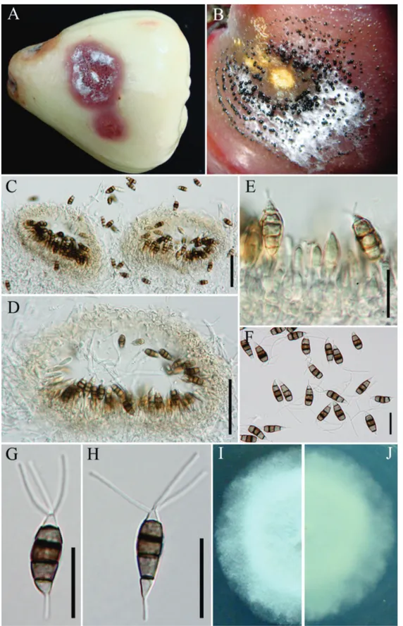
Initially small, circular, black, slightly sunken spots appear on fruits of Syzygium samarangense, which
later expand outwards on the surface of the fruit (Figure 1). Following storage these spots enlarge rapidly, become sunken, and result in a soft decay of the fruit flesh. Raised masses of black conidia arranged in concentric bands (up to 1–1.5 cm diam.) develop on the surface of the lesions and are surrounded by white mycelium. In most cases, spots coalesce and form large irregular rotting areas with lesions extending into the pulp. The disease appears the year round.
Pathogenicity testing
To comply with Koch’s postulates, lesions resembling initial symptoms were observed after 8 days on pin-pricked fruits inoculated with a conidial suspension. No symptoms were observed on fruits inoculated with a conidial suspension without pin-pricking. No symptoms were observed in the control fruits. The taxon re-isolated from diseased fruits was identical with the original isolates. The experiment was carried out using five replicates and was repeated three times with the same results.
Phylogeny
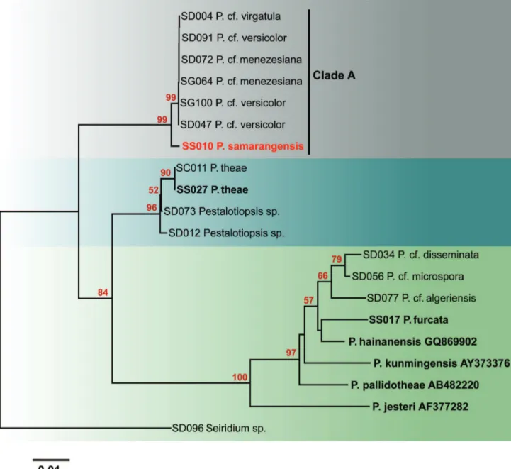
Sequences of nineteen stains of Pestalotiopsis are
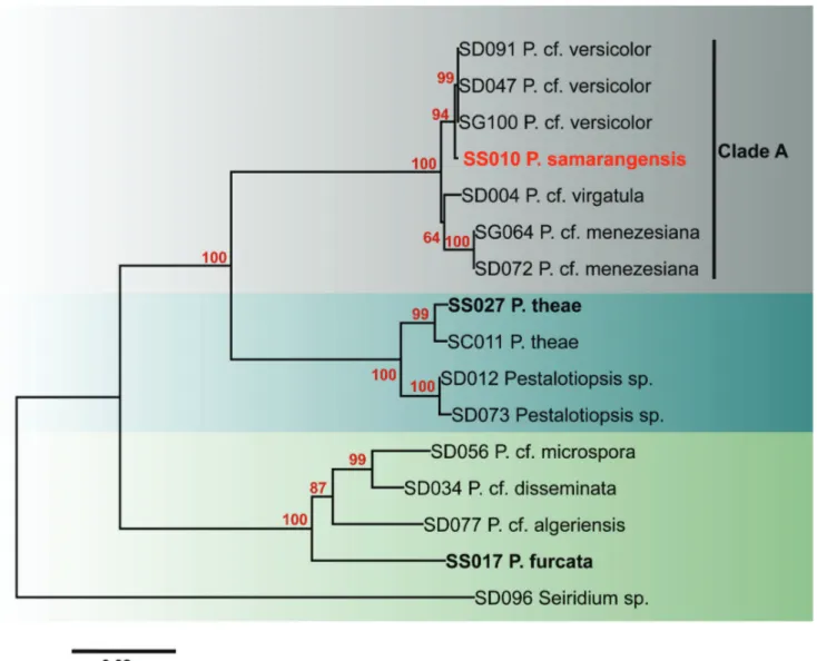
included in the ITS phylogram (Figure 2) including one new sequence generated in this study, six type or epitype strains, and 12 strains used in our previous study. In the ITS phylogram, seven strains (SD004, SD047, SD072, SD091, SG064, SG100 and SS010) cluster in Clade A with strong bootstrap support (100%). However, the strain isolated from wax apple (SS010) is separated from the other six strains. In the combined phylogram (Figure 3), the seven strains again clustered with strong bootstrap support (100%). Three strains of Pestalotiopsis cf. versicolor form
a strongly supported subclade (99%), while the wax apple strain is separated from this subclade. Two strains of P. cf. meneziana isolated from tea also cluster in Clade A with
100% bootstrap support. Both, ITS and multigene combined phylograms (Figures 2,3) show that the sequences obtained from Pestalotiopsis sp. on wax apple (SS010) are different
from sequences obtained from strains SD004, SD047,
SD072, SD091, SG064, and SG100. Therefore, the species represented by the SS010 strain from wax apple apparently is different from the Pestalotiopsis species included in the
analysis and might correspond to a species new to science.
Taxonomy
Pestalotiopsis samarangensis Maharachchikumbura &
K.D. Hyde, sp. nov.
MycoBank: MB 800178 Fig 1 A–J
Etymology: The specific epithet is based on the host species, from which the fungus was isolated.
Conidiomata acervuli, in concentric bands, confluent,
erumpent when mature, rounded to oval in outline, epidermal to superficial in origin, basal stroma and lateral wall 2–4 cells thick; cells hyaline to pale brown, textura angularis, 100–350 µm wide, 80–150 deep (Figure 1 B–C).
Conidiophores correspond to conidiogenous cells arising
within the acervuli. Conidiogenous cells discrete, simple,
short, filiform (Figure 1 E). Conidia 18–21 × 6.5–7.5 µm
(x = 20 × 7 µm), fusiform to ellipsoid, broadly clavate, straight to slightly curved, 4-septate, versicoloured; basal cell conical, hyaline, thin and smooth-walled, 3.5–4.8 µm long (x = 4 µm); apical cell 2.5–4.6 µm long (x = 3.4 µm), conical, hyaline, thin- and smooth-walled; three median cells together 12.8–13.8 µm long (x = 13.5 µm), with thick verruculose walls, dark brown, the second cell from base pale brown, 4.3–5.3 µm long (x = 4.8 µm); third cell darker brown, 3.7–5 µm long (x = 4.1 µm; the fourth cell darkest, 4.5–5.3 µm (x = 4.9 µm); three apical appendages 12–18 µm long (x = 15 µm), tubular, without terminal inflation, arising from the upper portion of the apical cell; single basal appendage, 3.5–5.2 µm long, filiform (Figure 1 F–H).
Colonies on PDA reaching 7 cm diam after 6 days at 25ºC, edge entire, whitish aerial mycelium, fruiting-bodies black, gregarious; reverse of culture white (Figure 1 I–J).
Habitat/Distribution: Known to cause fruit rot on
Syzygium samarangense in Thailand.
Material examined: THAILAND, Chiang Mai Province, Chiang Mai, on fruits of Syzygium samarangense,
20 January 2010, S.S.N. Maharachchikumbura S200110b (MFLU 12-0133; holotype) - ex-type culture MFLUCC 12-0233; ibid.,15 May 2011, S.S.N. Maharachchikumbura
S200511 (MFLU 12-0134); Chiang Rai Province, Chiang Rai, 15 September 2011, S.S.N. Maharachchikumbura S150911 (MFLU 12-0135) (Table 1).
As the names of Pestalotiopsis taxa downloaded
from GenBank have been shown to be unreliable (Maharachchikumbura et al., 2011) we prefer to label the downloaded taxa as Pestalotiopsis cf. menezesiana, P. cf. versicolor,and P. cf. virgatula to prevent future confusion.
FIGURE 1 - A. Pestalotiopsis samarangensis (holotype). B. Fruit rot of wax apple C.D. Acervular
FIGURE 2 - Neighbor-joining phylogram generated from ITS gene sequence analysis of type and putatively named Pestalotiopsis species
in GenBank. Data were analyzed with random addition sequence, unweighted parsimony and treating gaps as missing data. Seiridium sp.
is placed as the outgroup taxon, ex-type sequences are written in bold.
therefore we use cf. (similar to the available species, but not certainly identified as this species = compare with) until more species have been epitypified.
DISCUSSION
Previously, Pestalotiopsis eugeniae (Thüm.) S.
Kaneko has been described from wax apple fruit (Lan, 2001).
Pestalotiopsis eugeniae differs from P. samarangensis
by concolourous conidia. Pestalotiopsis samarangensis
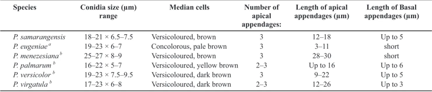
also has overlapping morphology with P. versicolor and
P. virgatula (Table 2). However, P. samarangensis differs
from P. versicolor and P. virgatula by molecular data and by shorter apical appendages (Guba, 1961; Nag Raj, 1993).
Pestalotiopsis samarangensis is somewhat similar in
morphology to P. palmarum (Cooke) Steyaert), which was
isolated in India from coconut. In the original description, Cooke (1876) did not indicate the range of conidial dimensions for P. palmarum, but only the average conidial
size as 15 × 5 µm, whereas in P. samarangensis conidia
measure on average 20 × 7 µm, thus definitely different. According to Guba (1961), the conidia of P. palmarum
FIGURE 3 - Neighbor-joining phylogram of Pestalotiopsis spp. generated from combined sequences of ITS, β-tubulin, and tef1. Seiridium
sp. is the outgroup taxon; ex-type sequences are written in bold.
species attacking members of the family Myrtaceae may be specific since the essential oils of the family are likely to exert a selection on pathogens capable to attack these plants (Lee et al., 2008). In this family, scab disease of Psidium guajava caused by P. clavispora (G.F. Atk.) Steyaert,
P. microspora (Speg.) G.C. Zhao & N. Li, P. psidii (Pat.)
Mordue, and P. disseminata (Thum.) Steyaert (Keith et al.,
2006); bark lesions of Eucalyptus globulus stems by P. guepinii
(Desm.) Steyert (Alonso et al., 2009); a leaf spot of Anogeissus latifolia caused by P. versicolor (Speg.) Steyaert (Agarwal and Ganguli 1959), and leaf spot of Eucalyptus camaldulensis by P. mangiferae (Henn.) Steyaert (El-Sayed et al., 1985) are known. Pestalotiopsis disseminata, P. guepinii, P. microspora,
and P. psidii have concolorous median cells. P. samarangensis
is distinct from all these species by versicoloured median cells. It is distinct from P. clavispora (conidia 18–26 × 6.5– 8.5 µm; apical appendages 17–31 µm)and P. mangiferae
(conidia 22–26 × 8–11µm; apical appendages 17–31 µm) by smaller conidia (18–21 × 6.5–7.5 µm) and shorter apical appendages (12–18 µm) (Guba, 1961; Nag Raj, 1993). We therefore introduce a new species to accommodate this taxon which causes a disease of wax apple.
Species of Pestalotiopsis are not well resolved in the
ITS tree, however, ITS, β-tubulin, and tef1 combined gene analysis successfully resolved the species.The combined phylogenetic tree comprises three ex-type strains and a further twelve putatively named species (due to their economically importance) from our studies. Only for six species of Pestalotiopsis, ex-type strains are available; P. furcata, P. hainanensis, P. jesteri, P. kunmingensis, P. pallidotheae and P.theae. Thus there is a need to epitypify
other Pestalotiopsis species (Maharachchikumbura et al.,
2011). Epitypification in Pestalotiopsis spp. is, however,
host-TABLE 1 - Isolates and sequences used in the phylogenetic study
*Acronyms: CGMCC= China General Microbiological Culture Collection, MFLUCC= Mae Fah Luang University Culture Collection; **SD =
sample number 2, SG = sample number 1, SS =sample number 3
TABLE 2 -Comparison of conidia of Pestalotiopsis samarangensis and other similar species
aBPI 406804 b Guba (1961)
specific, and material of morphologically similar species taken from the same host and geographical location may actually belong to a different species to what was originally described. Maharachchikumbura et al. (2011) showed that many Pestalotiopsis species had been previously
named according to host association, and that only a small number of characteristics are available to differentiate species. Therefore, identification of Pestalotiopsis to
species level is presently difficult and many sequences for
Pestalotiopsis spp. deposited in GenBank are likely to be
wrongly named. Because of this unreliability of sequences in GenBank we are referring these taxa as Pestalotiopsis
cf. menezesiana, P. cf. versicolor,and P. cf. virgatula for
the time being. Correct species identification is essential
in plant-pathogenic genera (Rossman & Palm-Hernández, 2008). Plant pathologists need to be able to confidently name causal agents in Pestalotiopsis, so that effective
quarantine measures can be put in place to prevent entry of unwanted diseases into a country (Maharachchikumbura et al., 2011). Only wounded or damaged fruits have developed disease symptoms indicating that careful handling of the fruit during transportation and storage can help prevent P. samarangensis fruit rot.
ACKNOWLEDGMENTS
Research Council of Thailand (grant for Pestalotiopsis
No: 55201020008), and Mae Fah Luang University (grant for Pestalotiopsis No: 55101020004) for supporting this
research. Appreciation is extended to the U.S. National Fungus Collections for providing type herbarium material.
REFERENCES
Agarwal GP, Ganguli G (1959) A leaf spot disease of Anogeissus latifolia Wall. due to Pestalotiopsis versicolor (Speg.) Steyaert. Current Science 28:295-296.
Alonso R, Tiscornia S, Alfenas AC, Bettucci L (2009) Fungi associated to bark lesions of Eucalyptus globulus stems in
plantations from Uruguay. Revista Árvore 33:591-597.
Chomnunti P, Schoch CL, Aguirre-Hudson B, Ko Ko TW, Hongsanan S, Jones EBG, Kodsueb R, Phookamsak R, Chukeatirote E, Bahkali AH, Hyde KD (2011) Capnodiaceae.
Fungal Diversity 51:103-134.
Cooke MC (1876) Some Indian fungi. Grevillea 4:114-118. Das Ranjana, Chutia M, Das K, Jha DK (2010) Factors affecting sporulation of Pestalotiopsis disseminata causing grey blight disease of Persea bombycina Kost., the primary food plant of
muga silkworm. Crop Protection 29:963-968.
El-Sayed, AB, Salem MA, Seif-El-Din AA, Omar AA, Mikhail SH (1985) Reaction of Eucalyptus species to Pestalotiopsis mangiferae in Egypt. Australian Forest Research 15:463-468. Glass NL, Donaldson GC (1995) Development of primer sets designed for use with the PCR to amplify conserved genes from filamentous ascomycetes. Applied and Environmental Microbiology 61:1323-1330.
Guba EF (1961) Monograph of Pestalotia and Monochaetia.
Cambridge MA, USA. Harvard University Press.
Guo LD, Hyde KD, Liew ECY (2000) Identification of endophytic fungi from Livistona chinensis (Palmae) using morphological and molecular techniques. New Phytologist147:617-630.
Hu HL, Jeewon R, Zhou DQ, Zhou TX, Hyde KD (2007) Phylogenetic diversity of endophytic Pestalotiopsis species
in Pinus armandii and Ribes spp.: evidence from rDNA and β-
tubulin gene phylogenies. Fungal Diversity 24:1-22.
Hyde KD, Fröhlich J (1995) Mycosphaerella palmicola associated
with leaf spots of Cocos nucifera in Australia, Iran Jaya and Papua New Guinea. Mycological Research 99:704-706.
Janick J, Paull RE (2008) The encyclopedia of fruit & nuts. Cambridge UK. Cambridge University Press.
Kaushik CD, Thakur DP, Chand JN (1972) Parasitism and control of Pestalotia psidii causing cankerous disease of ripe guava fruits.
Indian Phytopathology 25:61-64.
Keith LM, Velasquez ME, Zee FT (2006) Identification and characterization of Pestalotiopsis spp. causing scab disease of
guava, Psidium guajava in Hawaii. Plant Disease 90:16-23. Ko Ko T, Stephenson SL, Bahkali AH, Hyde KD (2011) From morphology to molecular biology: can we use sequence data to identify fungal endophytes? Fungal Diversity 50:113-120. Korsten L, De Jager ES, De Villers EE, Lourens A, Kotze JM, Wehner FC (1995) Evaluation of bacterial epiphytes isolated
from avocado leaf and fruit surfaces for biocontrol of avocado postharvest diseases. Plant Disease 79:1149.
Lan CC (2001) Diseases survey and integrated control of diseases on forcing culture on wax-apple fruit trees. Research Bulletin of Kaohsiung District Agricultural Improvement Station 13:20-29. Lee YS, Kim J, Shin SC, Lee SG, Park IK (2008) Antifungal activity of Myrtaceae essential oils and their components against three phytopathogenic fungi. Flavour and Fragrance Journal 23:23-28.
Lingappa BT, Lingappa Y (1965) Effects of nutrients on self-inhibition of germination of conidia of Glomerella cingulata.
Journal of General Microbiology41:67-75.
Maharachchikumbura SSN, Guo LD, Chukeatirote E, Bahkali AH, Hyde KD (2011) Pestalotiopsis – morphology, phylogeny,
biochemistry and diversity. Fungal Diversity 50:167-187. Maharachchikumbura SSN, Guo LD, Cai L, Chukeatirote E, Wu WP, Sun X, Crous PW, Bhat DJ, McKenzie EHC, Bahkali AH, Hyde KD (2012) A multi-locus backbone tree for Pestalotiopsis,
with a polyphasic characterization of 14 new species. Fungal Diversity 56:95-129.
Nag Rag TR (1993) Coelomycetous anamorphs withappendage bearing conidia. Waterloo Ontario, Canada. Mycologue Publications.
O’Donnell K, Cigelnik E (1997) Two divergent intragenomic rDNA ITS2 types within a monophyletic lineage of the fungus
Fusarium are non orthologous. Molecular Phylogenetics and
Evolution 7:103-116.
Phoulivong S, Cai L, Chen H, McKenzie EHC, Abdelsalam K, Chukeatirote E, Hyde KD (2010) Colletotrichum gloeosporioides
is not a common pathogen on tropical fruits. Fungal Diversity 44:33-43.
Rehner SA (2001) Primers for elongation factor 1-alpha (EF1-alpha). Available at: http://ocid.nacse.org/research/deephyphae/ EF1primer.pdf. Accessed on February 25, 2012.
Rossman AY, Palm-Hernández ME (2008) Systematics of plant pathogenic fungi: why it matters. Plant Disease 92:1376-1386. Saitou N, Nei M (1987) The neighbor-joining method: A new method for reconstructing phylogenetic trees. Molecular Biology and Evolution 4:406-425.
Sangchote S, Farungsang U, Farungsang N (1998) Pre and postharvest infection of rambutan by pathogens and effect on postharvest treatments. In: Coates LM, Hofman PJ, Johnson GI (Eds.) Disease control and storage life extension in fruits. ACIAR Proceeding No. 81:87-91.
Shü ZH, Tirtawinata R, Meon Z, Thanarut C (2008) Wax apple production in selected tropical Asian countries. Acta Horticulturae 773:161-164.
Srisaard A (2003) Chom-Phu. Bangkok Thailand. Naka Intermedia.
Tamura K, Dudley M, Nei M, KumarS (2007) MEGA4: Molecular evolutionary genetics analysis (MEGA) software version 4.0. Molecular Biology and Evolution 24:1596-1599.
Ullasa BA, Rawal RD (1989) Occurrence of a new post-harvest disease of mango due to Pestalotiopsis glandicola. Acta
Horticulturae 231:540-543.
Vara-Ubol S, Chambers E, Kongpensook V, Oupadissakoon C, Yenket R, Retiveau A (2006) Determination of the sensory characteristics of rose apples cultivated in Thailand. Journal of Food Science 71:547-552.
TPP-2012-0013 - Received 11 May 2012 - Accepted 21 December 2012 Section Editor: Meike Piepenbring
Xu L, Kusakari S, Hosomi A, Toyoda H, Ouchi A (1999) Postharvest disease of grape caused by Pestalotiopsis species.