Inte grins in vascular de ve lo pm e nt
Howard Hughes Medical Institute and Center for Cancer Research, Department of Biology, Massachusetts Institute of Technology, Cambridge, MA, USA
R.O . Hynes, B.L. Bader and K. Hodivala-Dilke
Abstract
Many growth factors and their protein kinase receptors play a role in regulating vascular development. In addition, cell adhesion mol-ecules, such as integrins and their ligands in the extracellular matrix, play important roles in the adhesion, migration, proliferation, survival and differentiation of the cells that form the vasculature. Some integrins are known to be regulated by angiogenic growth factors and studies with inhibitors of integrin functions and using strains of mice lacking specific integrins clearly implicate some of these molecules in vascu-logenesis and angiogenesis. However, the data are incomplete and sometimes discordant and it is unclear how angiogenic growth factors and integrin-mediated adhesive events cooperate in the diverse cell biological processes involved in forming the vasculature. Consider-ation of the results suggests working hypotheses and raises questions for future research directions.
Co rre spo nde nce
R.O . Hynes
Howard Hughes Medical Institute and Center for Cancer Research Department of Biology
Massachusetts Institute of Technology Cambridge, MA 02139
USA
Fax: + 1-617-253-8357 E-mail: [email protected]
Presented at the 5th Brazilian Symposium on Extracellular Matrix - SIMEC, Angra dos Reis, RJ, Brasil, September 7-10, 1998.
The present address of B.L. Bader is Department of Proteinchemistry, Max-Planck-Institute for Biochemistry, Am Klopferspitz 18a, D-82152 Martinsried near Munich, Germany.
Research supported by the Howard Hughes Medical Institute and by the USPHS National Institutes of Health (grants PO HL41484 and RO 1-CA17007). Fellowship support was provided by the Deutsche Forschungs-gemeinschaft (B.L. Bader), the Human Frontiers Science Program (K. Hodivala-Dilke) and the Dystrophic Epidermolysis Bullosa Research Association (K. Hodivala-Dilke).
Received November 11, 1998 Accepted December 14, 1998
Ke y wo rds
·Vasculogenesis
·Angiogenesis
·Integrins
·Knockout mice
Intro ductio n
The vascular system is one of the earliest organs to form during development. In mam-mals, both the extraembryonic vasculature in the yolk sac and the embryonic vascula-ture, comprising the major vessels and the primitive heart, develop soon after implanta-tion. The processes of vascular development have commonly been divided into vasculo-genesis, the generation of the vessels de novo from mesodermally derived angioblasts,
and angiogenesis, the formation of vessels as sprouts or offshoots of a preexisting vas-cular tree (1-7). In truth the situation is much more complex. The initial yolk sac vascula-ture does indeed form from fusion of blood islands in a process of de novo vasculogen-esis and the major vessels, such as dorsal aorta and heart, arise by aggregation of angioblasts, to give vessels where none pre-exist. However, the subsequent elaboration of these initial vasculatures involves
produc-tion of side vessels by at least two different mechanisms; sprouting (5) and splitting (in-tussusception; 8-10). The resulting vascular plexuses are then remodeled to differentiate large from small vessels and arterial from venous vasculature and the endothelial tubes become variously invested with accessory cells (pericytes, smooth muscle cells, etc.). The vasculature in different organs is clearly different in many different ways. Examples such as the high endothelial venules of lymph nodes, the fenestrated endothelium of the glomerulus and the extremely tight blood-brain barrier are well known but there exist many other variations in different organs (4).
The ro le s o f gro wth facto rs and the ir re ce pto rs
number of factors. Central among these are various growth factors; vascular endothelial growth factor (VEGF), basic fibroblast growth factor (FGF-2), TGFß, angiopoietins, neuregulin and platelet-derived growth fac-tor (PDGF) and their corresponding recep-tors (5,7,11-15). FGF-2, VEGF and angio-poietins act on endothelial cells by binding to tyrosine kinase receptors, whereas PDGF and neuregulin are produced by endothelial cells and act to recruit and organize acces-sory cells, again by acting on tyrosine kinase receptors on those cells. The list of growth factors and receptors known to be involved in control of blood vessel development is growing fast. Potential involvement of Notch-Jagged signalling in angiogenesis (16) and the recent demonstration that ephrin-B2 and
its counter receptor, Eph-B4, are involved in determining the distinction between venous and arterial development (17,18) are two cases in point and clearly there are others to be discovered.
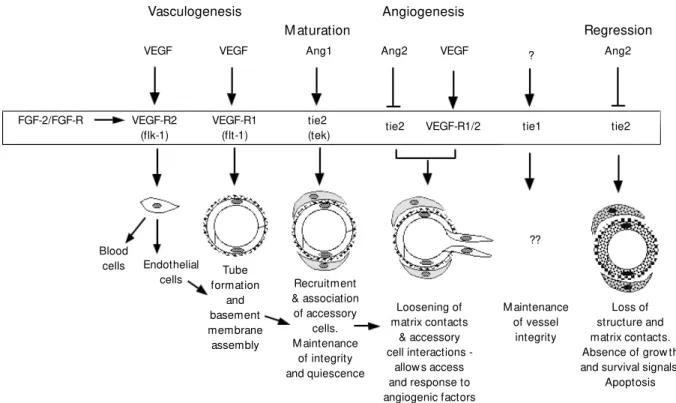
Various analyses, most notably those us-ing gene ablation methods to generate mice lacking specific factors or their receptors, have provided initial insights into the roles played by these different signalling systems and a rough sequence of inductive interac-tions can be formulated (Figure 1; 5,7,14). Thus, VEGF, acting through two different receptors, first controls the initial determina-tion of angioblasts and subsequently their ability to assemble into tubes. However, prior action of FGF-2 appears necessary to induce the expression of VEGF receptors in the
Figure 1 - Regulation of vessel formation. Diagram depicts the various phases of vascular development, the multiple grow th factors impinging on endothelial cells (basic fibroblast grow th factor (FGF-2), vascular endothelial grow th factor (VEGF), angiopoietins) and their receptors. Factors and ligands are arranged according to the processes they affect, based on inferences draw n from the phenotypes of genetically modified mice. Thus, VEGF acting through VEGF-R2 (flk-1) affects the “ birth” of endothelial cells, w hereas VEGF acting through VEGF-R1 (flt-1) instead affects tube formation and VEGF collaborating w ith angiopoietins affects recruitment of accessory cells and angiogenesis. What is unclear, and is not show n here, is how these ligand-receptor combinations affect the morphogenetic events involving cell-matrix and cell-cell adhesion. It is in this part of these processes that integrins and their ligands may play a role (see text). M odified from an original diagram by Hanahan (14).
Vasculogenesis Angiogenesis
M aturation Regression
VEGF VEGF Ang1 Ang2 VEGF Ang2
FGF-2/FGF-R VEGF-R2 (flk-1)
VEGF-R1 (flt-1)
tie2
(tek) tie2 VEGF-R1/2 tie1 tie2
Loss of structure and matrix contacts. Absence of grow th and survival signals.
Apoptosis M aintenance
of vessel integrity Loosening of
matrix contacts & accessory cell interactions
-allow s access and response to angiogenic factors Recruitment
& association of accessory
cells. M aintenance
of integrity and quiescence Tube
formation and basement membrane assembly Endothelial
cells Blood
cells
?
endothelial precursors. Subsequently, angi-opoietins acting on tie receptors affect fur-ther development of the vasculature prob-ably including interactions between endo-thelial and accessory cells. PDGF, TGFß and neuregulin signaling and ephrin/Eph inter-actions contribute further to the differentia-tion of different vessel types (12,17-19).
The ro le s o f ce ll adhe sio n mo le cule s
This rapidly developing understanding of the hierarchy of controls affecting vascu-lar development takes one only so far. We still need to understand how the factors and their receptor-mediated signals actually pro-duce vessels, inpro-duce branching and endothe-lial-accessory cell interactions and yield the array of different vessel types found in a mature animal. At the cell biological level, these events clearly require control of cell proliferation and survival, various cell mi-grations and cell adhesive events, basement membrane assembly and remodeling and stable interactions between cells and with the extracellular matrices around them. Cell-cell adhesion molecules such as cadherins are believed to play important roles and, indeed, gene ablation studies clearly impli-cate both N-cadherin (20) and VE-cadherin (21) in early steps of vessel formation. How-ever, we will focus here on a different family of cell adhesion receptors, the integrins, and their involvement in vascular development and remodeling.
Integrins are a family of heterodimeric cell surface receptors, which mediate adhe-sion of cells to extracellular matrix proteins and sometimes to other cells (22). In mam-mals, around two dozen integrins are known and endothelial cells can express at least five or six different ones (23). Cell surface ex-pression of integrins can be controlled by various growth factors, including, notably, VEGF (see below). In addition to mediating cell adhesion to, and cell migration on, a variety of extracellular matrix molecules
rel-evant to vascular development (fibronectin (FN), collagens, laminins, vitronectin, von Willebrand factor, thrombospondin, osteo-pontin, fibrinogen, entactin/nidogen), inte-grins also mediate intracellular signalling events involving various protein kinases, small GTPases, etc. (24-28) and these in turn control aspects of cytoskeletal organization and cell motility (29-31), and also regulate cell cycle progression, apoptosis and gene expression (32,33). Therefore, integrins oc-cupy a central position in any consideration of vascular development; they are regulated by growth factors known to control the pro-cess, they mediate exactly those cell biologi-cal processes (adhesion, migration, prolif-eration, survival and differentiation) needed to organize a vasculature and they are ex-pressed by the cells involved (endothelial cells, pericytes, smooth muscle cells). There is, in fact, a large and growing body of evidence implicating various integrins and integrin ligands in vascular development (23,34-36). However, it is not clear exactly which integrins are the most important nor exactly what each of them does. In this brief article, we will review the relevant results (Table 1) and discuss the many unresolved questions.
that monoclonal antibodies or peptides that selectively bind avß3 or avß5 can inhibit vasculogenesis during early quail embryo development and angiogenesis in the chicken chorioallantoic membrane (CAM) both dur-ing normal development (37) and in response to FGF-2 or VEGF (37,39) or tumor im-plants (37,41). They have also shown inhibi-tion of angiogenesis in response to tumor implants on human skin transplants to mice (38) and during neovascularization in the murine retina (39) and another group has provided corroborative data in the latter sys-tem (42). Cheresh and colleagues (43) have further shown that different angiogenic stimuli apparently rely on either avß3 (FGF-2, TNFa) or avß5 (VEGF, phorbol esters) and have shown that avß3 can bind the matrix metalloprotease (MMP-2) in a fash-ion that contributes to an invasive response and to angiogenesis (44,45). These results
have stimulated a lot of interest, not least because of the potential use of blocking reagents for therapy of a variety of disorders including tumor angiogenesis and blindness caused by retinal neovascularization.
Senger et al. (46) have reported similar antiangiogenic responses using antibodies directed against a1ß1 and a2ß1 integrins, which are also markedly upregulated by VEGF. Furthermore, Drake et al. (47) had earlier shown that antibodies to avian ß1 integrin interfere with dorsal aorta vasculo-genesis. These results suggest that integrins other than av integrins play significant roles in vascular development.
Results on mouse strains lacking specific integrins also implicate several different integrins in vasculogenesis and angiogene-sis (see Table 1). Ablation of a5ß1 integrin (48) or its ligand, fibronectin (49,50), causes major disruptions in development of
extraem-Table 1 - Integrins and their ligands in vascular development.
VEGF, Vascular endothelial grow th factor; FGF-2, basic fibroblast grow th factor; VCAM -1, vascular cell adhesion molecule.
Integrin Ligands Observations References
a1ß1 Collagens - Induced by VEGF 46
a2ß1 Laminins - Antibodies to those integrins block VEGF-induced angiogenesis in mouse skin - a1 knockout; viable; a2 knockout, not done 52 - a1ß1 is upregulated after vascular injury 68
a4ß1 Fibronectin - a4 knockout and VCAM -1 knockout show defects 69-71 VCAM -1 in formation of coronary vessels
a5ß1 Fibronectin - Antibodies to ß1 integrins block vasculogenesis in quail embryo 47 - a5 knockout show s defective vasculogenesis in yolk sac and embryo 48 - FN knockout show s even more severe defects in vasculogenesis 49,50
avß3 Vitronectin - Induced by VEGF, FGF-2, etc. 37,38,41,45
avß5 Fibrinogen - Antibodies and peptides block angiogenesis at many 43
avß1 Osteopontin sites in response to VEGF, FGF-2, tumors, etc.
Thrombospondin - Antibodies and peptides block vasculogenesis in quail embryo 40 von Willebrand factor - Retinal neovascularization is also inhibited by such agents 39,42 Plus others (see text) - av knockout show s extensive vasculogenesis and angiogenesis, 53
although cerebral vasculature is defective
- ß3 knockout show s normal vascular development including retinal vessels 59 Other integrins Other ligands - M any other knockouts of integrins or their ligands show 36,72
bryonic (yolk sac) and embryonic (heart, aorta) vasculature. In both cases, endothelial cells do differentiate, that is, the VEGF/ VEGF-R2-mediated induction of angioblasts is intact. However, absence of either a5ß1 or fibronectin disrupts vessel formation in a fashion somewhat reminiscent of the defects seen in embryos lacking VEGF-R1 (flt-1; 51). Clearly interactions of endothelial cells with FN play an important role in these early steps and there exists a distinct possibility that there is regulation of a5ß1 expression or function by VEGF/VEGF-R1 or that this signalling system cooperates with the a 5ß1-FN-regulated responses. This result conforms with the inhibition of early vascular devel-opment by anti-ß1 antibodies (47).
In contrast with this concordance be-tween antibody blocking and genetic abla-tion results, some other studies show less convergence. Although antibody blockade of a1ß1 and a2ß1 integrins blocks angio-genesis in the CAM (46), ablation of the a1 gene yields viable, fertile animals with no evidence of vascular defects (52). Since a1ß1 and a2ß1 both act as collagen and laminin receptors, it is possible that they serve over-lapping and to some extent redundant roles. Unfortunately the a2-knockout is not yet available. Time will tell whether there is indeed a conflict here between the immuno-logical and genetic approaches. However, it is already clear that ablation of the av inte-grin gene yields results that are difficult to reconcile with the results of av-inhibitors (53). av-null mouse embryos develop an apparently normal yolk sac and early embry-onic vasculature (53) in marked contrast with the blockade of quail dorsal aorta formation (40) or chicken chorioallantoic angiogenesis (37) by antibodies directed against avß3. Granted that the systems employed in these studies are different, the two sets of data differ greatly in their implications for the importance of av integrins in early vascular development. Indeed, 20% of av-null em-bryos develop to term and are born alive,
although they die promptly (53). There is extensive vasculogenesis and angiogenesis in most organs and tissues in the absence of all five av integrins. Although av-null em-bryos consistently develop defects in their brain vasculature, the basic endothelial pro-cesses of proliferation, migration, tube for-mation and branching, and basement mem-brane assembly all occur. Furthermore, there is no evidence for increased apoptosis of a v-null endothelial cells in contrast with the effects of blockade of av-integrins by anti-bodies or peptides (41). Again, the vascular systems under study are different but clearly the implications of the results differ signifi-cantly.
The intracerebral vasculature in av-null embryos is not normal; it becomes distended and eventually ruptures leading to cerebral hemorrhage (53). This result is somewhat reminiscent of the defects occurring in PDGF-B-null embryos, which are thought to be due to failure of immigration of pericytes along the cerebral vessels (54), raising the possi-bility that the av-null defect arises from a failure in pericyte recruitment to the vessels. However, the av-null defects initiate rather too early for this to be the sole cause and it remains to be discovered what exactly are the av-dependent processes unique to this vascular bed. Vascular defects in angiopoie-tin-1 or tie 2 knockout mice (19,55,56) also show some resemblances to those in av-null and PDGF-B-null embryos raising the possi-bility that these various genes may cooperate somehow in the assembly of a normal vascu-lature.
viable and fertile and show no obvious de-fects in their vascular development. How-ever, much remains to be investigated and it is possible that further analyses of these null strains, or of double mutants generated from them, may reveal dependence of angiogene-sis on one or more of these integrins, as might be expected from the av inhibition data. To date, the ß3-null mice have been investigated for defects in postnatal retinal angiogenesis and there are no major defects (59). However, the effects of perturbations such as hyperoxia or hypoxia or of combina-tions of ß-mutacombina-tions have yet to be studied and these mice should prove very useful both for such studies and for analyses of angiogenesis after wound healing, in response to tumors, etc.
The apparent discrepancy between the antibody and peptide blocking data and the genetic analyses of av integrins is enhanced by consideration of the phenotypes of mice lacking individual extracellular matrix pro-teins that are ligands for av integrins. Apart from the embryonic lethal phenotype of FN-null embryos (49,50) which is most likely a consequence of its interactions with a5ß1 (48,60), most other mouse strains lacking av integrin ligands are viable and fertile and have, so far, shown no evidence of vascular defects, although, as for the ß integrin-null mice discussed above, more detailed analy-ses are needed. Nonetheless, it is striking that mice lacking vitronectin (61), tenascin-C (62), osteopontin (63), fibrinogen (64) von Willebrand factor (65) or thrombospon-din-1 (66) are all viable.
What is one to make of the apparent discrepancies between genetics and inhibi-tion studies? As we have noted, some of the discrepancies may simply reflect the fact that inferences are being drawn from some-what different systems, in which case further analyses may show that the initial discrepan-cies are apparent rather than real. However, it is also possible that the two different
Figure 2 - Interplay of grow th factor-receptor signals and cell adhesion receptors. The figure depicts an endothelial cell in the center, expressing various integrins (w hose major ligands are noted), w hich are implicated in one or more aspects of vasculogenesis or angiogenesis (see text) and receptors for basic fibroblast grow th factor (FGF-2), vascular endothelial grow th factor (VEGF), and angiopoietins. The latter are produced by various cells in the vicinity of developing vessels and those cells in turn receive stimulation by factors secreted by the endothelial cells (e.g., platelet-derived grow th factor (PDGF) and neuregulin) that act on receptors on the accessory cells. The latter cells also express and use integrins (data not show n). Thus, there is a tw o-w ay “ conversation” betw een endothelial cells and their neighbors and adhesion events involving either or both. Also show n are interactions involving eph/ephrin family members, the Notch pathw ay and cadherins (see text). At the bottom are show n various cell biological events w hich must be appropriately controlled to yield vessels of different types. The challenge is to define the interplay among the various receptors and ligands and the contributions made by each to the cell biology of vasculogenesis and angiogenesis.
Co nclusio ns and future pro spe cts
So the current situation is as follows (Figure 2). We know of a variety of growth factors and receptors which are clearly im-plicated in controlling vasculogenesis and angiogenesis (Figure 1), although exactly what they all do is not yet clear. Most par-ticularly we do not know how they do what they do; that is, what are the intermediate molecules which they control? That is where integrins and their ligands come in. Some of these molecules clearly are regulated by VEGF and the like; others may be as well.
Integrins and their ligands clearly do play important roles in the cell biological subrou-tines necessary for vessel development (ad-hesion, migration, proliferation, survival, dif-ferentiation, matrix formation) but it is un-clear exactly which ones are most important in the different processes. Indeed the an-swers to those questions may differ depend-ing on the vascular bed or the angiogenic stimulus. It may well be, indeed it seems likely, that there is more than one form of angiogenesis. It could be that yolk sac vascu-lature relies primarily on a5ß1-FN interac-tions and less, or not at all, on av integrins,
FGF-2
(bFGF) VEGF
PDGF-R
Ang1 Ang2
ERB-2
neuregulin
Ephrin
Notch
Cadherins Cadherins
tie1 tie2
(tek) PDGF
VEGF-R2 & VEGF-R1 (flk-1) (flk-1) FGF-R
a1b1 a2b1 a5b1 avb3 avb5
Collagens Laminins
Fibronectin Fibronectin Vitronectin Fibrinogen Osteopontin von Willebrand factor
Thrombospondin
EPH Jagged ?
PROLIFERATION SURVIVAL DIFFERENTIATION
whereas retinal or tumor vasculatures may be more dependent on av integrins and their ligands. More detailed studies of the expres-sion patterns, regulation and functions of different integrins and their ligands in re-sponse to different angiogenic growth fac-tors are clearly necessary. Vessel develop-ment and remodeling involve multiple cell biological processes that need to be well coordinated to yield a functional vascula-ture. It stands to reason that such a complex process, involving as it does, several
differ-ent cell types acting in concert, would re-quire regulation by multiple adhesive pro-teins. It will be a fascinating challenge to unravel the regulatory networks and coordi-nated functions of all these players (Figure 2). The potential yield from a detailed under-standing of these processes is significant both in terms of the underlying biology and in terms of opportunities for intervention in diseases involving dysregulation of vessel growth.
Re fe re nce s
1. Noden DM (1989). Embryonic origins and assembly of blood vessels. American Re-view s of Respiratory Disease, 140: 1097-1103.
2. Folkman J & Shing Y (1992). Angiogene-sis. Journal of Biological Chemistry, 267: 10931-10934.
3. Risau W (1991). Vasculogenesis, angio-genesis and endothelial cell differentia-t ion. In: Feinberg RN, Sherer GK & Auerbach R (Editors), The Development of the Vascular System. Issues in Bio-medicine, 14: 58-68.
4. Risau W (1995). Differentiation of endo-thelium. FASEB Journal, 9: 926-933. 5. Risau W (1997). M echanisms of
angio-genesis. Nature, 386: 671-674.
6. Risau W & Flamme I (1995). Vasculogen-esis. Annual Review s of Cell and Devel-opmental Biology, 11: 73-91.
7. Beck Jr L & D’Amore PA (1997). Vascular development: cellular and molecular regu-lation. FASEB Journal, 11: 365-373. 8. Patan S, Haenni B & Burri PH (1996).
Implementation of intussusceptive mi-crovascular grow th in the chicken chorio-allantoic membrane (CAM ). 1. Pillar for-mation by folding of the capillary w all.
M icrovascular Research, 51: 80-98. 9. Patan S, M unn LL & Jain RK (1996).
Intus-susceptive microvascular grow th in a hu-man colon adenocarcinoma xenograft: a novel mechanism of tumor angiogenesis.
M icrovascular Research, 51: 260-272. 10. Patan S, Haenni B & Burri PH (1997).
Implementation of intussusceptive micro-vascular grow th in the chicken chorioal-lantoic membrane (CAM ). 2. Pillar forma-tion by capillary fusion. M icrovascular Re-search, 53: 33-52.
11. M ustonen T & Alitalo K (1995).
Endotheli-al receptor tyrosine kinases involved in angiogenesis. Journal of Cell Biology, 129: 895-898.
12. Folkman J & D’Amore PA (1996). Blood vessel formation: w hat is its molecular basis? Cell, 87: 1153-1155.
13. Breier G & Risau W (1996). The role of vascular endothelial grow th factor in blood vessel formation. Trends in Cell Biology, 6: 454-456.
14. Hanahan D (1997). Signaling vascular mor-phogenesis and maintenance. Science, 277: 48-50.
15. Korpelainen EI & Alitalo K (1998). Signal-ing angiogenesis and lymphangiogenesis.
Current Opinion in Cell Biology, 10: 159-164.
16. Zimrin AB, Pepper M S, M cM ahon GA, Nguyen F, M ontesano R & M aciag T (1996). An antisense oligonucleotide to the Notch ligand Jagged enhances fibro-blast grow th factor-induced angiogenesis
in vitro. Journal of Biological Chemistry, 271: 32499-32502.
17. W ang HU, Chen ZF & Anderson DJ (1998). M olecular distinction and angio-genic interaction betw een embryonic ar-teries and veins revealed by ephrin-B2 and its receptor Eph-B4. Cell, 93: 741-753.
18. Yancopoulos GD, Klagsbrun M & Folkman J (1998). Vasculogenesis, angiogenesis, and grow th factors: ephrins enter the fray at the border. Cell, 93: 661-664. 19. Radice G, Rayburn H, M atsunam i H,
Knudsen KA, Takeichi M & Hynes RO (1997). Developmental defects in mouse embryos lacking N-cadherin. Develop-mental Biology, 181: 64-78.
20. Suri C, Jones PF, Patan S, Bartunkova S, M aisonpierre PC, Davis S, Sato TN &
Yancopoulos GD (1996). Requisite role of angiopoietin-1, a ligand for the TIE2 re-ceptor, during embryonic angiogenesis.
Cell, 87: 1171-1180.
21. Vittet D, Buchou T, Scheitzer A, Dejana E & Huber P (1997). Targeted null-mutation in the vascular endothelial-cadherin gene impairs the organization of vascular-like structures in embryoid bodies. Proceed-ings of the National Academy of Sciences, USA, 94: 6273-6278.
22. Hynes RO (1992). Integrins: versatility, modulation, and signaling in cell adhesion.
Cell, 69: 11-25.
23. Luscinskas FW & Law ler J (1994). Inte-grins as dynamic regulators of vascular function. FASEB Journal, 8: 929-938. 24. Clark EA & Brugge JS (1995). Integrins
and signal transduction pathw ays: the road taken. Science, 268: 233-239. 25. Schw artz M A, Schaller M D & Ginsberg
M H (1995). Integrins: emerging para-digms of signal transduction. Annual Re-view s of Cell andDevelopmental Biology, 11: 549-599.
26. Yamada KM & M iyamoto S (1995). Inte-grin transmembrane signaling and cyto-skeletal control. Current Opinion in Cell Biology, 7: 681-689.
27. Clark EA & Hynes RO (1997). M eeting report. 1997 Keystone Symposium on Sig-nal Transduction by Cell Adhesion Recep-tors. BBA Review s on Cancer, 1333: R9-R16.
28. Giancotti FG (1997). Integrin signaling: specificity and control of cell survival and cell cycle progression. Current Opinion in Cell Biology, 9: 691-700.
De-velopmental Biology, 12: 463-518. 30. Huttenlocher A, Sandborg RR & Horw itz
AF (1995). Adhesion in cell migration, 7: 697-706.
31. Lauffenburger DA & Horw itz AF (1996). Cell migration: a physically integrated mo-lecular process. Cell, 84: 359-369. 32. Assoian RK (1997). Anchorage-dependent
cell cycle progression. Journal of Cell Bi-ology, 136: 1-4.
33. Frisch SM & Ruoslahti E (1997). Integrins and anoikis. Current Opinion in Cell Biol-ogy, 9: 701-706.
34. Varner JA, Brooks PC & Cheresh DA (1995). The integrin alpha V beta 3: angio-genesis and apoptosis. Cell Adhesion and Communication, 3: 367-374.
35. Strömblad S & Cheresh DA (1996). Cell adhesion and angiogenesis. Trends in Cell Biology, 6: 462-468.
36. Hynes RO & Bader BL (1997). Targeted mutations in integrins and their ligands: their implications for vascular biology.
Thrombosis and Haemostasis, 78: 83-87. 37. Brooks PC, Clark RA & Cheresh DA (1994). Requirement of vascular integrin alpha v beta 3 for angiogenesis. Science, 264: 569-571.
38. Brooks PC, St rom blad S, Klem ke R, Visscher D, Sarkar FH & Cheresh DA (1995). Antiintegrin alpha v beta 3 blocks human breast cancer grow th and angio-genesis in human skin. Journal of Clinical Investigation, 96: 1815-1822.
39. Friedlander M , Theesfeld CL, Sugita M , Fruttiger M , Thomas M A, Chang S & Cheresh DA (1996). Involvement of in-tegrins alpha v beta 3 and alpha v beta 5 in ocular neovascular diseases. Proceedings of the National Academy of Sciences, USA, 93: 9764-9769.
40. Drake CJ, Cheresh DA & Little CD (1995). An antagonist of integrin alpha v beta 3 prevents maturation of blood vessels dur-ing embryonic neovascularization. Journal of Cell Science, 108: 2655-2661. 41. Brooks PC, M ontgomery AM , Rosenfeld
M , Reisfeld RA, Hu T, Klier G & Cheresh DA (1994). Integrin alpha v beta 3 antago-nists promote tumor regression by induc-ing apoptosis of angiogenic blood ves-sels. Cell, 79: 1157-1164.
42. Hammes HP, Brow nlee M , Jonczyk A, Sutter A & Preissner KT (1996). Subcuta-neous injection of a cyclic peptide antago-nist of vitronectin receptor-type integrins inhibits retinal neovascularization. Nature M edicine, 2: 529-533.
43. Friedlander M , Brooks PC, Shaffer RW, Kincaid CM , Varner JA & Cheresh DA (1995). Definition of tw o angiogenic
path-w ays by distinct alpha v integrins. Sci-ence, 270: 1500-1502.
44. Brooks PC, Stromblad S, Sanders LC, von Schalscha TL, Aim es RT, St et ler-Stevenson WG, Quigley JP & Cheresh DA (1996). Localization of matrix metallo-proteinase M M P-2 to the surface of inva-sive cells by interaction w ith integrin al-pha v beta 3. Cell, 85: 683-693.
45. Brooks PC, Silletti S, von Schalscha TL, Friedlander M & Cheresh DA (1998). Dis-ruption of angiogenesis by PEX, a non-catalytic metalloproteinase fragment w ith integrin binding activity. Cell, 92: 391-400. 46. Senger DR, Claffey KP, Benes JE, Perruzzi CA, Sergiou AP & Detmar M (1997). An-giogenesis promoted by vascular endo-thelial grow th factor: regulation through alpha1beta1 and alpha2beta1 integrins.
Proceedings of the National Academy of Sciences, USA, 94: 13612-13617. 47. Drake CJ, Davis LA & Little CD (1992).
Antibodies to ß1-integrins cause alter-ations of aortic vasculogenesis, in vivo.
Developmental Dynamics, 193: 83-91. 48. Yang JT, Rayburn H & Hynes RO (1993).
Embryonic mesodermal defects in alpha 5 integrin-deficient mice. Development, 119: 1093-1105.
49. George EL, Georges-Labouesse EN, Patel-King RS, Rayburn H & Hynes RO (1993). Defects in mesoderm, neural tube and vascular development in mouse em-bryos lacking fibronectin. Development, 119: 1079-1091.
50. George EL, Baldw in HS & Hynes RO (1997). Fibronectins are essential for heart and blood vessel morphogenesis but are dispensable for initial specification of pre-cursor cells. Blood, 90: 3073-3081. 51. Fong GH, Rossant J, Gertsenstein M &
Breitman M L (1995). Role of the Flt-1 re-ceptor tyrosine kinase in regulating the assembly of vascular endothelium. Na-ture, 376: 66-70.
52. Gardner H, Kreidberg J, Koteliansky V & Jaenisch R (1996). Deletion of integrin a1 by homologous recombination permits normal murine development but gives rise to a specific deficit in cell adhesion. De-velopmental Biology, 175: 301-313. 53. Bader BL, Rayburn H, Crow ley D & Hynes
RO (1998). Extensive vasculogenesis, an-giogenesis and organogenesis precede lethality in mice lacking all av integrins.
Cell (in press).
54. Lindahl P, Johansson BR, Leveen P & Betsholtz C (1997). Pericyte loss and mi-croaneurysm formation in PDGF-B-defi-cient mice. Science, 277: 242-245. 55. Dumont DJ, Gradw ohl G, Fong GH, Puri
M C, Gert senst ein M , Auerbach A & Breitman M L (1994). Dominant-negative and targeted null mutations in the endo-thelial receptor tyrosine kinase, tek, re-veal a critical role in vasculogenesis of the em bryo. Genes and Developm ent, 8: 1897-1909.
56. Sato TN, Tozaw a Y, Deutsch U, Wolburg-Buchholz K, Fujiw ara Y, Gendron-M aguire M , Gridley T, Wolburg H, Risau W & Qin Y (1995). Distinct roles of the receptor ty-rosine kinases Tie-1 and Tie-2 in blood vessel formation. Nature, 376: 70-74. 57. Hodivala-Dilke KM , DiPersio CM ,
KreidbergJA & Hynes RO (1998). Novel roles for a3ß1 integrin as a regulator of cytoskeletal assembly and as a transdomi-nant inhibitor of integrin receptor function in mouse keratinocytes. Journal of Cell Biology, 142: 1357-1369.
58. Huang XZ, Wu JF, Cass D, Erle DJ, Corry D, Young SG, Farese Jr RV & Sheppard D (1996). Inactivation of the integrin beta 6 subunit gene reveals a role of epithelial integrins in regulating inflammation in the lung and skin. Journal of Cell Biology, 133: 921-928.
59. Hodivala-Dilke KM , M cHugh K, Tsakiris DA, Rayburn H, Crow ley D, Ullm an-Culleré M , Ross FP, Coller BS, Teitelbaum S & Hynes RO (1999). ß3-integrin-defi-cient mice: a model for Glanzmann throm-basthenia show ing placental defects and reduced survival. Journal of Clinical Inves-tigation (in press).
60. Goh KL, Yang JT & Hynes RO (1997). M esodermal defects and cranial neural crest apoptosis in alpha5 integrin-null em-bryos. Development, 124: 4309-4319. 61. Zheng X, Saunders TL, Cam per SA,
Samuelson LC & Ginsburg D (1995). Vitro-nectin is not essential for normal mam-malian development and fertility. Proceed-ings of the National Academy of Sciences, USA, 92: 12426-12430.
62. Saga Y, Yagi T, Ikaw a Y, Sakakura T & Aizaw a S (1992). M ice develop normally w ithout tenascin. Genes and Develop-ment, 6: 1821-1831.
63. Liaw L, Birk DE, Ballas CB, Whitsitt JS, Davidson JM & Hogan BLM (1998). Al-tered w ound healing in mice lacking a functional osteopontin gene (spp1). Jour-nal of Clinical Investigation, 101: 1468-1478.
64. Suh TT, Holm bäck K, Jensen NJ, Daughterty CC, Small K, Simon DI, Potter SS & Degen JL (1995). Resolution of spontaneous bleeding events but failure of pregnancy in fibrinogen-deficient mice.
65. Denis CC, M ethia N, Frenette PS, Rayburn H, Ullm an-Culleré M , Hynes RO & Wagner DD (1998). A mouse model of severe von Willebrand disease: defects in hemostasis and thrombosis. Proceedings of the National Academy of Sciences, USA, 95: 9524-9529.
66. Law ler J, Sunday M , Thibert V, Duquette M , George EL, Rayburn H & Hynes RO (1998). Thrombospondin-1 is required for normal murine pulmonary homeostasis and its absence causes pneumonia. Jour-nal of Clinical Investigation, 101: 982-992. 67. Diaz-Gonzalez F, Forsyth J, Steiner B & Ginsberg M H (1996). Transdominant
inhi-bition of integrin function. M olecular Biol-ogy of the Cell, 7: 1939-1951.
68. Gotw als PJ, Chi-Rosso G, Lindner V, Yang J, Ling L, Faw ell SE & Koteliansky VE (1996). The a1ß1 integrin is expressed during neointima formation in rat arteries and mediates collagen matrix reorganiza-tion. Journal of Clinical Investigation, 97: 2469-2477.
69. Yang JT, Rayburn H & Hynes RO (1995). Cell adhesion events mediated by alpha 4 integrins are essential in placental and car-diac development. Development, 121: 549-560.
70. Kw ee L, Baldw in S, Stew art CS, Buck C &
Labow M A (1995). Defective develop-ment of the embryonic and extraembry-onic circulatory systems in vascular cell adhesion molecule (VCAM -1) deficient mice. Development, 121: 489-503. 71. Gurtner GC, Davis V, Li H, M cCoy M J,
Sharp A & Cybulsky M I (1995). Targeted disruption of the murine VCAM -1 gene: essential role of VCAM -1 in chorioallan-toic fusion and placentation. Genes and Development, 9: 1-14.