ISSN 1414-431X
www.bjournal.com.br
www.bjournal.com.br
Volume 45 (12) 1102-1340 December 2012
Braz J Med Biol Res, December 2012, Volume 45(12) 1255-1261
10.1590/S0100-879X2012007500143
doi:
Pamidronate for the treatment of osteoporosis secondary to chronic
cholestatic liver disease in Wistar rats
F.A. Pereira, R. Mattar, I. Facincani, H.L.A. Defino, L.N.Z. Ramalho, V. Jorgetti, J.B. Volpon and
F.J.A. de Paula
Institutional Sponsors
The Brazilian Journal of Medical and Biological Research is partially financed by
Faculdade de Medicina de Ribeirão Preto Campus
Ribeirão Preto
Explore High - Performance MS Orbitrap Technology In Proteomics & Metabolomics
analiticaweb.com.br S C I E N T I F I C
BIOMEDICAL SCIENCES
AND
Pamidronate for the treatment of osteoporosis
secondary to chronic cholestatic liver
disease in Wistar rats
F.A. Pereira
1, R. Mattar
1†, I. Facincani
2, H.L.A. Defino
3, L.N.Z. Ramalho
4,
V. Jorgetti
5, J.B. Volpon
3and F.J.A. de Paula
11Departamento de Clínica Médica, Faculdade de Medicina de Ribeirão Preto,
Universidade de São Paulo, Ribeirão Preto, SP, Brasil
2Departamento de Pediatria e Neonatologia, Faculdade de Medicina de Ribeirão Preto,
Universidade de São Paulo, Ribeirão Preto, SP, Brasil
3Departamento de Biomecânica, Medicina e Reabilitação do Aparelho Locomotor,
Faculdade de Medicina de Ribeirão Preto, Universidade de São Paulo, Ribeirão Preto, SP, Brasil
4Departamento de Patologia, Faculdade de Medicina de Ribeirão Preto,
Universidade de São Paulo, Ribeirão Preto, SP, Brasil
5Departamento de Nefrologia, Faculdade de Medicina, Universidade de São Paulo, São Paulo, SP, Brasil
Abstract
Osteoporosis is a major complication of chronic cholestatic liver disease (CCLD). We evaluated the efficacy of using disodium
pamidronate (1.0 mg/kg body weight) for the prevention (Pr) or treatment (Tr) of cholestasis-induced osteoporosis in male Wistar rats: sham-operated (Sham = 12); bile duct-ligated (Bi = 15); bile duct-ligated animals previously treated with pamidronate before and 1 month after surgery (Pr = 9); bile duct-ligated animals treated with pamidronate 1 month after surgery (Tr = 9). Rats were
sacrificed 8 weeks after surgery. Immunohistochemical expression of IGF-I and GH receptor was determined in the proximal
growth plate cartilage of the left tibia. Histomorphometric analysis was performed in the right tibia and the right femur was used
for biomechanical analysis. Bone material volume over tissue volume (BV/TV) was significantly affected by CCLD (Sham = 18.1 ± 3.2 vs Bi = 10.6 ± 2.2%) and pamidronate successfully increased bone volume. However, pamidronate administered in
a preventive regimen presented no additional benefit on bone volume compared to secondary treatment (BV/TV: Pr = 39.4 ± 12.0; Tr = 41.2 ± 12.7%). Moreover, the force on the momentum of fracture was significantly reduced in Pr rats (Sham = 116.6 ± 23.0; Bi = 94.6 ± 33.8; Pr = 82.9 ± 22.8; Tr = 92.5 ± 29.5 N; P < 0.05, Sham vs Pr). Thus, CCLD had a significant impact on
bone histomorphometric parameters and pamidronate was highly effective in increasing bone mass in CCLD; however, preven-tive therapy with pamidronate has no advantage regarding bone fragility.
Key words: Hepatic osteodystrophy; Osteoporosis pathogenesis; Growth factors; Biomechanics; Animal models; Pamidronate
Introduction
Correspondence: F.J.A. de Paula, Departamento de Clínica Médica, Faculdade de Medicina de Ribeirão Preto, USP,
Av. Bandeirantes, 3900, 14049-900 Ribeirão Preto, SP, Brasil. Fax: +55-16-3633-6695. E-mail: [email protected]
†In memoriam
Received May 12, 2012. Accepted August 31, 2012. Available online September 14, 2012. Published December 17, 2012. Hepatic osteodystrophy is a generic term used to identify
bone diseases associated with the complex metabolic distur -bance linked to liver diseases, especially chronic cholestatic liver disease (CCLD) (1). Osteoporosis is the major bone complication of CCLD. Multiple factors contribute to bone loss in CCLD and all can be easily recognized at the end stage of chronic liver disease (malnutrition, hypogonadism,
vitamin D deficiency, insulin-like growth factor-I (IGF-I) defi -ciency, and cholestasis) (2). However, decreased production
of endocrine IGF-I by the liver (3,4), as well as paracrine/ autocrine IGF-I expression in the bone microenvironment
(5), has been shown to be an early crucial determinant of bone impairment in CCLD.
Bisphosphonates are potent inhibitors of bone
resorp-tion, usually indicated for patients with clinically significant
1256 F.A. Pereira et al.
Rheumatology, it is also indicated for the primary prevention
of glucocorticoid-induced osteoporosis (6). In this context, attempts to extend the list of diseases, which would benefit
from primary prevention with bisphosphonate treatment must be counterbalanced by considering an array of peculiarities and potentially negative issues: a) the mechanism behind bone loss, i.e., decreased bone formation or increased bone
resorption, b) concerns exist about how long bone remodeling could artificially be inhibited by anti-resorptive drugs without serious impairment of bone repairing capacity (7), and c) the
effectiveness of pharmacologic therapy in the prevention of
osteoporosis in young individuals (8).
Bone mineral density assessment represents a landmark in the clinical investigation and diagnosis of osteoporosis. However, this surrogate end point represents a partial evalua-tion of bone strength and an increase in bone mineral density does not necessarily mean a reduction of fracture risk.
Ani-mal models, especially of rodents, have made an extensive
contribution to the advancement of the understanding of the mechanistic role of several factors in the etiopathogenesis of osteoporosis (9,10). Thus, translational research in mice and rats is a fundamental tool for the development of new drugs and the design of new treatment schemes. However, an important limitation of the study of osteoporosis in small
animal models is the lack of fracture due to bone insufficiency
(11). Biomechanical tests have been a reasonable option to
circumvent this limitation and are used extensively for the experimental analysis of bone strength (12,13).
In this scenario, the major objective of the present study
was to evaluate the beneficial effect of disodium pamidronate
(pamidronate) as primary prevention therapy for hepatic osteodystrophy in CCLD. Rats submitted to bile duct ligation (BDL) surgery received pamidronate before and 1 month after surgery (primary prevention) or only 1 month after surgery. The results obtained with these two groups were compared to those obtained with the sham-operated group and another group submitted only to BDL. Biochemical evaluation of mineral metabolism and liver function was performed, the
expression of IGF-I and of growth hormone receptor (GHR)
in the growth plate was evaluated in all animals and bone histomorphometry of the tibia and biomechanical assays of the femur were also analyzed.
Material and Methods
All experimental designs and procedures were approved by the Animal Research Ethics Committee of the Faculdade de Medicina de Ribeirão Preto, USP. Forty-five 2-month-old
male Wistar rats, housed individually at 25°C on a 12:12-h light-dark cycle, were used in this study. They were strictly pair-fed a laboratory diet provided by Nuvital (Nuvilab CR1;
Brazil) containing 22% protein, 53% carbohydrate, and 4.5% lipid. Animals had free access to water. Rats sub -jected to BDL surgery were used as reference. The study was comprised of 4 groups of male Wistar rats weighing
150-175 g: a) sham-operated rats (Sham, N = 12); b) rats
subjected to bile duct ligation surgery (Bi, N = 15); c) rats subjected to bile duct ligation surgery plus pamidronate as primary prevention therapy administered before and 1 month after the procedure (Pr, N = 9); d) rats subjected to bile duct ligation surgery plus pamidronate as conventional therapy, administered 1 month after the procedure (Tr, N = 9). The protocol was repeated 3 times, each comprised of 4 rats in the Sham group and 6 rats in the groups submitted to BDL. Three rats in the Bi group and 9 rats in the Pr and Tr groups died during the period of observation. All groups were followed for 2 months after surgery; pamidronate was injected intraperitoneally at the dose of 1.0 mg/kg
body weight. Vehicle (0.9% sodium chloride) was injected
in Sham and Bi groups. At the end of the second month, all rats submitted do BDL showed clinical manifestations of cholestatic disease.
Bile duct ligation technique
Anesthesia was induced by intraperitoneal injection of
2.5% tribromoethanol (Aldrich, USA; 250 mg/kg body mass).
A median laparotomy was performed and the common bile
duct was exposed and doubly ligated as described previ
-ously. In Sham rats, the procedure consisted of exposure
of the liver hilum, which was gently mobilized with a sterile cotton applicator. After surgery, the BDLgroups were
in-jected intramuscularly once a week with vitamin K (8 mg/kg
body weight). Sham rats were injected with an equivalent
volume of saline infusion (0.9% sodium chloride).
Biochemical analysis
Blood collections were performed at baseline (2 mL
from the tail) and at sacrifice when 7 mL blood was drawn
from the left ventricle. The samples were maintained on ice and then centrifuged at 4°C. Serum aliquots were immedi-ately used for the determination of albumin, total calcium, inorganic phosphorus, bilirubin, alanine aminotransferase (ALT), and aspartate aminotransferase (AST).
Biomechanical evaluation
Biomechanical tests. Frozen femora were thawed at
room temperature for 2 h before testing. Mechanical tests were performed on intact right femora using a destructive three-point bending procedure. The femur was placed supine on two round bars at a distance of 20 mm in a
mechanical testing machine (10,000 N, EMIC, Brazil) and deflected by a notched bar on the opposite side of the bone.
The descending speed of the notched bar was 1 mm/min.
Maximal force and stiffness were determined from force/ deflection plots.
Histological assessment
weight) was injected intraperitoneally 7 and 2 days before sacrifice. The right tibia was removed and dehydrated in ethanol, infiltrated and embedded without demineralization in methyl methacrylate. Undecalcified sections were cut at
a thickness of 5 µm and mounted unstained for dynamic measurements. Consecutive sections were stained with toluidine blue to quantify bone cells. The
histomorpho-metric indexes are reported according to the standardized
nomenclature recommended by the American Society of Bone and Mineral Research (14).
Bone specimens for immunohistochemical analysis
were processed as previously described (5). Briefly, bone
samples were sectioned at the growth plate at a thickness
of 4 µM from paraffin blocks containing representative
samples and mounted on poly-L-lysine-coated slides.
Nonspecific protein binding was blocked with normal
serum (Novostain Universal Super ABC Kit; Novocastra Laboratories, UK) for 30 min. The sections were then
incubated with primary monoclonal antibodies specific for IGF-I (1:200; American Diagnostic, USA) and GHR (1:100, American Diagnostic). Next, the slides were incubated with the avidin-biotin-peroxidase complex (Novostain Universal Super ABC Kit), counterstained with Harris hematoxylin,
dehydrated, and mounted with Permount (Biomeda, USA). As negative controls, all specimens were incubated with a control antibody under identical conditions. The prepara-tions for each marker were evaluated at random in at least
10 representative high-power fields (40X magnification),
and graded according to their intensities and percentage of positive cells. Cells were designated as positive when they displayed a distinct brown cytoplasm staining.
Semi-quantitative expression was scored as follows: 0, no stained
cells; +, fewer than 10% positive cells; ++, 10-50% positive cells; +++, >50% positive cells.
Statistical analysis
Data are reported as means ± SEM. Statistical signifi -cance was determined using ANOVA followed by the Tukey test for multiple comparisons, and the Pearson correlation
coefficient was employed for the analysis of correlation between parametric variables using the GraphPad Prism software (GraphPad Prism, USA)
Results
There was no significant difference in body weight be
-tween groups either at baseline (Sham = 164.3 ± 3.5; Bi = 164.9 ± 2.7; Pr = 165.3 ± 2.3; Tr = 166.3 ± 1.9 g) or at the end of the experiment (Sham = 483.6 ± 55.3; Bi = 458.7 ± 60.4; Pr = 438.8 ± 62.4; Tr = 448.8 ± 68.4 g). All groups exhibited significant weight gain during the 2 months of observation (P < 0.001).
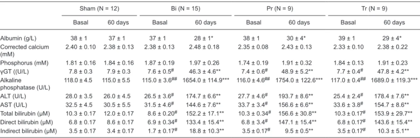
Sham-operated animals exhibited no variation in se
-rum albumin levels during the experiment, while the other
groups showed decreased levels of serum albumin at the
end of the study. At sacrifice, the three groups submitted
to BDL showed lower serum levels of albumin than the
Sham group, P < 0.01 (Table 1), whereas hepatic enzymes
[gamma-glutamyl transpeptidase (γGT), alkaline phos
-phatase, ALT, and AST] and bilirubin increased significantly
in these 3 groups compared to the Sham group (Table 1).
No significant differences in the levels of hepatic enzymes
were observed between the groups submitted to surgery for ligation of the bile duct, i.e., hepatic enzymes were similar
Table 1. Biochemical evaluation of rats under basal conditions and at 60 days after sham surgery (Sham), bile duct ligation (BDL) sur-gery (Bi), BDL sursur-gery plus pamidronate prevention therapy (Pr, basal and 30 days after sursur-gery), and BDL sursur-gery plus pamidronate therapy (Tr) 30 days after surgery.
Sham (N = 12) Bi (N = 15) Pr (N = 9) Tr (N = 9)
Basal 60 days Basal 60 days Basal 60 days Basal 60 days
Albumin (g/L) 38 ± 1 37 ± 1 37 ± 1 28 ± 1* 38 ± 1 30 ± 4* 39 ± 1 29 ± 4*
Corrected calcium (mM)
2.40 ± 0.10 2.38 ± 0.13 2.38 ± 0.13 2.48 ± 0.18 2.35 ± 0.08 2.43 ± 0.13 2.33 ± 0.10 2.38 ± 0.22
Phosphorus (mM) 1.81 ± 0.16 1.84 ± 0.16 1.87 ± 0.19 1.97 ± 0.26 1.74 ± 0.19 1.91 ± 0.32 1.84 ± 0.13 1.91 ± 0.23 γGT ((U/L) 7.8 ± 0.3 7.9 ± 0.3 7.6 ± 0.5# 46.3 ± 4.6** 7.4 ± 0.6# 48.9 ± 5.2** 7.7 ± 0.4# 47.8 ± 4.2**
Alkaline phosphatase (U/L)
118.0 ± 4.5 115.0 ± 5.5 115.0 ± 3.6## 1654.0 ± 114.9*** 116.0 ± 4.6## 1754.0 ± 122.6*** 117.0 ± 0.4## 1689.0 ± 119.3***
ALT (U/L) 28.0 ± 3.5 26.0 ± 4.5 26.5 ± 3.6# 174.7 ± 6.6** 27.7 ± 4.6# 193.7 ± 8.6** 25.4 ± 2.4# 178.4 ± 7.6**
AST (U/L) 32.5 ± 4.5 30.5 ± 5.5 31.5 ± 4.6# 144.6 ± 7.6** 33.7 ± 3.4# 156.6 ± 6.6** 33.6 ± 3.8# 154.7 ± 8.6**
Total bilirubin (µM) 10.3 ± 0.17 12.0 ± 0.17 8.6 ± 0.20# 152.2 ± 17.1** 10.3 ± 0.34# 156.6 ± 30.8** 10.3 ± 0.17# 153.9 ± 29.7**
Direct bilirubin (µM) 6.8 ± 0.17 8.6 ± 0.17 6.9 ± 0.34# 133.4 ± 15.4** 6.8 ± 3.4# 147.1 ± 15.4** 6.8 ± 0.17# 143.6 ± 15.4**
Indirect bilirubin (µM) 3.5 ± 0.17 3.4 ± 0.17 1.7 ± 0.17# 18.8 ± 10.3** 3.5 ± 0.17# 9.5 ± 0.5** 3.5 ± 0.17# 10.3 ± 5.1**
Data are reported as means ± SEM. γGT = gamma-glutamyl transpeptidase; ALT = alanine aminotransferase; AST = aspartate aminotrans
-ferase. *P < 0.01, 60 days Bi, Pr, Tr compared to 60 days Sham; **P < 0.001, 60 days Bi, Pr, Tr compared to 60 days Sham; #P < 0.001,
basal Bi, Pr, Tr compared to 60 days Bi, Pr, Tr, respectively; ***P < 0.0001, 60 days Bi, Pr, Tr compared to 60 days Sham; ##P < 0.0001,
1258 F.A. Pereira et al.
in animals treated with vehicleor pamidronate. There were
no significant differences in serum levels of corrected total
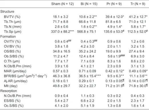
calcium and phosphorus between groups (Table 1). CCLD affected negatively bone microarchitecture; 2 months after BDL, bone material volume over tissue volume (BV/TV) and trabecular thickness were decreased, whereas trabecular separation was enhanced in the Bi group. On the other hand, pamidronate reversed all of these altera-tions and both groups subjected to preventive (Pr) and
secondary (Tr) pamidronate treatment showed significantly greater BV/TV and trabecular thickness and significantly
lower trabecular separation than the Sham and Bi groups
(Table 2). Bone formation parameters were not significantly
affected by cholestasis. However, pamidronate induced a reduction in osteoblast number (e.g., osteoblast surface/
bone surface: Sham = 11.2 ± 4.7; Bi = 12.4 ± 8.4; Pr = 5.6 ± 1.9; Tr = 6.9 ± 3.0%; P< 0.05, Bi vs Pr) and in osteoblast
activity (e.g., bone formation rate/bone surface: Sham =
46.3 ± 36.8; Bi = 36.5 ± 15.4; Pr = 9.5 ± 6.3; Tr = 11.1 ± 3.6
µm2·(µm3)-1·day-1; P< 0.05, Sham vs Pr, Sham vs Tr, Bi vs
Pr). The bone resorption in Bi was similar to that observed in Sham. However, pamidronate induced high suppression of the number of osteoblasts and of osteoblastic activity in the tibiae (Table 2).
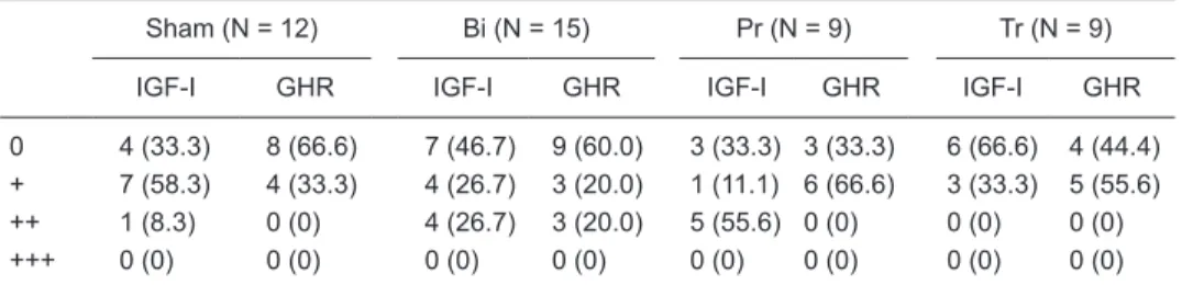
There were no significant differences in IGF-I or GHR expression in the tibial growth plate cartilage of the four
groups (Table 3).
Table 4 shows that the maximum force necessary to fracture bone was not significantly higher in the Sham
group compared to both the group with obstructed bile duct without treatment and to the group with obstructed bile duct receiving pamidronate as treatment. In the cholestasis group and in the cholestasis group receiving pamidronate treatment the lowest force necessary to induce fracture
Table 2. Histomorphometric evaluation of the proximal tibial metaphysis at 60 days after sham sur -gery (Sham), bile duct ligation (BDL) sur-gery (Bi), BDL sur-gery plus pamidronate prevention therapy (Pr,basal and 30 days after surgery), and BDL surgery plus pamidronate therapy (Tr)30 days after surgery.
Sham (N = 12) Bi (N = 15) Pr (N = 9) Tr (N = 9)
Structure
BV/TV (%) 18.1 ± 3.2 10.6 ± 2.2** 39.4 ± 12.0* 41.2 ± 12.7*
Tb.Th (µm) 71.7 ± 8.8 66.6 ± 11.8 81.8 ± 6.5 71.0 ± 12.1 Tb.N (/mm) 2.6 ± 0.6 1.6 ± 0.2** 4.9 ± 1.4* 5.8 ± 1.7* Tb.Sp (µm) 337.0 ± 88.2*** 566.8 ± 75.1 135.6 ± 53.0# 112.5 ± 52.0#
Formation
OV/TV (%) 0.6 ± 0.4## 0.4 ± 0.3## 0.9 ± 0.6 1.2 ± 0.6
OV/BV (%) 3.8 ± 1.8 4.2 ± 3.0 2.0 ± 1.1 3.2 ± 1.5
OS/BS (%) 34.8 ± 16.5 35.2 ± 24.2 19.0 ± 9.9 27.4 ± 9.4
Ob.S/BS (%) 11.2 ± 4.7 12.4 ± 8.4 5.6 ± 1.9### 6.9 ± 3.0
O.Th (µm) 7.7 ± 1.7 7.1 ± 0.9 8.3 ± 1.6 8.6 ± 2.0 N.Ob/B.Pm (/mm) 3.9 ± 1.6 4.1 ± 2.1 2.3 ± 0.9 3.1 ± 1.3 MAR (µm/day) 0.57 ± 0.3 0.71 ± 0.09 0.42 ± 0.12+ 0.49 ± 0.09+ BFR/BS (µm2·(µm3)-1·day-1) 46.3 ± 36.8 36.5 ± 15.4+++ 9.5 ± 6.3++ 11.1 ± 3.6++
Aj.AR (µm/day) 0.18 ± 0.1 0.29 ± 0.1 0.13 ± 0.05¶ 0.14 ± 0.07¶
Mlt (day) 49.8 ± 29.7 32.2 ± 22.7 71.2 ± 31.9¶ 71.8 ± 30.0¶
Resorption
N.Oc/B.Pm (/mm) 0.9 ± 0.4 1.1 ± 0.3 0.3 ± 0.2 0.4 ± 0.3
ES/BS (%) 5.4 ± 2.7 6.6 ± 2.2 2.0 ± 1.5 2.3 ± 1.7
Oc.S/BS (%) 4.1 ± 2.0 5.1 ± 1.9 1.3 ± 0.8 1.6 ± 1.4
Data are reported as means ± SEM. BV = bone volume; TV = tissue volume; Tb.th = trabecular thick -ness; Tb.N = trabecular number; Tb.sp = trabecular separation; OV = osteoid volume; OS = osteoid surface; BS = bone surface; Ob.S = osteoblast surface; O.Th = osteoid thickness; N.Ob = osteoblast number; B.Pm = bone perimeter; MAR = mineral apposition rate; BFR = bone formation rate; Aj.AR =
adjusted apposition rate; Mlt = mineralization lag time; N.Oc = osteoclast number; ES = eroded sur
-face; Oc.S = osteoclast surface. *P < 0.001, Pr and Tr compared to Sham; **P < 0.001, Bi compared to Pr and TR; ***P < 0.001, Sham compared to Bi, Pr and TR; #P < 0.001, Pr and TR compared to
Bi; ##P < 0.05, Sham and Bi compared to Tr; ###P < 0.05, Pr compared to Bi; +P < 0.05, Pr and Tr
compared to Bi; ++P < 0.05, Pr and Tr compared to Sham; +++P < 0.05, Bi compared to Pr; ¶P < 0.05,
was decreased to about the same level, 18.9 and 20.7%, respectively, compared to control. In
parallel, in the group that received pamidronate as a preventive scheme the force on the
momen-tum of fracture was even more reduced (29.0%, P < 0.05).
Discussion
The present study showed that pamidronate used for primary prevention therapy, administered immediately before and 1 month after surgery-induced cholestasis, enhances bone mass. How-ever, primary prevention therapy had no additional
advantage in comparison to conventional treatment (i.e., therapy initiation after established bone disease, 1 month after cholestasis). The improvement in bone microstructure of animals treated by primary prevention therapy was similar
to the benefit observed in the group in which pamidronate
treatment was started only after established bone disor-der. Additionally, the force required to fracture bone from animals treated with primary prevention was lower than that required to fracture bone from animals treated with secondary prevention therapy.
Bisphosphonates decrease fracture risk in large part by reducing the rate of bone remodeling and associated microarchitectural bone deterioration as well as by increas-ing bone mass. Bone remodelincreas-ing is the mechanism by which bone repairs microdamage and delivers calcium into the circulation (15,16). Bone formation rates estimated by tetracycline labeling are reduced in patients on bisphospho-nates. Most patients on bisphosphonates show reductions in remodeling to the range seen in healthy premenopausal
women (17). The primary end-point required for Food and
Drug Administration (FDA) approval of therapies for the
treatment of postmenopausal osteoporosis is significant
reduction in incident morphometric vertebral fractures over
3 years compared with placebo (18). The extensive experi
-ence accumulated in osteoporosis treatment involves post-menopausal women, showing severe densitometric bone loss or established osteoporosis, which means a previous history of fracture (19-21).
Currently, glucocorticoid-induced osteoporosis (GIO)
is the sole clinical condition for which the use of bispho-sphonates is advocated for primary prevention (6). This
approach is based on the pathophysiology of GIO, namely
the occurrence of two distinct phases of bone loss, the
first involving a fast bone remodeling rate and the second involving a slow process of bone loss. In GIO, the early
use of a potent antiresorptive drug prevents the increment of bone resorption activity and bone loss. Although some studies have suggested that osteoporosis in CCLD is due to a combination of decreased bone formation and acceler-ated bone resorption (22,23), most studies describe a more conspicuous impairment of bone formation (24,25). Studies on hepatic osteodystrophy in humans have indicated that
the most striking change in the profile of biochemical mark -ers of bone remodeling is the reduction of osteocalcin, i.e.,
reduction in the activity of bone formation (26,27). These
data are supported by results obtained in histomorphometric
evaluation in experimental models of CCLD (5) as well as in humans (28,29).
In a previous study on 2-month-old Wistar rats, we
Table 3. Insulin-like growth factor-I (IGF-I) and growth hormone receptor (GHR) expression in the proximal tibial metaphysis at 60 days after sham surgery (Sham), bile duct ligation (BDL) surgery (Bi),
BDL surgery plus pamidronate prevention therapy (Pr,basal and 30 days after surgery), and BDL surgery plus pamidronate therapy (Tr)30 days after surgery.
Sham (N = 12) Bi (N = 15) Pr (N = 9) Tr (N = 9)
IGF-I GHR IGF-I GHR IGF-I GHR IGF-I GHR
0 4 (33.3) 8 (66.6) 7 (46.7) 9 (60.0) 3 (33.3) 3 (33.3) 6 (66.6) 4 (44.4)
+ 7 (58.3) 4 (33.3) 4 (26.7) 3 (20.0) 1 (11.1) 6 (66.6) 3 (33.3) 5 (55.6)
++ 1 (8.3) 0 (0) 4 (26.7) 3 (20.0) 5 (55.6) 0 (0) 0 (0) 0 (0)
+++ 0 (0) 0 (0) 0 (0) 0 (0) 0 (0) 0 (0) 0 (0) 0 (0)
Data are reported as number of animals with percent in parentheses. (0) = no stained cells; (+) = fewer than 10% positive cells; (++) = 10-50% positive cells; (+++) = >50% positive cells.
Table 4. Biomechanical evaluation of the femur at 60 days after sham surgery (Sham), bile duct ligation (BDL) surgery (Bi), BDL surgery plus pamidronate prevention therapy (Pr, basal and 30 days after surgery), and BDL surgery plus pamidronate therapy (Tr) 30 days after surgery.
Sham (N = 12) Bi (N = 15) Pr (N = 9) Tr (N = 9)
Length (mm) 39.4 ± 3.1 37.5 ± 3.3 39.0 ± 1.4 37.5 ± 2.6 Ultimate load (N) 116.6 ± 23.0 94.6 ± 33.8 82.9 ± 22.8* 92.5 ± 29.5 Stiffness (N/mm) 129.0 ± 77.8 90.3 ± 52.4 67.2 ± 1.5 72.6 ± 7.6
Data are reported as means ± SEM. *P < 0.05, Pr compared to Sham
1260 F.A. Pereira et al.
have shown that four weeks after BDL surgery there are clear signs of osteoporosis determined by bone
histomor-phometry, expressed as decreased BV/TV and osteoblast
number (5). In the present study, the previous evidence was replicated with a more severe pattern after 2 months
of cholestasis. Additionally, we observed the efficiency of
pamidronate in reversing the process of bone loss. Animals
treated with pamidronate 1 month after BDL exhibited in -creased bone volume (BV/TV), as well as high trabecular thickness and trabecular number. On the other hand, his-tomorphometric data showed that there was no additional
benefit when pamidronate was used for primary prevention,
i.e., the improvement of bone structure was of the same magnitude as that observed when treatment was started only after established bone disease. Furthermore, the bone capacity to withstand mechanical stress was decreased in femur specimens from animals subjected to primary prevention treatment compared to those obtained from animals treated with the secondary regimen. Similar to our study, there are data showing that distinct bisphosphonates are able to prevent trabecular osteopenia in ovariectomy,
a classical experimental model of osteoporosis (13,30).
These studies observed improvement in the ultimate force for bone fracture only in lumbar spine bone and, similar to
our results, no effect was verified in long bones of animals
subjected to bisphosphonate treatment (13,30,31). Kippo et al. (31) suggested that in the rat the amount of cortical
bone and external bone dimensions would contribute more
to the structural strength of the femur than the amount and microarchitecture of trabecular bone. In addition to those studies, the present investigation showed that pamidronate used as preventive treatment may impair bone strength in rats submitted to surgically induced cholestasis. Femur from cholestatic rats submitted to preventive pamidronate treatment showed a decreased ultimate load for fracture and a trend to reduced stiffness. These results agree with previous data showing an inverse correlation between intraosseous concentration of pamidronate and ultimate load at failure and stiffness (32).
Other histomorphometric parameters indicate impair-ment of bone remodeling activity in the tibia from BDL rats subjected to preventive pamidronate therapy. Alteration in skeletal mineralization and decreased bone repairing capacity were demonstrated by an increased time required for mineralization and a concomitant decrement in the
num-ber of osteoclasts and on surface resorption area in BDL animals treated with pamidronate. Although the number of osteoblasts was not decreased in pamidronate-treated
animals, these animals exhibited a lower bone formation
rate. Taken together, these results indicate impaired turn-over and accumulation of microcracks due to suppressed osteoclast-mediated bone resorption.
No difference in IGF-I or GHR expression in growth
cartilage was detected between animals treated with both pamidronate regimens and vehicle. Most likely, this
occur-rence reflects the animals’ age since no diffeoccur-rence was also
observed between sham-operated animals and the other
three groups. In a previous study, we detected a significant decrease in IGF-I and GHR expression in 3-month-old
Wistar rats submitted to BDL surgery. It is well known that
the age-related decline in the expression of IGF-I is part of
the maturation process (33). Thus, the different results are
probably due to the distinct ages of the experimental rats. Liver-derived and autocrine/paracrine IGF-I are both crucial
components for bone mass development and maintenance
(34-36). In the present study, the serum levels of IGF-I were not measured, but liver-derived IGF-I is characteristically
decreased in cholestatic diseases.
Therefore, the present study shows that pamidronate is highly effective in inducing an apparent improvement in
bone microstructure in an experimental model of CCLD.
The data also show that early pharmacological treatment used as primary prevention does not represent a better ap-proach. Histomorphometric evaluation demonstrated that cholestatic animals submitted to a primary or secondary
treatment regimen exhibit the same bone volume. However,
bone strength tested by biomechanical assays showed a better capacity to resist mechanical stress in rats treated with a secondary regimen.
Acknowledgments
Rinaldo Mattar participated in the study design and made an important contribution to the development of this investigation. We thank Sebastião L. Brandão Filho, Sebastião Assis, Carlos Alberto Moro, Auristela de Melo Martins, Adalberto Vallada Verceze, Mauricio Rodrigues de Arantes, and Roni Charles Fabbris for technical help and
laboratory assistance. Research supported by FAPESP (#06/56890-0), CNPq, and FAEPA.
References
1. Lopez-Larramona G, Lucendo AJ, Gonzalez-Castillo S,
Tenias JM. Hepatic osteodystrophy: An important matter for consideration in chronic liver disease. World J Hepatol 2011;
3: 300-307.
2. Gasser RW. Cholestasis and metabolic bone disease - a clini -cal review. Wien Med Wochenschr 2008; 158: 553-557.
3. de Albuquerque Taveira AT, Fernandes MI, Galvao LC,
Sawamura R, de Mello V, de Paula FJ. Impairment of bone mass development in children with chronic cholestatic liver disease. Clin Endocrinol 2007; 66: 518-523.
of hepatic osteodystrophy in children and adolescents with chronic cholestatic liver disease. Braz J Med Biol Res 2010;
43: 1127-1134.
5. Pereira FA, Facincani I, Jorgetti V, Ramalho LN, Volpon JB,
Dos Reis LM, et al. Etiopathogenesis of hepatic osteodystro -phy in Wistar rats with cholestatic liver disease. Calcif Tissue Int 2009; 85: 75-83.
6. Grossman JM, Gordon R, Ranganath VK, Deal C, Caplan
L, Chen W, et al. American College of Rheumatology 2010 recommendations for the prevention and treatment of glu-cocorticoid-induced osteoporosis. Arthritis Care Res 2010; 62: 1515-1526.
7. Bachrach LK, Ward LM. Clinical review 1: Bisphosphonate
use in childhood osteoporosis. J Clin Endocrinol Metab
2009; 94: 400-409.
8. Thornton J, Ashcroft D, O’Neill T, Elliott R, Adams J, Rob -erts C, et al. A systematic review of the effectiveness of strategies for reducing fracture risk in children with juvenile idiopathic arthritis with additional data on long-term risk of fracture and cost of disease management. Health Technol Assess 2008; 12: iii-xiv, 1.
9. Rosen CJ. Building bones by knocking down genes. Nat Med 2012; 18: 202-204.
10. Horton JA, Bariteau JT, Loomis RM, Strauss JA, Damron TA. Ontogeny of skeletal maturation in the juvenile rat. Anat Rec
2008; 291: 283-292.
11. Turner AS. Animal models of osteoporosis - necessity and limitations. Eur Cell Mater 2001; 1: 66-81.
12. Peng Z, Tuukkanen J, Zhang H, Jamsa T, Vaananen HK. The mechanical strength of bone in different rat models of
experimental osteoporosis. Bone 1994; 15: 523-532. 13. Kippo K, Hannuniemi R, Isaksson P, Lauren L, Osterman
T, Peng Z, et al. Clodronate prevents osteopenia and loss
of trabecular connectivity in estrogen-deficient rats. J Bone Miner Res 1998; 13: 287-296.
14. Parfitt AM, Drezner MK, Glorieux FH, Kanis JA, Malluche H,
Meunier PJ, et al. Bone histomorphometry: standardization of nomenclature, symbols, and units. Report of the ASBMR Histomorphometry Nomenclature Committee. J Bone Miner Res 1987; 2: 595-610.
15. Burr DB. Targeted and nontargeted remodeling. Bone 2002; 30: 2-4.
16. Li J, Mashiba T, Burr DB. Bisphosphonate treatment sup-presses not only stochastic remodeling but also the targeted repair of microdamage. Calcif Tissue Int 2001; 69:
281-286.
17. Eriksen EF, Melsen F, Sod E, Barton I, Chines A. Effects of
long-term risedronate on bone quality and bone turnover in women with postmenopausal osteoporosis. Bone 2002; 31: 620-625.
18. Miller PD. Anti-resorptives in the management of osteopo -rosis. Best Pract Res Clin Endocrinol Metab 2008; 22:
849-868.
19. Black DM, Cummings SR, Karpf DB, Cauley JA, Thompson
DE, Nevitt MC, et al. Randomised trial of effect of alen
-dronate on risk of fracture in women with existing vertebral fractures. Fracture Intervention Trial Research Group. Lan-cet 1996; 348: 1535-1541.
20. Reginster J, Minne HW, Sorensen OH, Hooper M, Roux
C, Brandi ML, et al. Randomized trial of the effects of rise-dronate on vertebral fractures in women with established
postmenopausal osteoporosis. Vertebral Efficacy with Rise
-dronate Therapy (VERT) Study Group. Osteoporos Int 2000;
11: 83-91.
21. Black DM, Delmas PD, Eastell R, Reid IR, Boonen S, Cau -ley JA, et al. Once-yearly zoledronic acid for treatment of postmenopausal osteoporosis. N Engl J Med 2007; 356:
1809-1822.
22. Crawford BA, Kam C, Donaghy AJ, McCaughan GW. The
heterogeneity of bone disease in cirrhosis: a multivariate analysis. Osteoporos Int 2003; 14: 987-994.
23. Monegal A, Navasa M, Guanabens N, Peris P, Pons F,
Martinez de Osaba MJ, et al. Osteoporosis and bone min-eral metabolism disorders in cirrhotic patients referred for orthotopic liver transplantation. Calcif Tissue Int 1997; 60:
148-154.
24. Capra F, Casaril M, Gabrielli GB, Stanzial A, Ferrari S, Gan
-dini G, et al. Plasma osteocalcin levels in liver cirrhosis. Ital J Gastroenterol 1991; 23: 124-127.
25. Guanabens N, Pares A, Marinoso L, Brancos MA, Piera C, Serrano S, et al. Factors influencing the development of
metabolic bone disease in primary biliary cirrhosis. Am J Gastroenterol 1990; 85: 1356-1362.
26. Chen CC, Wang SS, Jeng FS, Lee SD. Metabolic bone disease of liver cirrhosis: is it parallel to the clinical severity of cirrhosis? J Gastroenterol Hepatol 1996; 11: 417-421.
27. Goral V, Simsek M, Mete N. Hepatic osteodystrophy and
liver cirrhosis. World J Gastroenterol 2010; 16: 1639-1643.
28. Jorge-Hernandez JA, Gonzalez-Reimers CE, Torres-Ramir
-ez A, Santolaria-Fernand-ez F, Gonzal-ez-Garcia C,
Batista-Lopez JN, et al. Bone changes in alcoholic liver cirrhosis. A histomorphometrical analysis of 52 cases. Dig Dis Sci 1988;
33: 1089-1095.
29. Chappard D, Plantard B, Fraisse H, Palle S, Alexandre C, Riffat G. Bone changes in alcoholic cirrhosis of the liver. A
histomorphometric study. Pathol Res Pract 1989; 184:
480-485.
30. Toolan BC, Shea M, Myers ER, Borchers RE, Seedor JG, Quartuccio H, et al. Effects of 4-amino-1-hydroxybutylidene
bisphosphonate on bone biomechanics in rats. J Bone Miner Res 1992; 7: 1399-1406.
31. Kippo K, Hannuniemi R, Lauren L, Peng Z, Isaksson P, Virtamo T, et al. Clodronate prevents bone loss in aged ovariectomized rats. Calcif Tissue Int 1997; 61: 151-157.
32. Yang KH, Won JH, Yoon HK, Ryu JH, Choo KS, Kim JS. High concentrations of pamidronate in bone weaken the mechani-cal properties of intact femora in a rat model. Yonsei Med J
2007; 48: 653-658.
33. Okuda S, Myoui A, Ariga K, Nakase T, Yonenobu K, Yo-shikawa H. Mechanisms of age-related decline in insulin-like growth factor-I dependent proteoglycan synthesis in rat intervertebral disc cells. Spine 2001; 26: 2421-2426.
34. Courtland HW, Elis S, Wu Y, Sun H, Rosen CJ, Jepsen KJ, et al. Serum IGF-1 affects skeletal acquisition in a temporal and compartment-specific manner. PLoS One 2011; 6: e14762.
35. Elis S, Courtland HW, Wu Y, Rosen CJ, Sun H, Jepsen KJ, et al. Elevated serum levels of IGF-1 are sufficient to
establish normal body size and skeletal properties even in
the absence of tissue IGF-1. J Bone Miner Res 2010; 25:
1257-1266.
36. Olson LE, Ohlsson C, Mohan S. The role of GH/IGF-I-mediated mechanisms in sex differences in cortical bone