ISSN 1414-431X
www.bjournal.com.br
www.bjournal.com.br
Volume 45 (12) 1102-1340 December 2012
Braz J Med Biol Res, December 2012, Volume 45(12) 1150-1156
10.1590/S0100-879X2012007500159
doi:
Effect of hepatocyte growth factor and angiotensin II on rat
cardiomyocyte hypertrophy
Ai-Lan Chen, Cai-Wen Ou, Zhao-Chu He, Qi-Cai Liu, Qi Dong and Min-Sheng Chen
Institutional Sponsors
The Brazilian Journal of Medical and Biological Research is partially financed by
Faculdade de Medicina de Ribeirão Preto Campus
Ribeirão Preto
Explore High - Performance MS Orbitrap Technology In Proteomics & Metabolomics
analiticaweb.com.br S C I E N T I F I C
BIOMEDICAL SCIENCES
AND
Effect of hepatocyte growth factor and
angiotensin II on rat cardiomyocyte
hypertrophy
Ai-Lan Chen
1, Cai-Wen Ou
2, Zhao-Chu He
1, Qi-Cai Liu
3, Qi Dong
4and
Min-Sheng Chen
51Department of Cardiology, The First Affiliated Hospital of Guangzhou Medical University, Guangzhou, China
2TheFourth Affiliated Hospital of Guangzhou Medical University, Guangzhou, China
3Experimental Medical Research Center, Guangzhou Medical University, Guangzhou, China
4Department of Physiology, Guangzhou Medical University, Guangzhou, China
5Guangzhou Key Laboratory of Cardiovascular Disease, Guangzhou Institute of Cardiovascular Disease,
The Second Affiliated Hospital of Guangzhou Medical University, Guangzhou, China
Abstract
Angiotensin II (Ang II) plays an important role in cardiomyocyte hypertrophy. The combined effect of hepatocyte growth factor
(HGF) and Ang II on cardiomyocytes is unknown. The present study was designed to determine the effect of HGF on cardiomyo
-cyte hypertrophy and to explore the combined effect of HGF and Ang II on cardiomyo-cyte hypertrophy. Primary cardiomyo-cytes
were isolated from neonatal rat hearts and cultured in vitro. Cells were treated with Ang II (1 µM) alone, HGF (10 ng/mL) alone,
and Ang II (1 µM) plus HGF (10 ng/mL) for 24, 48, and 72 h. The amount of [3H]-leucine incorporation was then measured to
evaluate protein synthesis. The mRNA levels of β-myosin heavy chain and atrial natriuretic factor were determined by real-time PCR to evaluate the presence of fetal phenotypes of gene expression. The cell size of cardiomyocytes was also studied. Ang
II (1 µM) increased cardiomyocyte hypertrophy. Similar to Ang II, treatment with 1 µM HGF promoted cardiomyocyte hyper
-trophy. Moreover, the combination of 1 µM Ang II and 10 ng/mL HGF clearly induced a combined pro-hypertrophy effect on cardiomyocytes. The present study demonstrates for the first time a novel, combined effect of HGF and Ang II in promoting
cardiomyocyte hypertrophy.
Key words: Angiotensin II; Hepatocyte growth factor; Cardiomyocyte; Hypertrophy
Introduction
Correspondence: Min-Sheng Chen, Guangzhou Key Laboratory of Cardiovascular Disease, Guangzhou Institute of Cardiovascular Disease, The Second Affiliated Hospital of Guangzhou Medical University, Guangzhou 510260, China. Fax: +86-20-813-40448. E-mail: [email protected] and/or [email protected]
Received February 27, 2012. Accepted August 5, 2012. Available online October 15, 2012. Published December 17, 2012.
Myocardial hypertrophy is defined as a thickening of the myocardium, which results in a decrease in the size of the chamber of the heart, including the left and right ventricles, and is an important risk factor for subsequent cardiac morbidity and mortality. The pathophysiology and role of cardiomyocyte hypertrophy in heart disease have been intensively investigated; however, to date they are only partially understood. Cardiac hypertrophic growth is the primary responsive mechanism by which the heart reduces stress on the ventricular wall. Conventional views suggest that cardiomyocyte hypertrophy is a compensatory response to increased hemodynamic overload, which leads to cardiac disease. However, recent findings in genetic animal models of myocardial hypertrophy as well as human studies have
revealed support for a molecular basis whereby either compensatory or maladaptive forms of hypertrophy exist, and only the latter lead to cardiac failure (1). In the case of myocardial infarction, hypertrophic responses occur in cardiomyocytes in the surviving portion of the ventricle, and ventricular dilatation follows as a result of re-organization of the myocardium, including cardiomyocytes and mesen-chymal cells in combination (2,3). To execute this response, the cardiomyocytes are stimulated by neurohumoral factors and subsequent intracellular reactive cascade systems. All of these processes entail an increase in protein synthesis as well as the size and architectural rearrangement within individual cardiomyocytes.
renin-The combined effects of HGF and Ang II on rat cardiomyocytes 1151
angiotensin system, is believed to be one of the most important regulators of the initiation of a positive feedback regulation of the cardiac hypertrophic response (4). In cul-tured cardiomyocytes, Ang II was shown to directly induce the gene expression of β-myosin heavy chain (β-MHC) and atrial natriuretic factor (ANF) (4) and also increase protein synthesis and protein content (5) through the activation of angiotensin receptor I. More detailed mechanisms have been described in recent years, including involvement with mitochondrial oxidative stress (6), autophagy (7,8), p70-S6 protein kinase (9), nitric oxide (10), focal adhesion kinase (11), and AMP-activated protein kinase (12,13). In fact, in addition to Ang II, large numbers of intracellular or extracel -lular factors affectcardiomyocyte hypertrophy in vivo (14). Moreover, the influence of these factors on Ang II-induced cardiomyocyte hypertrophy is largely unknown.
In the present study, we hypothesized that there may be crosstalk between Ang II and hepatocyte growth factor (HGF) in cardiomyocyte hypertrophy. HGF is a paracrine cellular growth, motility, and morphogenic factor. It was initially isolated from fibroblasts and was shown to stimu -late the motility of epithelial cells and to have a wide range of effects on many biological processes (15). Specifically, HGF acts primarily on epithelial cells, endothelial cells, and hematopoietic progenitor cells (16). It has been shown to have a major role in embryonic organ development, adult organ regeneration, and wound healing (16). In recent years, the impact of HGF on the heart or cardiomyocytes has been gradually uncovered. HGF protects the heart from ischemic/reperfusion injury, attenuates cardiac remodeling, improves angiogenesis, and induces endothelial progenitor cell mobilization (16). In our study, we first determined the effects of a single treatment of Ang II or HGF on cardiomyo -cytes and then examined whether the combined treatment of Ang II and HGF differed from each factor administered individually.
Material and Methods
Animals and reagents
Newborn Sprague-Dawley rats (18-20 g) were supplied by the Animal Center of Guangzhou Medical University. Rats were housed and used in accordance with our institutional guidelines for animal care and the Guide for Animal Care of the National Institutes of Health. Dulbecco’s modified Eagle’s medium (DMEM), fetal bovine serum (FBS), and TRIzol were purchased from Gibco BRL (USA). Trypsin was obtained from Amersco (USA). Alpha-sarcomeric actin was purchased from Santa Cruz Biotechnology (USA). The SYBR®GreenPCR Master Mix was obtained from Applied
Biosystems (USA). The Thermo ScriptRT-PCR kit was purchased from Invitrogen (USA). [3H]-leucine (Leu) was
obtained from the China Institute of Atomic Energy (China) and recombinant Ang II and HGF were purchased from Sigma (Germany).
Primary cardiomyocyte culture
Rat neonatal ventricular cardiomyocytes were prepared as previously described, with some modifications (17). The cells were suspended in DMEM containing 10% FBS and 0.1 mM bromodeoxyuridine and plated on either glass coverslips or polystyrene-treated Petri dishes. The cells were cultured at 37°C in a 5% CO2 incubator for 72 h. Pulsations of the
cardiomyocytes were observed with an inverted biological microscope (18). In addition, the cardiomyocytes were fixed and stained by immunohistochemical methods using an anti-α-sarcomeric actin antibody (19). The purity of the cardiomyocytes was approximately 95%.
Groups and treatments
The cardiomyocyte medium was changed to serum-free DMEM for 24 h. The cardiomyocytes were then treated with different concentrations of Ang II (0.01-10 µM) and HGF (1, 10, 100 ng/mL) for 48 h in order to explore the most appropriate concentrations of each factor.
Based on these experiments, we identified an optimal treatment regimen. Cardiomyocytes were divided into four groups: Control group (0.1 M PBS), HGF-treated group (10 ng/mL), Ang II-treated group (1 µM), and HGF (10 ng/mL) plus Ang II (1 µM)-treated group. The cells were treated with these agents for 24, 48, and 72 h.
[3H]-Leu incorporation assay
Tritiated leucine ([3H]-Leu) incorporation studies were
performed as previously described (20) to investigate the effects of the treatments on total protein synthesis. Cells were seeded on 24-well plates (1 x 10⁵ cells/well) and treated with HGF (10 ng/mL) or Ang II (1 µM) or HGF (10 ng/mL) plus Ang II (1 µM), as described in the preceding section. Cells were then pulsed with [3H]-Leu (104 Bq/mL)
for 12 h before the end of the treatment. At the end of the incubation, the cells were washed three times with ice-cold PBS and then disrupted by the addition of 200 µL 0.1% so -dium dodecyl sulfate and 0.1 N NaOH. The solubilized cell lysates (100 µL) were added to 5 mL scintillation fluid. The incorporation of [3H]-Leu into the protein was determined
by scintillation counting. Incorporation is reported as the ratio: [3H]-Leu incorporation/number of cells.
RNA extraction and real-time PCR analysis
(two-step method) (24). Finally, a melting curve analysis was performed from 60° to 85°C. Data were evaluated with the Applied Biosystems software. The following prim-ers designed with the Primer Express Software (Applied Biosystems) were used: ANF: 5’-AAA GCA AAC TGA GGG CT-3’ (sense) and 5’-GGG ATC TTT TGC GAT CT-3’ (an -tisense); β-MHC: 5’-TGC AGT TAA AGG TGA AGG C-3’ (sense) and 5’-CAG GGC TTC ACA GGC AT-3’ (antisense); glyceraldehyde 3-phosphate dehydrogenase (GAPDH): 5’-CGG AGT CAA 5’-CGG ATT TGG TGG TAT-3’ (sense) and 5’-AGC CTT CTC CAT GGT GGT GAA GAC-3’ (antisense). GAPDH was used as an internal control and the amount of target was defined by the 2∆∆Ct method (25).
Measurement of cell size
Cardiomyocytes were seeded on 24-well plates and cultured for 48 h. After treatment with HGF (10 ng/mL), Ang II (1 µM), or HGF (10 ng/mL) plus Ang II (1 µM) for 12 h, the cells were digested with EDTA-trypsin (0.25%) for 5 min, 10% FBS was used to terminate the reaction and the average cell size was determined using the Image J soft -ware (http://rsb.info.nih.gov/ij/download.html) as previously described (26). The average cell size was determined by observation of 150 cells (50 cells/well x 3 wells).
Statistical analysis
Data are reported as means ± SD. The differences were evaluated by a two-tailed Student t-test (2 groups) or one-way analysis of variance (ANOVA) followed by the Tukey post hoc test (3 or more groups) using the SPSS software (version 9.0, USA) (27). Statistical significance was set at P < 0.05.
Results
Effects of Ang II on [3H]-Leu incorporation and on ANF
and β-MHC mRNA levels in primary cardiomyocytes
As shown in Table 1, Ang II (0.01 µM) caused a slight but significant increase in [3H]-Leu incorporation compared
to control (1494.58 ± 65.00 vs 1188.83 ± 72.20 cpm/well,
respectively; P < 0.05). However, at this concentration of
Ang II, the mRNA levels of ANF and β-MHC in primary cardiomyocytes were not changed.
When Ang II was administered at a higher concentra-tion (0.1 µM), the [3H]-Leu incorporation increased to 1820
cpm/well, which was approximately 1.5-fold higher than the control group (P < 0.01). In addition, this concentration of Ang II also significantly enhanced the mRNA levels of ANF (1.2-fold, P < 0.01) and β-MHC (1.75-fold, P < 0.01).
When the Ang II concentration was increased to 1 µM, the amount of [3H]-Leu incorporation increased
approxi-mately 2-fold compared to the control group (2374.08 ± 49.34 vs 1188.83 ± 72.20 cpm/well, respectively; P < 0.01),
while the mRNA levels of ANF and β-MHC increased by 44% (P < 0.01) and 85% (P < 0.01), respectively.
The highest concentration of Ang II tested (10 µM) in-creased the [3H]-Leu incorporation only slightly (2414.67 ±
56.70 vs 1188.83 ± 72.20 cpm/well, respectively; P < 0.01).
Moreover, at this concentration of Ang II, the mRNA levels of ANF and β-MHC only increased by 1.65-fold and 2.08-fold, respectively. Therefore, we chose 1 µM as the optimal concentration of Ang II for subsequent experiments.
Effects of HGF on [3H]-Leu incorporation and on ANF
and β-MHC mRNA levels in primary cardiomyocytes
We sought to determine the effects of different con-centrations of HGF on [3H]-Leu incorporation as well as
on mRNA levels of ANF and β-MHC in primary cardio -myocytes. As shown in Table 2, administration of 1 ng/mL HGF significantly increased the [3H]-Leu incorporation in
cardiomyocytes to 2430.47 ± 72.65 cpm/well, which was approximately 2.2-fold higher than the control group (P < 0.01). Moreover, HGF at this concentration also significantly (P < 0.01) enhanced the mRNA levels of ANF (2.33-fold) and β-MHC (2.14-fold).
When the HGF concentration was increased to 10 ng/mL, the [3H]-Leu incorporation was higher compared
to control (3848 ± 71.57 vs 1113.45 ± 62.58 cpm/well,
respectively; P < 0.01). Moreover, the ANF and β-MHC mRNA levels were also increased (3.32- and 2.81-fold, respectively; P < 0.01). Interestingly, the highest concentra -tion of HGF (100 ng/mL) did not further increase [3H]-Leu
Table 1. Effects of different concentrations of Ang II on [3H]-Leu incorporation, and mRNA levels of
ANF and β-MHC in primary cardiomyocytes.
Ang II (µM) [3H]-Leu incorporation (cpm/well) Relative ANF mRNA Relative β-MHC mRNA
Control 1188.83 ± 72.20 1 ± 0 1 ± 0
0.01 1494.58 ± 65.00* 1.07 ± 0.09 1.17 ± 0.06
0.1 1821.33 ± 54.65* 1.20 ± 0.02* 1.75 ± 0.03*
1 2374.08 ± 49.34* 1.44 ± 0.08* 1.85 ± 0.13*
10 2414.67 ± 56.70* 1.65 ± 0.04* 2.08 ± 0.14*
Data reported as means ± SD for N = 6/concentration performed in triplicate. Ang II = angiotensin II; ANF
The combined effects of HGF and Ang II on rat cardiomyocytes 1153
incorporation or ANF and β-MHC mRNA levels compared to the lower concentration of HGF (10 ng/mL). Therefore, we chose 10 ng/mL as the optimal concentration of HGF in subsequent experiments.
Effects of HGF in combination with Ang II on [3H]-Leu
incorporation and on ANF and β-MHC mRNA levels
in primary cardiomyocytes
We next investigated whether HGF and Ang II had combined effects on cardiomyocytes. Ang II and HGF in combination induced a significant increase in [3H]-Leu
incorporation and in ANF mRNA and β-MHC mRNA levels (Figure 1A-C). Interestingly, the HGF-induced cardiomyo -cyte hypertrophy was even greater than Ang II-induced hypertrophy (Figure 1A-C).
Compared to the HGF (10 ng/mL) or Ang II (1 µM) groups, the combined treatment of HGF (10 ng/mL) and Ang II (1 µM) increased [3H]-Leu incorporation even further at
24, 48, and 72 h (Figure 1A; P < 0.01). Similarly, combined treatment of cardiomyocytes with HGF (10 ng/mL) and Ang II (1 µM) significantly upregulated the mRNA levels of ANF (Figure 1B) and β-MHC (Figure 1C) compared to separate treatment with HGF (10 ng/mL) or Ang II (1 µM) alone at these time points.
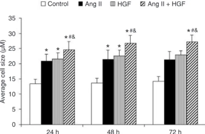
Effect of HGF and Ang II on the cell size of primary cardiomyocytes
We also determined the effect of HGF and Ang II on cardiomyocyte cell size. Compared to the control group, both the HGF (10 ng/mL)-treated group and Ang II (1 µM)-treated group showed an increase in cell size (Figure 2). The combination treatment of HGF and Ang II increased the cell size of cardiomyocytes further (Figure 2).
Discussion
This study was the first to directly assess whether there was a combined effect of HGF and Ang II on cardiomyocyte hypertrophy. Based on the increase in [3H]-Leu incorporation, the increased ANF and β-MHC
mRNA expression and the increase in cell size in pri-mary cardiomyocytes, we concluded that HGF and Ang II have a combined effect on neonatal cardiomyocyte
Figure 1. Effects of Ang II (1 µM), HGF (10 ng/mL), and HGF
(10 ng/mL) plus Ang II (1 µM) on [3H]-Leu incorporation (A), the
mRNA levels of ANF (B) and the mRNA levels of β-MHC (C) in
primary cardiomyocytes. N = 8 per group. The experiments were repeated three times. HGF = hepatology growth factor; Ang II = angiotensin II; ANF = atrial natriuretic factor; β-MHC = β-myosin
heavy chain. *P < 0.05 vs control; #P < 0.05 vs HGF; &P < 0.05 vs
Ang II (one-way ANOVA).
[
H]-Leu incorporation
3
6000
5000
4000
3000
2000
1000
0
*
*
#&*
#&
*
*
*
*
#&*
*
24 h 48 h 72 h
Relative ß-MHC mRNA
6
5
4
3
2
1
0
*
*
#&*
*
*
*
#&*
*
24 h 48 h 72 h
*
#&Control Ang II HGF Ang II + HGF
Relative ANF mRNA
(fold)
6
5
4
3
2
1
0
*
*
#&*
*
*
*
#&*
*
24 h 48 h 72 h
*
#&A
B
C
Table 2. Effects of different concentrations of HGF on [3H]-Leu incorporation, and mRNA levels of
ANF and β-MHC in primary cardiomyocytes.
HGF (ng/mL) [3H]-Leu incorporation (cpm/well) Relative ANF mRNA Relative β-MHC mRNA
Control 1113.45 ± 62.58 1 ± 0 1 ± 0
1 2430.47 ± 72.65* 2.33 ± 0.04* 2.14 ± 0.03*
10 3848 ± 71.57* 3.32 ± 0.08* 2.81 ± 0.07*
100 3256 ± 68.34* 3.01 ± 0.01* 2.42 ± 0.06*
Data are reported as means ± SD for N = 6/concentration performed in triplicate. HGF = hepatology
growth factor; ANF = atrial natriuretic factor; β-MHC = β-myosin heavy chain. *P < 0.05 vs control
hypertrophy. In addition, we also demonstrated for the first time that HGF alone can potently induce cultured neonatal cardiomyocyte hypertrophy.
In this study, we first found that HGF could induce car -diomyocyte hypertrophy. To our surprise, the HGF-induced cardiomyocyte hypertrophy was even greater than Ang II-induced hypertrophy (Figure 1A-C). The presence of the HGF receptor on cardiomyocytes was first confirmed by Akiyama et al. (28). Subsequently, several independent reports have demonstrated a protective effect of HGF on myocardial infarction in vivo (29,30). However, as yet
the underlying mechanisms for the pro-hypertrophy of cardiomyocytes are not clear. Moreover, the results of
in vivo experiments cannot exclude the possibility that
HGF treatment stimulates cardiac fibroblasts (31) and thereby releases factors that induce hypertrophy of the surrounding cardiomyocytes. Pathological cardiomyocyte hypertrophy is characterized not only by an increase in cardiomyocyte size, but also by cardiomyocyte gene reprogramming, as shown by the enhanced expression of fetal phenotypes of genes, such as skeletal α-actin, β-MHC, and ANF (32). In our study, we evaluated cell hypertrophy by determining [3H]-Leu incorporation and
the average cell size as well as ANF and β-MHC mRNA levels. Based on these various parameters, we have clearly shown that HGF can directly induce cardiomyo -cyte hypertrophy. It should be noted that Nakamura et al. (33) demonstrated an interference of administered HGF on cardiomyopathy remodeling in hamster hearts. The authors found a direct negative effect of HGF on the transcript levels of ANF. In the present study, we showed an inductive effect of HGF on ANF levels, in agreement with a previous report (34). We considered that this dis -parity might be due to the species difference.
Based on these findings, we sought to determine how this pro-hypertrophy effect of HGF may occur. Since the most common cause of cardiomyocyte hypertrophy is hypertension, and since an increased Ang II level in hypertension is a critical pro-hypertrophy trigger, we next explored whether there might be an association between HGF and Ang II in cardiomyocytes. Importantly, we found a combined effect of HGF and Ang II on cardiomyocyte hypertrophy. The roles of Ang II in the regulation of the cardiovascular system under normal and pathological conditions have been well documented, and it has been well accepted that Ang II is a direct pro-hypertrophy factor in cardiomyocytes. Moreover, there is evidence that Ang II and related proteins such as interleukin-1β and tumor necrosis factor-α, play key roles in cardiac fibroblast growth and collagen deposition following myocardial infarction (35). In addition, heparin, a highly sulfated glycosaminoglycan and widely used injectable antico -agulant, was found to potently inhibit Ang II-mediated cardiomyocyte hypertrophy (36). The interleukin-6 family of cytokines was also reported to contribute to Ang
II-induced cardiomyocyte hypertrophy (37). However, to the best of our knowledge, there has been no report to date on the combined effect of Ang II and any other protein on cardiomyocyte hypertrophy. More importantly, since cardiomyocyte hypertrophy is related to the pathophysi-ological mechanisms of many cardiovascular diseases, our findings in this study may bring new focus on HGF in treating cardiovascular diseases.
One limitation of this study is that the molecular mechanisms underlying the combined effect of Ang II and HGF on cardiomyocyte hypertrophy remain to be elucidated. It has been reported that post-infarction HGF gene therapy resulted in substantial cardiomyocte hyper -trophy at the edges of the infarcted tissue, accompanied by the overexpression of the HGF receptor (c-Met), which is a transmembrane tyrosine kinase through which HGF activates the Ras-Raf-MEK-ERK signaling pathway, thereby contributing to myocardial hypertrophy (38). The c-Met is also a receptor for the scatter factor (SF) (39). The activation of c-Met by HGF/SF can elicit a variety of cellular responses including proliferation, migration, invasion, and branching morphogenesis. These pro -cesses are associated with several signaling pathways such as PI3K/Akt, Src, and STAT3 (39). Moreover, these signaling pathways also contribute to Ang II-induced hypertrophy. Aoki et al. (40) have shown that the ERK pathway has an important role in Ang II-induced cardiac hypertrophy. Moreover, the PI3K/Akt, Src, and STAT3 pathways have also been reported to be vital controllers in Ang II-induced cardiac hypertrophy. Therefore, it may be difficult to determine the exact underlying molecular mechanisms of the combined effect of HGF and Ang II on cardiomyocyte hypertrophy. The convergence point between HGF and Ang II needs to be further elucidated in future experiments.
Figure 2. Effects of Ang II (1 µM), HGF (10 ng/mL), and HGF (10
ng/mL) plus Ang II (1 µM) on average cell size. N = 8 per group. The experiments were repeated three times. HGF = hepatology
growth factor; Ang II = angiotensin II. *P < 0.05 vs control; #P <
0.05 vs HGF; &P < 0.05 vs Ang II (one-way ANOVA).
A
verage cell size (µM)
35
30
25
20
15
10
5
0
24 h
* *
*
#&*
#&* *
*
#&48 h 72 h
The combined effects of HGF and Ang II on rat cardiomyocytes 1155
Our study has provided the first evidence that HGF and Ang II administered in combination induce cardiomyocyte hypertrophy. This finding was supported by the changes ob -served in protein synthesis, the presence of fetal phenotypes of gene expression (ANF and β-MHC), an increase in cell size, and ultrastructural changes of neonatal cardiomyo-cytes. These results may help to improve the management and treatment of patients with cardiac hypertrophy.
Acknowledgments
Research supported by the National Natural Science Foundation of China (Grant #30570759), the Natural Science Foundation of Guangdong Province, China (#S201101004269) and the Foundation of Guangzhou Municipal Health Bureau Scientific Research and Education Managment System (#201102A213125).
References
1. Lips DJ, deWindt LJ, van Kraaij DJ, Doevendans PA. Mo
-lecular determinants of myocardial hypertrophy and failure:
alternative pathways for beneficial and maladaptive hyper
-trophy. Eur Heart J 2003; 24: 883-896.
2. Li Q, Li B, Wang X, Leri A, Jana KP, Liu Y, et al. Overex
-pression of insulin-like growth factor-1 in mice protects from
myocyte death after infarction, attenuating ventricular dila
-tion, wall stress, and cardiac hypertrophy. J Clin Invest 1997; 100: 1991-1999.
3. Orlic D, Kajstura J, Chimenti S, Jakoniuk I, Anderson SM,
Li B, et al. Bone marrow cells regenerate infarcted myocar-dium. Nature 2001; 410: 701-705.
4. Sadoshima J, Izumo S. Molecular characterization of an-giotensin II-induced hypertrophy of cardiac myocytes and
hyperplasia of cardiac fibroblasts. Critical role of the AT1
receptor subtype. Circ Res 1993; 73: 413-423.
5. Wada H, Zile MR, Ivester CT, Cooper G, McDermott PJ. Comparative effects of contraction and angiotensin II on
growth of adult feline cardiocytes in primary culture. Am J Physiol 1996; 271: H29-H37.
6. Dai DF, Rabinovitch P. Mitochondrial oxidative stress medi
-ates induction of autophagy and hypertrophy in angiotensin-II treated mouse hearts. Autophagy 2011; 7: 917-918. 7. Gottlieb RA, Mentzer RM Jr, Linton PJ. Impaired mitophagy
at the heart of injury. Autophagy 2011; 7: 1573-1574.
8. Zois CE, Giatromanolaki A, Sivridis E, Papaiakovou M, Kainulainen H, Koukourakis MI. “Autophagic flux” in normal
mouse tissues: focus on endogenous LC3A processing.
Autophagy 2011; 7: 1371-1378.
9. Jonassen AK, Sack MN, Mjos OD, Yellon DM. Myocardial
protection by insulin at reperfusion requires early administra
-tion and is mediated via Akt and p70s6 kinase cell-survival
signaling. Circ Res 2001; 89: 1191-1198.
10. Landmesser U, Engberding N, Bahlmann FH, Schaefer A, Wiencke A, Heineke A, et al. Statin-induced improvement
of endothelial progenitor cell mobilization, myocardial neo
-vascularization, left ventricular function, and survival after experimental myocardial infarction requires endothelial nitric
oxide synthase. Circulation 2004; 110: 1933-1939. 11. Taylor JM, Rovin JD, Parsons JT. A role for focal adhesion
kinase in phenylephrine-induced hypertrophy of rat ventricu
-lar cardiomyocytes. J Biol Chem 2000; 275: 19250-19257. 12. Timmers L, Sluijter JP, Verlaan CW, Steendijk P, Cramer
MJ, Emons M, et al. Cyclooxygenase-2 inhibition increases mortality, enhances left ventricular remodeling, and impairs
systolic function after myocardial infarction in the pig. Circu-lation 2007; 115: 326-332.
13. Xie Z, He C, Zou MH. AMP-activated protein kinase modu
-lates cardiac autophagy in diabetic cardiomyopathy.
Au-tophagy 2011; 7: 1254-1255.
14. Rohini A, Agrawal N, Koyani CN, Singh R. Molecular targets
and regulators of cardiac hypertrophy. Pharmacol Res 2010; 61: 269-280.
15. Funakoshi H, Nakamura T. Hepatocyte growth factor: from
diagnosis to clinical applications. Clin Chim Acta 2003; 327:
1-23.
16. Morishita R, Aoki M, Yo Y, Ogihara T. Hepatocyte growth
factor as cardiovascular hormone: role of HGF in the patho
-genesis of cardiovascular disease. Endocr J 2002; 49:
273-284.
17. Simpson P, McGrath A, Savion S. Myocyte hypertrophy in neonatal rat heart cultures and its regulation by serum and
by catecholamines. Circ Res 1982; 51: 787-801.
18. Mellor KM, Reichelt ME, Delbridge LM. Autophagy anoma
-lies in the diabetic myocardium. Autophagy 2011; 7:
1263-1267.
19. Przyklenk K, Undyala VV, Wider J, Sala-Mercado JA, Got
-tlieb RA, Mentzer RM Jr. Acute induction of autophagy as a novel strategy for cardioprotection: getting to the heart of
the matter. Autophagy 2011; 7: 432-433.
20. Huang Z, Li J, Jiang Z, Qi Y, Tang C, Du J. Effects of ad-renomedullin, C-type natriuretic peptide, and parathyroid
hormone-related peptide on calcification in cultured rat vas
-cular smooth muscle cells. J Cardiovasc Pharmacol 2003; 42: 89-97.
21. Dong LW, Hou YJ, Tan YX, Tang L, Pan YF, Wang M, et al.
Prognostic significance of Beclin 1 in intrahepatic cholangio
-cellular carcinoma. Autophagy 2011; 7: 1222-1229. 22. Cottam EM, Maier HJ, Manifava M, Vaux LC,
Chandra-Schoenfelder P, Gerner W, et al. Coronavirus nsp6 proteins
generate autophagosomes from the endoplasmic reticulum
via an omegasome intermediate. Autophagy 2011; 7:
1335-1347.
23. Wang P, Xu TY, Guan YF, Su DF, Fan GR, Miao CY. Perivas
-cular adipose tissue-derived visfatin is a vas-cular smooth
muscle cell growth factor: role of nicotinamide mononucle-otide. Cardiovasc Res 2009; 81: 370-380.
24. Wang P, Zhang RY, Song J, Guan YF, Xu TY, Du H, et al. Loss of AMP-activated protein kinase-alpha2 impairs the insulin-sensitizing effect of calorie restriction in skeletal
muscle. Diabetes 2012; 61: 1051-1061.
25. Yang J, Zhao Y, Ma K, Jiang FJ, Liao W, Zhang P, et al. Deficiency of hepatocystin induces autophagy through an
mTOR-dependent pathway. Autophagy 2011; 7: 748-759. 26. Wang P, Xu TY, Guan YF, Tian WW, Viollet B, Rui YC, et al.
ischemic stroke through SIRT1-dependent adenosine mono
-phosphate-activated kinase pathway. Ann Neurol 2011; 69:
360-374.
27. Trocoli A, Mathieu J, Priault M, Reiffers J, Souquere S, Pier
-ron G, et al. ATRA-induced upregulation of Beclin 1 prolongs the life span of differentiated acute promyelocytic leukemia
cells. Autophagy 2011; 7: 1108-1114.
28. Akiyama Y, Ashizawa N, Seto S, Ohtsuru A, Kuroda H, Ito M, et al. Involvement of receptor-type tyrosine kinase gene
families in cardiac hypertrophy. J Hypertens 1999; 17: 1329-1337.
29. Ueda H, Nakamura T, Matsumoto K, Sawa Y, Matsuda H, Nakamura T. A potential cardioprotective role of hepatocyte
growth factor in myocardial infarction in rats. Cardiovasc Res 2001; 51: 41-50.
30. Jin H, Yang R, Li W, Ogasawara AK, Schwall R, Eberhard
DA, et al. Early treatment with hepatocyte growth factor
im-proves cardiac function in experimental heart failure induced
by myocardial infarction. J Pharmacol Exp Ther 2003; 304:
654-660.
31. Taniyama Y, Morishita R, Nakagami H, Moriguchi A, Sa
-konjo H, Shokei K, et al. Potential contribution of a novel antifibrotic factor, hepatocyte growth factor, to prevention of
myocardial fibrosis by angiotensin II blockade in cardiomyo
-pathic hamsters. Circulation 2000; 102: 246-252.
32. Kim S, Iwao H. Molecular and cellular mechanisms of an
-giotensin II-mediated cardiovascular and renal diseases. Pharmacol Rev 2000; 52: 11-34.
33. Nakamura T, Matsumoto K, Mizuno S, Sawa Y, Matsuda H, Nakamura T. Hepatocyte growth factor prevents tissue
fibrosis, remodeling, and dysfunction in cardiomyopathic
hamster hearts. Am J Physiol Heart Circ Physiol 2005; 288: H2131-H2139.
34. Roggia C, Ukena C, Bohm M, Kilter H. Hepatocyte growth factor (HGF) enhances cardiac commitment of differentiating
embryonic stem cells by activating PI3 kinase. Exp Cell Res
2007; 313: 921-930.
35. Ho CY. Hypertrophic cardiomyopathy. Heart Fail Clin 2010; 6: 141-159.
36. Akimoto H, Ito H, Tanaka M, Adachi S, Hata M, Lin M, et al. Heparin and heparan sulfate block angiotensin II-induced
hypertrophy in cultured neonatal rat cardiomyocytes. A
pos-sible role of intrinsic heparin-like molecules in regulation of
cardiomyocyte hypertrophy. Circulation 1996; 93: 810-816. 37. Sano M, Fukuda K, Kodama H, Pan J, Saito M, Matsuzaki
J, et al. Interleukin-6 family of cytokines mediate angiotensin
II-induced cardiac hypertrophy in rodent cardiomyocytes. J Biol Chem 2000; 275: 29717-29723.
38. Li Y, Takemura G, Kosai K, Yuge K, Nagano S, Esaki M, et al.
Postinfarction treatment with an adenoviral vector express
-ing hepatocyte growth factor relieves chronic left ventricular
remodeling and dysfunction in mice. Circulation 2003; 107: 2499-2506.
39. Gao CF, Vande Woude GF. HGF/SF-Met signaling in tumor
progression. Cell Res 2005; 15: 49-51.
40. Aoki H, Richmond M, Izumo S, Sadoshima J. Specific role
of the extracellular signal-regulated kinase pathway in an
![Table 2. Effects of different concentrations of HGF on [ 3 H]-Leu incorporation, and mRNA levels of ANF and β-MHC in primary cardiomyocytes.](https://thumb-eu.123doks.com/thumbv2/123dok_br/15811838.651441/5.918.470.803.379.968/table-effects-different-concentrations-incorporation-levels-primary-cardiomyocytes.webp)