Yeast
CUP1
protects HeLa cells against
copper-induced stress
X.X. Xie
1,2,3*, Y.F. Ma
1,2*, Q.S. Wang
1,2, Z.L. Chen
1,2, R.R. Liao
1,2and Y.C. Pan
1,21Department of Animal Sciences, School of Agriculture and Biology, Shanghai Jiao Tong University, Shanghai, China 2Shanghai Key Laboratory of Veterinary Biotechnology, Shanghai, China 3College of Biological and Environmental Engineering, Zhejiang University of Technology, Hangzhou, China
Abstract
As an essential trace element, copper can be toxic in mammalian cells when present in excess. Metallothioneins (MTs) are small, cysteine-rich proteins that avidly bind copper and thus play an important role in detoxification. YeastCUP1is a member of theMTgene family. The aim of this study was to determine whether yeastCUP1could bind copper effectively and protect cells against copper stress. In this study, CUP1expression was determined by quantitative real-time PCR, and copper content was detected by inductively coupled plasma mass spectrometry. Production of intracellular reactive oxygen species (ROS) was evaluated using the 2’,7’-dichlorofluorescein-diacetate (DCFH-DA) assay. Cellular viability was detected using the 3-(4,5-dimethylthiazol-2-yl)-2,5-diphenyltetrazolium bromide assay, and the cell cycle distribution of CUP1 was analyzed by fluorescence-activated cell sorting. The data indicated that overexpression of yeastCUP1in HeLa cells played a protective role against copper-induced stress, leading to increased cellular viability (Po0.05) and decreased ROS production (Po0.05). It was also
observed that overexpression of yeastCUP1reduced the percentage of G1 cells and increased the percentage of S cells, which suggested that it contributed to cell viability. We found that overexpression of yeastCUP1protected HeLa cells against copper stress. These results offer useful data to elucidate the mechanism of theMTgene on copper metabolism in mammalian cells.
Key words: Yeast; Overexpression; Copper stress; Viability; ROS
Introduction
Copper (Cu) is a very important intracellular trace element (1) that is required for a number of biological activities as an indispensable catalytic cofactor of many enzymes (2). However, Cu overload may initiate oxidative stress owing to redox reactions that can generate reactive oxygen species (ROS), and the accumulation of ROS will initiate oxidative damage to many biological targets (3). Metallothioneins (MTs) are ubiquitous low molecular weight peptides in eukaryotes that exhibit high Cu-binding capacity by virtue of their unusual amino acid composi-tions (4,5). Mammalian MTs contain a large amount (30%) of cysteine (Cys) residues, which are involved in the binding of Cu (5,6). In addition, MTs may function as intracellular antioxidants to protect cells against excessive amounts of Cu ions (3,5,7,8). MTs also play important roles in Cu homeostasis, including regulating both absorption and storage of Cu; thus they can be described as storage proteins (5).
Yeast CUP1, a member of the MT gene family, encodes a Cys-rich protein and accounts for Cu binding
in the yeast Saccharomyces cerevisiae. The ability to bind Cu is correlated with overproduction of Cu chelation, which is determined by the number of copies of the CUP1 gene and subsequent mRNA expression (9–11); therefore, high CUP1 expression
levels result in increased Cu-binding capacity (10,12). Phylogenetically, yeast and mammalian MTs have highly divergent primary sequences (4). However, they share identical functional sequence motifs of X-Cys or Cys-X-X-Cys, which are precisely conserved and are involved in Cu binding (9,13).
To investigate the role of a foreign MT gene on inhibition of Cu-induced stress in mammalian cells, we took advantage of the yeast CUP1 gene for further studies. Here, the yeastCUP1gene was transfected into HeLa cells and a stable cell line was established. By overexpression ofCUP1, its role in protecting cells against Cu-induced stress was evaluated. Ourfindings provided essential data to elucidate the role of theMTgene on Cu metabolism in mammalian cells.
Correspondence: Yuchun Pan:[email protected].
*These authors contributed equally to this study.
Material and Methods
Cell model and viability assessment
To select the optimal Cu-His concentration, which was produced from CuSO45H2O and histidine (Sigma-Aldrich,
USA) as described (14,15), HeLa cells were seeded onto 96-well plates at a density of 2104cells/well. After 24 h, Cu-His at different concentrations (25, 50, 100, 200, 400, 600, 800, and 1000mM) was added to the wells and
incubated for 24 h (3). As a negative control, cells were treated with phosphate-buffered saline (PBS). The cells were washed twice with PBS to remove Cu-His, and cell viability was examined using the 3-(4,5-dimethylthiazol-2-yl)-2,5-diphenyltetrazolium bromide (MTT) reduction assay. The cells were incubated with 20mL of MTT stock solution (5 mg/mL) at 37°C for 4 h, and 150mL of dimethyl sulfoxide
were added to formazan crystals for 20 min at room temperature. Absorbance was determined using a micro-plate reader (Ticen, Switzerland) at a wavelength of 490 nm. The percentage of viable cells was presented relative to the absorbance obtained from the negative control cells, which were not exposed to Cu stress, as described by Teo et al. (16).
The relative cellular viability was evaluated using the MTT assay, as described earlier, after the cells were exposed to Cu-His at a concentration of 200, 400, 600, 800, or 1000mM for 6, 24, 48, 72, and 96 h.
Quantification of intracellular Cu
In the following experiments, the cells stably expres-sing the CUP1 protein were named test cells, and the cells expressing empty vectors were used as controls. Equal concentrations of the control and test cells were seeded onto 35-mm dishes, incubated for 48 h, and then exposed for 48 h to growth medium, which was supplemented with a Cu-His complex at 10 or 100mM.
For the experiment, the cells were washed twice before Cu treatment, and the incubation medium was changed every 3 days.
After treatment, the growth medium was removed, the cells were washed twice with PBS, and then centrifuged at 8000gfor 5 min. Next, the cells were repelleted, dissolved in 500mL nitric acid (Merck KGaA, Germany), and
digested in boiling water for at least 2 h. After filtration, Cu content was determined by inductively coupled plasma mass spectrometry (ICP-MS; 7500 Series ICP-MS system; Agilent Technologies, Inc., USA). Each digested sample volume was standardized to 5 mL.
Cell cycle analysis
The control and test cells, at equal concentrations, were seeded onto a 35-mm dish, incubated for 24 h, then cultured in DMEM supplemented with 0.5% fetal calf serum for 96 h to arrest cells at the G0/G1 phase (17). Then the cells were exposed to 100mM Cu-His for 4, 8,
16, or 24 h, treated with PBS at each incubation time and
used as a loading control. For cell cycle analysis, attached cells were collected, washed twice with PBS, andfixed in 70% cold ethanol at 4°C for 24 h. After fixation, ethanol was removed and propidium iodide (PI) buffer (20mg/mL
of RNase A and 20mg/mL of PI in PBS; Sigma-Aldrich) was added. After 30 min of incubation, the cell cycle profile was analyzed using a FACSCalibur (Becton Dickinson and Company, USA). Data were collected from at least 10,000fluorescent cells per sample and analyzed using Coulter System software (Becton Dickinson and Company).
Detection of intracellular ROS
The control and test cells grown on 35-mm dishes were treated with Cu-His at 200, 400, 600, 800, or 1000mM for 48 h, and the production of intracellular ROS
was evaluated using the DCFH-DA (2’,7’-dichlorofl uores-cein-diacetate) assay (18). After treatment, the cells were incubated with DCFH-DA probes for 30 min, then washed twice with PBS. Dichlorofluorescein (DCF) fluorescence was read at an excitation wavelength of 485 nm and emission wavelength of 528 nm using a fluorescence microplate reader (Bio-TEK Instuments, Inc., USA).
Statistical analysis
Variables of at least three separate experiments were tested and the results are reported as means±SE. Variable differences were compared using the t-test and analysis of variance using the SPSS version 16.0 statistical software (USA). Po0.05 was considered to be significant.
Results
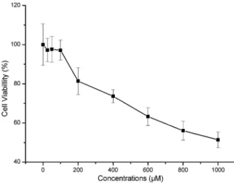
Concentrations of Cu-His
As shown in Figure 1, Cu-His effectively inhibited the cytoactivity of HeLa cells with an obvious loss of approximately 20–50% cell viability when Cu-His was
introduced into the cells at different concentrations (200, 400, 600, 800, or 1000mM), indicating that the cells
were under Cu stress, and Cu-His at concentrations under 100mM was not cytotoxic to the cells. No obvious
dead cells were observed when Cu-His was at the highest concentration of 1000mM. The concentrations
over 100mM were used for further experiments on Cu stress.
Intracellular Cu content
(Table 1). The results indicated that yeast CUP1 over-expression could bind Cu effectively in HeLa cells and increased intracellular Cu content.
Cell viability analysis
After incubation, cell viability was analyzed using the MTT assay to compare the average absorbance of the test cells with that of the control cells. A comparison of the relative viability of the cells after treatment with Cu-His at different concentrations is shown in Figure 2, A-E. The results demonstrated that the viability of the test cells was significantly greater than that of the control cells (Po0.05) after treatment with Cu-His at 200, 400, and 600mM
(Figure 2, A-C), and the differences were also significant (Po0.01) after treatment with Cu-His at 800 and 1000mM
(Figure 2, D and E). Comparatively, the test cells appeared to have a greater viability at all incubation times, supporting a protective role against excess Cu. Hence, yeastCUP1may allow the cells to bind more Cu, resulting in an increase in the intracellular antioxidative ability to protect the cells against excessive amounts of Cu, as reported by Richards (7).
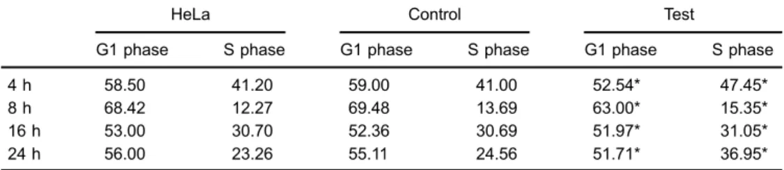
CUP1-mediated cell cycle
Based on the above results, using FACS we further investigated whether the cell cycle was mediated by yeast CUP1. Cell cycle analysis showed a high level of cycle synchronization, and the cells were mostly arrested at G0/G1 phase after 96 h of serum starvation. However, a decreased proportion of cells was in the G1 phase (Po0.01) and an increased proportion of the test cells was in the S phase (Po0.01) relative to the control cells when incubated with Cu-His at 100mM for 4, 8, 16, and 24 h (Table 2), but no significant difference was observed between HeLa and control cells (P40.01). The same was
also observed between the HeLa cells, control and test cells when incubated with PBS for all incubation times (P40.01; Table 3).
Intracellular ROS
Considering the damage to the cells upon treatment over the range of high Cu concentrations, we detected Figure 1.Percentage of viable of HeLa cells at different Cu-His
concentrations. Cellular viability was analyzed using the MTT assay. Cu-His at different concentrations (200, 400, 600, 800, and 1000mM) inhibited cell viability by approximately 20%-50% (Po0.01,t-test), but not for concentrations under 100mM (n=8) (P40.05). The results were reported relative to the response of the negative control cells. MTT, 3-(4,5-dimethylthiazol-2-yl)-2,5-diphenyltetrazolium bromide.
Table 1.Cellular copper content at different Cu-His concentrations.
Copper content 10mM 100mM
Control (mg/L) 10.71±2.2036 13.12±0.3515 Test (mg/L) 13.67±2.3005* 26.98±0.0849*
Control: cells expressing empty vectors; Test: cells stably expressing CUP1 protein. *Po0.01, compared to control (t-test).
ROS production as a measurement of Cu stress using the DCFH-DA assay. An increase in fluorescence intensity indicated an increase in intracellular ROS (18). The fluorescence intensity of the test cells was significantly lower than that of the control cells (Po0.05) after treatment with Cu-His at 200, 400, and 600mM, and the
differences were also significant (Po0.01) after treatment with Cu-His at 800 and 1000mM (Figure 3).
Discussion
The functions of MTs, such as storage of metal ions, metal detoxification, and oxidative scavenging, have been exten-sively studied (19), but the roles of MTs on intracellular anti-oxidant activity remain elusive. In the present study, our goal was to elucidate the role of yeastCUP1in Cu metabolism, as well as its functions on cellular Cu content, cell viability, cell cycling, and intracellular ROS. Cell lines that stably expressed yeastCUP1were used to assess whether yeastCUP1can bind Cu effectively and protect cells against Cu stress.
Our findings indicated that the expression of yeast CUP1was highly abundant in HeLa cells (Supplementary Figure S1). CUP1 possesses identical Cu-binding geometry with human MT (4), as shown in the HeLa cells. In the presence of Cu (100mM for different durations), the relative abundance of humanMTincreased with incubation times, in accordance with previous observations (15,19), whereas no increase inCUP1mRNA expression was observed (Supple-mentary Figure S2), becauseCUP1expression was initiated by the cytomegalovirus promoter of the pEGFP-N1 plasmid. Our results indicated thatMTplays an important role in the Cu-dependent induction of its own transcription, which was in agreement with the results of previous studies (15,20). At all incubation time points, expression of CUP1 mRNA was significantly greater than that of humanMTmRNA, suggest-ing that CUP1 played a dominant role in binding Cu compared to the humanMTgene.
MT is a primary Cu-binding protein under physiological conditions (21), and characterization of the MT-Cu complex Table 3.Proportion of cells in G1 and S phases after incubation with phosphate-buffered saline for different
times.
HeLa Control Test
G1 phase S phase G1 phase S phase G1 phase S phase
4 h 51.94 45.07 53.00 43.45 52.50 44.84
8 h 64.17 12.31 63.05 13.00 62.60 11.84
16 h 43.50 29.00 44.70 28.90 42.71 29.84
24 h 51.71 32.10 50.48 31.75 52.00 31.50
Control: cells expressing empty vectors; Test: cells stably expressing CUP1 protein. P40.01 (t-test). Table 2.Proportion of cells in G1 and S phases after incubation with Cu-His for different times.
HeLa Control Test
G1 phase S phase G1 phase S phase G1 phase S phase
4 h 58.50 41.20 59.00 41.00 52.54* 47.45*
8 h 68.42 12.27 69.48 13.69 63.00* 15.35*
16 h 53.00 30.70 52.36 30.69 51.97* 31.05*
24 h 56.00 23.26 55.11 24.56 51.71* 36.95*
Control: cells expressing empty vectors; Test: cells stably expressing CUP1 protein. *Po0.01 compared to HeLa and Control (t-test).
suggests that MT is beneficial for intracellular storage of Cu (15). It has been demonstrated that an increase in the content of cellular Cu is directly correlated with an increase in the amount of MT-Cu (22), and MT was involved in the process of Cu absorption and storage (5,19). In our experiments, the increase in cellular Cu content resulting from overexpression of yeast CUP1 demonstrated that CUP1possessed capabilities of cellular storage within the physiological range of Cu exposure. Additional evidence has shown that different cells exhibit increased Cu content in response to a gradual increase in Cu exposure (19,23), and a similar phenomenon was observed in our experiments.
Cu is a very important catalytic cofactor in many biological processes (1), and Cu deficiency compromises cellular antioxidant defense capability, thereby increasing cellular susceptibility to oxidative DNA damage (24). However, enhanced Cu can lead to cytotoxicity due to ROS formation (1). High levels of exogenous ROS directly inactivate protein phosphorylation and interfere with the balance of cellular kinase/phosphatase activity toward added enzymatic phos-phorylation events (25). Some nutrients reportedly provide protection against Cu-induced oxidative damage by acting as nonenzymatic antioxidants, such as vitamin C, vitamin E, and glutathione (26). Cu/Zn superoxide dismutase (SOD) and catalase are enzymes that efficiently eliminate ROS by catalyzing the breakdown of excess superoxide and H2O2,
and are involved in antioxidant defense (25). Upregulation of SOD and catalase expression leads to reduced ROS levels (27), which, in turn, seems to promote cellular viability, whereas increased ROS generation can suppress cellular activity by inhibiting activities of SOD and catalase, which protect cells against oxidative stress through the dismuta-tion of superoxide to O2 and H2O2 (27,28). Reducing
oxidative stress by nonenzymatic antioxidants as well as antioxidant enzymes could potentially reduce ROS forma-tion (29). Ourfindings indicated that overexpression of yeast CUP1 resulted in decreased intracellular ROS formation, which supports a protective role for MT (CUP1) in response to Cu excess by inhibiting ROS formation as nonenzymatic antioxidants, similar to thefindings of Tapia et al. (15).
It has been strongly suggested that MT protein content is directly associated with resistance to excess Cu
exposure in mammalian cells (19,23), which protects against Cu-dependent cytotoxicity by its antioxidant activity (30) and could eliminate ROS generated from Cu exposure (19), or primarily by its ability to bind Cu with high affinity. Thus, the multiple Cys residues in MT act as effective Cu chelators that react with ROS and can effectively protect the cell from Cu toxicity (31). Conditions correlated with Cu overload may lead to Cu-induced stress (19), which gives rise to the produc-tion of increased amounts of ROS capable of generating oxidative stress, because Cu can function as a transi-tion metal with redox cycling capacity (20). Here, the results of the MTT assay showed an increase in viability of the test cells compared to the control cells. Because of the close relationship between cell viability and the cell cycle (32), the cell cycle was further analyzed. Thus, the decreased proportions of G1 phase cells and the increased proportions of S phase cells suggest enhanced cellular viability (33,34). One reasonable explanation for this observation is the abundance of yeastCUP1produced in the test cells that likely bound the Cu, which stimulated an increase in cell viability, perhaps by ameliorating oxidative stress or reducing ROS production (1), because viability in cells lacking Cu/Zn-SOD can be complemented by MT overexpres-sion (28). In summary, our study provided essential insights into the physiological regulation of yeastCUP1 on binding Cu and blocking Cu-induced stress. We found that overexpression of yeastCUP1was beneficial to protect HeLa cells against Cu stress.
Supplementary Material
Click here to view [pdf].
Acknowledgments
The authors wish to express their gratitude to the members of our team, especially to He Meng and Tao Sun for their help in revising this article. Research supported by the National Transgenic Breeding Program (#2011ZX08009-003-006).
References
1. Gaetke LM, Chow CK. Copper toxicity, oxidative stress, and antioxidant nutrients.Toxicology2003; 189: 147-163, doi: 10.1016/s0300-483x(03)00159-8.
2. Liu XD, Thiele DJ. Yeast metallothionein gene expression in response to metals and oxidative stress.Methods1997; 11: 289-299, doi: 10.1006/meth.1996.0423.
3. Song MO, Li J, Freedman JH. Physiological and toxi-cological transcriptome changes in HepG2 cells exposed to copper.Physiol Genomics2009; 38: 386-401, doi: 10.1152/ physiolgenomics.00083.2009.
4. Ecker DJ, Butt TR, Sternberg EJ, Neeper MP, Debouck C, Gorman JA, et al. Yeast metallothionein function in metal ion detoxification.J Biol Chem1986; 261: 16895-16900. 5. Bremner I. Involvement of metallothionein in the hepatic
metabolism of copper.J Nutr1987; 117: 19-29, doi: 10.1016/ 0076-6879(91)05142-i.
7. Richards MP. Recent developments in trace element meta-bolism and function: role of metallothionein in copper and zinc metabolism.J Nutr1989; 119: 1062-1070, doi: 10.1007/ 978-3-642-68269-8_160.
8. Yen NT, Lin CS, Ju CC, Wang SC, Huang MC. Mitochondrial DNA polymorphism and determination of effects on repro-ductive trait in pigs. Reprod Domest Anim 2007; 42: 387-392, doi: 10.1111/j.1439-0531.2006.00797.x.
9. Karin M, Najarian R, Haslinger A, Valenzuela P, Welch J, Fogel S. Primary structure and transcription of an ampli-fied genetic locus: the CUP1 locus of yeast. Proc Natl Acad Sci U S A 1984; 81: 337-341, doi: 10.1073/ pnas.81.2.337.
10. Butt TR, Sternberg EJ, Gorman JA, Clark P, Hamer D, Rosenberg M, et al. Copper metallothionein of yeast, structure of the gene, and regulation of expression.Proc Natl Acad Sci U S A 1984; 81: 3332-3336, doi: 10.1073/ pnas.81.11.3332.
11. Fogel S, Welch JW, Cathala G, Karin M. Gene amplification in yeast: CUP1 copy number regulates copper resistance.
Curr Genet1983; 7: 347-355, doi: 10.1007/bf00445874. 12. Wimalarathna RN, Pan PY, Shen CH. Chromatin
reposi-tioning activity and transcription machinery are both recrui-ted by Ace1p in yeast CUP1 activation. Biochem Biophys Res Commun 2012; 422: 658-663, doi: 10.1016/j. bbrc.2012.05.047.
13. Jensen LT, Howard WR, Strain JJ, Winge DR, Culotta VC. Enhanced effectiveness of copper ion buffering by CUP1 metallothionein compared with CRS5 metallothionein in Sac-charomyces cerevisiae.J Biol Chem1996; 271: 18514-18519, doi: 10.1074/jbc.271.31.18514.
14. Kreuder J, Otten A, Fuder H, Tumer Z, Tonnesen T, Horn N, et al. Clinical and biochemical consequences of copper-histidine therapy in Menkes disease. Eur J Pediatr1993; 152: 828-832, doi: 10.1007/bf02073380.
15. Tapia L, Gonzalez-Aguero M, Cisternas MF, Suazo M, Cambiazo V, Uauy R, et al. Metallothionein is crucial for safe intracellular copper storage and cell survival at normal and supra-physiological exposure levels.Biochem J2004; 378: 617-624, doi: 10.1042/bj20031174.
16. Teo WZ, Khim Chang EL, Sofer Z, Pumera M. Cytotoxicity of halogenated graphenes.Nanoscale2013; 6: 1173-1180, doi: 10.1039/c3nr05275c.
17. Lima-Neto JF, Fernandes CB, Alvarenga MA, Golim MA, Landim-Alvarenga FC. Viability and cell cycle analysis of equinefibroblasts culturedin vitro.Cell Tissue Bank2010; 11: 261-268, doi: 10.1007/s10561-009-9131-6.
18. Cai X, Chen X, Wang X, et al. Pre-protective effect of lipoic acid on injury induced by H2O2in IPEC-J2 cells.Mol Cell Biochem2013; 1-9, doi: 10.1007/s11010-013-1595-9. 19. Kawai K, Liu SX, Tyurin VA, Tyurina YY, Borisenko GG,
Jiang JF, et al. Antioxidant and antiapoptotic function of metallothioneins in HL-60 cells challenged with copper nitrilotriacetate.Chem Res Toxicol2000; 13: 1275-1286, doi: 10.1021/tx000119l.
20. Suazo M, Hodar C, Morgan C, Cerpa W, Cambiazo V, Inestrosa NC, et al. Overexpression of amyloid precursor
protein increases copper content in HEK293 cells.Biochem Biophys Res Commun2009; 382: 740-744, doi: 10.1016/j. bbrc.2009.03.096.
21. Palmiter RD. The elusive function of metallothioneins.Proc Natl Acad Sci U S A 1998; 95: 8428-8430, doi: 10.1073/ pnas.95.15.8428.
22. Labadie GU, Beratis NG, Price PM, Hirschhorn K. Studies of the copper-binding proteins in Menkes and normal cultured skin fibroblast lysates.J Cell Physiol 1981; 106: 173-178, doi: 10.1002/jcp.1041060202.
23. Schilsky ML, Stockert RJ, Kesner A, Gorla GR, Gagliardi GS, Terada K, et al. Copper resistant human hepato-blastoma mutant cell lines without metallothionein induction overexpress ATP7B.Hepatology1998; 28: 1347-1356, doi: 10.1002/hep.510280525.
24. Pan Y, Loo G. Effect of copper deficiency on oxidative DNA damage in Jurkat T-lymphocytes.Free Radic Biol Med2000; 28: 824-830, doi: 10.1016/s0891-5849(00)00165-9. 25. Day RM, Suzuki YJ. Cell proliferation, reactive oxygen and
cellular glutathione. Dose-Response 2005; 3: 425, doi: 10.2203/dose-response.003.03.010.
26. Chow CK. Vitamin E and oxidative stress. Free Radic Biol Med1991; 11: 215-232, doi: 10.1016/0891-5849(91)90174-2. 27. Chang Q, Pan J, Wang X, Zhang Z, Chen F, Shi X. Reduced reactive oxygen species-generating capacity contributes to the enhanced cell growth of arsenic-transformed epithelial cells.Cancer Res2010; 70: 5127-5135, doi: 10.1158/0008-5472.can-10-0007.
28. Kommuguri UN, Bodiga S, Sankuru S, Bodiga VL. Copper deprivation modulates CTR1 and CUP1 expression and enhances cisplatin cytotoxicity in Saccharomyces cerevi-siae.J Trace Elem Med Biol2012; 26: 13-19, doi: 10.1016/j. jtemb.2011.12.001.
29. Valko M, Rhodes CJ, Moncol J, Izakovic M, Mazur M. Free radicals, metals and antioxidants in oxidative stress-induced cancer.Chem Biol Interact2006; 160: 1-40, doi: 10.1016/j. cbi.2005.12.009.
30. Lazo JS, Kuo SM, Woo ES, Pitt BR. The protein thiol metallothionein as an antioxidant and protectant against antineoplastic drugs. Chem Biol Interact 1998; 111-112: 255-262, doi: 10.1016/s0009-2797(97)00165-8.
31. Thornalley PJ, Vašák M. Possible role for metallothionein in protection against radiation-induced oxidative stress. Kinet-ics and mechanism of its reaction with superoxide and hydroxyl radicals.BBA-Protein Struct M1985; 827: 36-44, doi: 10.1016/0167-4838(85)90098-6.
32. Teodoro AJ, Oliveira FL, Martins NB, Maia GA, Martucci RB, Borojevic R. Effect of lycopene on cell viability and cell cycle progression in human cancer cell lines. Cancer Cell Int
2012; 12: 36, doi: 10.1186/1475-2867-12-36.
33. Li VC, Ballabeni A, Kirschner MW. Gap 1 phase length and mouse embryonic stem cell self-renewal.Proc Natl Acad Sci U S A2012; 109: 12550-12555, doi: 10.1073/pnas.1206740109. 34. Kern S, Eichler H, Stoeve J, Kluter H, Bieback K.
Com-parative analysis of mesenchymal stem cells from bone marrow, umbilical cord blood, or adipose tissue.Stem Cells