Universidade de Lisboa
Faculdade de Medicina de Lisboa
ROLE OF HISTONE H1 IN CHROMATIN AND GENE
EXPRESSION IN THE AFRICAN TRYPANOSOME:
BROAD SKILLS, SPECIFIC FUNCTIONS?
Ana Pena
Tese especialmente elaborada para a obtenção do grau de Doutor em
Ciências Biomédicas na Especialidade de Microbiologia e Parasitologia
Universidade de Lisboa Faculdade de Medicina de Lisboa
ROLE OF HISTONE H1 IN CHROMATIN AND GENE EXPRESSION IN THE AFRICAN TRYPANOSOME: BROAD SKILLS, SPECIFIC FUNCTIONS?
Ana Pena
Orientador: Dra. Luísa Figueiredo Co-orientador: Dr. Christian Janzen
Tese especialmente elaborada para a obtenção do grau de Doutor em Ciências Biomédicas na Especialidade de Microbiologia e Parasitologia
JÚRI Presidente:
Faculdade de Medicina – Universidade de Lisboa Professor Doutor José Melo Cristino
Vogais:
Universidade de Würzburg, Alemanha Professor Tim Nicolai Siegel, PhD
The Rockefeller University, EUA
Professora F. Nina Papavasiliou, Associate Professor
InstiMedicina Molecular – Faculdade de Medicina – Universidade de Lisboa Doutor Rui Miguel Prudêncio Cunha Pignatelli
Doutora Liliana Mâncio Silva
Doutora Luísa Miranda Figueiredo (orientadora) Faculdade de Medicina – Universidade de Lisboa Professor Doutor Sérgio Alexandre Fernandes de Almeida
A impressão desta dissertação foi aprovada pelo Conselho Científico da
Faculdade de Medicina de Lisboa em reunião de 22 de Setembro de 2015
TABLE OF CONTENTS
ACKNOWLEDGMENTS ... iv
ABBREVIATIONS ... v
RESUMO ... ix
SUMMARY ... xii
1 INTRODUCTION ... 1
1.1
TRYPANOSOMA BRUCEI ... 1
1.1.1 EARLY EVOLUTIONARY ORIGINS ... 1
1.1.2 AFRICAN TRYPANOSOMIASIS ... 3
1.1.3 A CHALLENGING LIFE - FROM MAMMAL TO FLY ... 5
1.1.4 ANTIGENIC VARIATION - THE POWER OF CHANGING ... 8
1.2
CHROMATIN... 12
1.2.1 GENOME ORGANIZATION... 12
1.2.2 THE LARGE VSG REPERTOIRE ... 13
1.2.3 CHROMATIN ORGANIZATION IN EUKARYOTES ... 16
1.2.4 CHROMATIN ORGANIZATION IN T. BRUCEI ... 18
1.2.5 EPIGENETIC REGULATORS IN T. BRUCEI ... 20
1.3
GENE EXPRESSION IN T. BRUCEI ... 24
1.3.1 TRANSCRIPTIONAL AND POST-TRANSCRIPTIONAL REGULATION ... 24
1.3.2 CONTROL OF BLOODSTREAM EXPRESSION SITES ... 30
1.3.3 CONTROL OF PROCYCLIN LOCI ... 38
1.4
HISTONE H1 ... 41
1.4.1 STRUCTURAL AND FUNCTIONAL DIVERSITY ... 41
2
AIMS ... 48
3
RESULTS ... 49
3.1
TRYPANOSOMA BRUCEI HISTONE H1 INHIBITS RNA POLYMERASE I
TRANSCRIPTION ... 49
3.1.1 AUTHOR CONTRIBUTIONS ... 49
3.1.2 RESULTS ... 50
3.1.2.1 T. brucei histone H1 family comprises three hypothetical protein classes ... 50
3.1.2.2 Depletion of H1 causes no considerable changes in parasite growth in culture ... 52
3.1.2.3 H1 compacts chromatin at different levels across the genome ... 55
3.1.2.4 H1 regulates expression of Pol I-transcribed genes ... 59
3.1.2.5 H1 inhibits transcription from silent BESs and procyclin sites ... 65
3.1.2.6 Depletion of H1 increases resistance to MMS-induced DNA damage . ... 68
3.1.3 MATERIALS AND METHODS ... 70
3.1.4 SUPPLEMENTARY FIGURES AND TABLES ... 80
3.2
HISTONE H1 IS IMPORTANT FOR T. BRUCEI FITNESS IN VIVO ... 92
3.2.1 AUTHOR CONTRIBUTIONS ... 92
3.2.2 RESULTS ... 92
3.2.2.1 H1 depletion impairs progression of acute infection in mice ... 92
3.2.2.2 In vivo, H1 depleted parasites adapt and regain fitness ... 95
3.2.2.3 H1 is important for T. brucei ability to evade the host immune system 97 3.2.3 MATERIALS AND METHODS ... 99
3.3
NEW METHOD FOR STUDYING TRANSCRIPTION IN T. BRUCEI –
IMPLEMENTATION OF 4-THIOURIDINE LABELING OF NASCENT
TRANSCRIPTS ... 102
3.3.1 AUTHOR CONTRIBUTIONS ... 102
3.3.2 RESULTS ... 105
3.3.2.1 Labeling efficiency ... 106
3.3.2.2 4sU transport across the membrane ... 107
3.3.2.3 4sU deviation by RNA editing ... 109
3.3.2.4 Protein contamination ... 110
3.3.2.5 RNA integrity ... 111
3.3.3 MATERIALS AND METHODS ... 113
4
GENERAL DISCUSSION ... 118
4.1
H1 condenses chromatin globally but has a more pronounced
effect at silent Pol I transcription units ... 118
4.2
H1 plays a role in DNA repair ... 126
4.3
H1 is important for parasite fitness in vivo ... 128
5
CONCLUDING REMARKS ... 133
6
REFERENCES ... 136
7
ANNEXES ... 160
iv
ACKNOWLEDGMENTS
I thank my supervisor Luísa Figueiredo for leading me in this big adventure and challenge that is doing a PhD. Thank you for accepting me in your lab, for being a close supervisor, attentively guiding me through the way. I am grateful to your encouragement and advises, scientific and for ‘life’ which made me grow as a better scientist and person.
I also thank my co-supervisor Christian Janzen, for always being available for scientific discussions and for having patience for the bureaucratic issues.
To my thesis committee: Dr. Miguel Godinho, Dr. Maria Mota and Dr. Ana Pamplona for the scientific input and for ‘teasing me’ in good ways. I am special grateful to Dr. Ana Pamplona. It was by your hand that I engaged in the world of Science and started the way that leaded me to the present moment in my career.
Words are very little to thank all the friends I have made at IMM. Your words, smiles, enthusiasm were my source of courage and positive thinking each day. I specially thank to my colleagues (present and former): Leonor, Idálio, Daniel, Mafalda, Sandra, Filipa, Fabien, Xico, Fábio, Margarida and Helena. Our lab is unique, and only we know why! A warm embrace also to our nice neighbours from Miguel Prudêncio lab, particularly to my dear and old friend Jorge Santos.
To my flatmate Eliana Real, for all of our intellectually elevated conversations, very futile but healthy gossiping and most of all for being such a good and present friend. To Maria and Idálio, for your help during the thesis writing and especially to Idálio for the support, care and understanding.
Finally, to my family and friends ‘since ever’ I am more grateful than words could say. Thank you for being proud of me, for believing me and for giving me your love.
v
ABBREVIATIONS
3’UTR 3’ untranslated region
4sU 4-thiouridine
4TU 4-thiouracil
5’UTR 5’ untranslated region
aa amino acid(s)
AAT animal African trypanosomiasis ActD actinomycin D
AmpR ampicilin resistance gene BARP brucei alanine-rich protein
base J β-D-glucosyl-hydroxymethyluracil
BES bloodstream expression site
Br-UTP 5-bromouridine-5’-triphophate BSDR blasticidin resistance gene
BSF bloodstream form
CDS coding sequence
ChIP chromatin immunoprecipitation CITFA class I transcription factor A
COIII cytochrome oxidase subunit III gene DCV Vybrant® DyeCycle™ Violet
dsRNA double-stranded RNA
EP procyclin with Glu-Pro repeats
ESAG expression site-associated gene
ESB expression site body
FACS fluorescence activated cell sorting
FAIRE formaldehyde-assisted isolation of regulatory elements
vi
GPEET procyclin with Gly-Pro-Glu-Glu-Thr repeats
GPI glycosylphosphatidylinositol
GRESAG gene related with ESAG GSEA gene set enrichment analysis
H1 histone H1
HAT human African trypanosomiasis HMGB high-mobility group box protein
HR homologous recombination
IC50 half maximal inhibitory concentration kDNA kinetoplast DNA
LUC luciferase gene
MACS magnetic-activated cell sorting MES metacyclic expression site MMS methyl methanesulphonate MNase Micrococcal nuclease
mRNA messenger RNA
MVSG metacyclic variant surface glycoprotein ND7 NADH dehydrogenase subunit 7 gene PAG procyclin-associated gene
PF procyclic form
PI propidium iodide
Pol I RNA polymerase I Pol II RNA polymerase II Pol III RNA polymerase III ppm parts per million
PTEN phosphatase and tensin homolog PTM post-translational modification
vii PURR puromycin resistance gene
qPCR quantitative real-time PCR
Rag2-/-c-/- recombinase activating gene 2 and common cytokine receptor chain double knock-out
RBP RNA-binding protein
rDNA ribosomal DNA
RNAi RNA interference RNA-Seq RNA sequencing rRNA ribosomal RNA siRNA small interfering RNA
SL spliced leader
snoRNA small nucleolar RNA snRNA small nuclear RNA
SSR strand-switch region
TbASF1A Trypanosoma brucei anti-silencing factor 1A
TbBDF3 Trypanosoma brucei bromodomain factor 3
TbCAF-1b Trypanosoma brucei chromatin assembly factor 1b
TbDAC1 Trypanosoma brucei histone deacetylase 1
TbDAC3 Trypanosoma brucei histone deacetylase 1
TbDOT1A Trypanosoma brucei disruptor of telomeric silencing 1A
TbDOT1B Trypanosoma brucei disruptor of telomeric silencing 1B
TbELP3b Trypanosoma brucei elongator protein 3b
TbFACT Trypanosoma brucei facilitates chromatin transcription complex
TbHAT2 Trypanosoma brucei histone acetyltransferase 2
TbHAT3 Trypanosoma brucei histone acetyltransferase 3
TbISWI Trypanosoma brucei imitation switch
TbKu70/Ku80 Trypanosoma brucei Ku heterodimer (70 kDa and 80 kDa
proteins)
TbMCM-BP Trypanosoma brucei mini-chromosome maintenance-binding
viii
TbNLP Trypanosoma brucei nucleoplasmin-like protein
TbNUP-1 Trypanosoma brucei lamin-like protein
TbORC1/CDC6 Trypanosoma brucei origin replication complex 1/cell division
cycle 6-like protein
TbRAP1 Trypanosoma brucei repressor/activator of protein 1
TbRPB5z Trypanosoma brucei RNA Pol I RPB5 variant subunit
TbRPB6z Trypanosoma brucei RNA Pol I RPB6 variant subunit
TbSIR2rp1 Trypanosoma brucei Silent information regulator 2-related
protein 1
TbTIF2 Trypanosoma brucei TRF-interacting factor 2
TbTRF Trypanosoma brucei telomeric repeat-binding factor
TbU3 Trypanosoma brucei high-affinity uracil transporter 3
TDB trypanosome dilution buffer
TDP-1 trypanosome DNA binding protein
TPE telomere position effect
tRNA transfer RNA
TSS transcription start site
TTS transcription termination site
UTP uridine-5'-triphosphate
VSG variant surface glycoprotein
ix
RESUMO
O Trypanosoma brucei é um protozoário unicelular que vive na corrente sanguínea e espaços intersticiais de um hospedeiro mamífero e que é transmitido pela mosca tsétsé, o seu vetor de transmissão. Em humanos, o T. brucei causa a doença do sono. O T brucei tem um ciclo de vida complexo no qual o parasita passa por vários estadios de desenvolvimento. Dentro do mamífero, o parasita vive sob a forma de tripomastigotas sanguíneos (bloodstream forms; BSFs), e após ser transmitido para a mosca tsé-tsé, diferencia-se em tripomastigotas procíclicos (procyclic forms; PFs). O parasita T. brucei é um organismo que divergiu cedo na evolução e que possui muitas características pouco usuais para um eucariota. A sua transcrição é maioritariamente constitutiva e executada pela RNA polimerase II (Pol II)., enquanto que a RNA polimerase I (Pol I) transcreve não só os RNAs ribossomais, mas também genes codificantes de proteínas. Entre os genes transcritos por Pol I encontram-se os genes que codificam as glicoproteínas variantes de superfície (variant surface glycoproteins; VSGs) e as prociclinas. As VSGs e as prociclinas são proteínas muito abundantes que revestem a superfície dos BSFs e dos PFs, respetivamente, e cuja expressão é estritamente regulada ao longo do desenvolvimento do parasita.
Nos BFSs, os genes das VSGs são transcritos em locais de expressão subteloméricos (bloodstream expression sites; BESs). Existem cerca de 15 BESs no genoma, contudo apenas um está transcricionalmente ativo em determinado momento. Os restantes BESs necessitam de ser mantidos silenciados por forma a manter a expressão monoalélica de um único BES. Para evitar o reconhecimento pelo sistema imunitário do hospedeiro, periodicamente os parasitas mudam o gene VSG expresso, através de um mecanismo denominado de variação antigénica. Nos BSFs o loci das prociclinas também está parcialmente reprimido por forma a evitar a substituição das VSGs por prociclinas à superfície. Existem evidências de que em T.
brucei alguns fatores epigenéticos regulam a ativação ou o silenciamento transcricional
de Pol I nos BESs e loci das prociclinas. Para além disso, existem algumas variantes de histonas e modificações pós-traducionais de histonas que estão associadas com os locais de iniciação e terminação da transcrição por Pol II.
x
No núcleo de células eucarióticas o DNA encontra-se enrolado em volta de um octâmero de histonas formando o nucleosoma, a unidade básica da cromatina. A histona H1 (H1) liga-se ao DNA que entra e sai do nucleosoma, estabilizando a sua estrutura e contribuindo para o estabelecimento de estruturas de cromatina de ordem mais elevada. Em T. brucei a H1 é codificada por pelo menos 5 genes que, de acordo com a sua sequência proteica esperada, podem agrupar-se em 3 classes. No meu trabalho de doutoramento, tive por objetivo investigar as funções da H1 na estrutura da cromatina, na expressão génica e na transcrição de T. brucei. Para além disso, também foi estudada a importância da H1 nas infeções in vivo de T. brucei no hospedeiro mamífero.
Foram realizados estudos funcionais em que se depletaram todas as classes de H1 por RNA de interferência (RNAi). Na presença de tetraciclina, RNAs de dupla-cadeia são produzidos que induzem a depleção dos RNAs mensageiros (mRNAs) de H1. Usando imunoprecipitação de chromatina (chromatin immunoprecipitation; ChIP) associada a histona H3 e formaldehyde-assisted isolation of regulatory elements (FAIRE), detetou-se uma descondensação global de cromatina ao longo do genoma nos parasitas depletados de H1. No entanto, esta descondensação foi mais proeminente nos promotores de BES silenciados e nos loci de prociclinas, apesar de mais ligeiramente nestes últimos. Curiosamente, a sequenciação de RNA (RNA-Seq) revelou que a abertura global da cromatina devida à depleção de H1 não estava associada a alterações generalizadas de expressão génica mas pelo contrário à desrepressão de um subgrupo específico de genes. De cerca de 9000 genes, apenas 26 passarm a ser significativamente sobreexpressos, maioritariamente genes de BESs silenciados (inclusive VSGs silenciados) e genes de loci de prociclinas. Eu adaptei e apliquei a marcação metabólica de RNA com 4-tiouridina (4sU) em T. brucei para purificar e quantificar os transcritos nascentes produzidos nos loci transcritos por Pol I e Pol II. A marcação com 4sU traz vantagens em relação à técnica classicamente utilizada de
nuclear run–on, por permitir a medição da transcrição de novo em células não
perturbadas. A quantificação de RNA nascente marcado com 4sU demonstrou que a H1 é necessária para reprimir a transcrição de Pol I nos BESs silenciados e nos loci de prociclinas, mas aparentemente não o é para os genes transcritos por Pol II.
xi No seu conjunto, os nossos resultados suportam um modelo em que a H1 induz a compactação global de cromatina, mas desempenha funções mais específicas no que respeita à expressão génica, nomeadamente a regulação transcricional de genes silenciados de Pol I. Este modelo é consistente com a noção de que a transcrição policistrónica por Pol II, que ocorre na maior parte do genoma de tripanossomas, é constitutiva e a maioria dos genes são controlados ao nível pós-transcricional. Pelo contrário, existem evidências de que os genes transcritos por Pol I são regulados ao nível transcricional, consistente com a função da H1 no silenciamento da iniciação/elongamento da transcrição por Pol I.
Outro fenótipo observado em parasitas depletados de H1 foi um aumento da resistência a danos no DNA induzidos por metanossulfonato de metilo (MMS). Esta observação poderá indicar que a H1 desempenha funções mais gerais ao suprimir a recombinação homóloga e, talvez, a instabilidade genómica.
Interessantemente, os parasitas de T. brucei depletados em H1 revelaram uma aparente perda de fitness durante infeções in vivo. Em murganhos infetados com parasitas depletados em H1, a progressão da parasitémia (percentagem de parasitas no sangue) foi mais lenta e os murganhos sucumbiram mais tarde. Curiosamente, este fenótipo foi completamente abolido em murganhos severamente imuno-comprometidos que não possuem linfócitos T, B e NK, demonstrando que a fitness de parasitas depletados em H1 é perturbada porque, de alguma forma, estes parasitas tornam-se mais suscetíveis ao sistema imunitário do hospedeiro.
Coletivamente os nossos resultados mostram que a H1 de T. brucei, um parasita que divergiu cedo na evolução, tem capacidades abrangentes porque é necessária para compactar a cromatina globalmente ao longo do genoma, mas em termos de transcrição desempenha funções específicas porque inibe a transcrição de um grupo restrito de genes, nomeadamente de loci silenciados transcritos por Pol I. A regulação de um grupo específio de genes mediada por H1 foi também observada noutros eucariotas superiores e inferiores, o que sugere que apesar de ser a família mais divergente de histonas, as suas funções foram conservadas ao longo da evolução.
xii
SUMMARY
Trypanosoma brucei is a unicellular protozoan parasite that lives in the
bloodstream and interstitial spaces of a mammalian host and that is transmitted by the tsetse, an insect vector. In humans, it causes sleeping sickness. T. brucei has a complex life cycle that progresses through different developmental stages. Inside the mammal, the parasite lives as bloodstream forms (BSFs), which differentiates differentiates into procyclic forms (PFs) upon transmission to the insect vector. T. brucei is an evolutionarily early-branching organism that possesses many unusual features for an eukaryote. Most of its transcription is constitutive and driven by RNA polymerase II (Pol II) and RNA polymerase I (Pol I) transcribes not only ribosomal RNA genes, but also protein-coding genes, including genes encoding variant surface glycoproteins (VSGs) and procyclins. These are highly abundant proteins that cover the surface of the BSFs and the PFs, respectively, and whose expression is tightly developmentally regulated.
In BSFs, VSGs are transcribed in subtelomeric bloodstream expression sites (BESs). There are ~15 BESs in the genome, however only one is fully transcriptionally active at a time. The remaining BESs need to be kept silent to maintain monoallelic expression of BES. To evade the host immune system, parasites switch the expressed
VSG from time to time by a mechanism called antigenic variation. In addition, in BSFs
procyclin loci are also partially repressed to prevent illegitimate VSG replacement for procyclins. Some epigenetic factors have been found to regulate Pol I transcriptional activation/silencing at BESs and procyclin loci. Besides, a number of histone variants and histone modifications seem to be associated with Pol II transcription initiation and termination.
Inside the eukaryotic nucleus, DNA is wrapped around an octamer of histones that forms the nucleosome, the basic unit of chromatin. Linker histone or histone H1 (H1) binds to the DNA entering and exiting the nucleosome, stabilizing its structure and contributing to the establishment of higher-order chromatin structures. In T. brucei, H1 is encoded by at least five genes and, based on the predicted protein sequence, they can be split in three classes. In my PhD work I aimed at analyzing the roles of H1 in
xiii chromatin structure, genome-wide gene expression and transcription in T. brucei. In addition, the importance of H1 for T. brucei mammalian infections in vivo was also addressed.
Functional studies were performed by simultaneously depleting all classes of H1 by RNA interference (RNAi). Using chromatin immunoprecipitation (ChIP) of histone H3 and formaldehyde-assisted isolation of regulatory elements (FAIRE), in H1-depleted parasites we detected a global chromatin opening across the genome, even though this happened at silent BESs promoters more prominently and procyclin loci, to a lesser extent. Curiously, RNA sequencing (RNA-Seq) revealed that global chromatin opening due to H1 depletion is not associated with widespread alterations in gene expression but rather with derepression of a specific subset of genes. Out of ~9,000 genes, only 26 were significantly upregulated, mostly from silent BESs (including silent
VSGs) and procyclin loci genes. I adapted and used 4-thiouridine (4sU) metabolic
labeling of RNA in T. brucei to purify and quantify nascent transcripts emanating from Pol I and Pol II-transcribed loci. 4sU-labeling has the advantage over classical nuclear run-on of measuring transcription de novo in unperturbed cells. Quantification of 4sU-labeled nascent RNA showed that H1 is necessary to repress Pol I transcription at silent BESs and procyclin loci but apparently not at Pol II genes.
Altogether our data strongly supports a model in which H1 induces global chromatin compaction, but in what concerns gene expression, H1 plays more specific functions, namely on transcriptional regulation of silent Pol I transcribed genes. This is consistent with the long-stand notion that polycistronic transcription by Pol II is constitutive and most genes are controlled essentially at the post-transcriptional level. On the other hand, Pol I genes are known to be regulated at the transcriptional level, consistent with a role of H1 in silencing Pol I transcription initiation/elongation.
An additional phenotype observed in H1-depleted parasites was its increased resistance to methyl methanoesulfonate (MMS)-induced DNA damage. This might indicate that H1 also plays a more general role such as suppressing homologous recombination and perhaps genomic instability.
xiv
Interestingly, H1-depleted T. brucei parasites revealed an apparent loss-of-fitness during in vivo infections. In mice infected with H1-depleted parasites, progression of parasitaemia (percentage of parasites in the blood) was delayed and mice succumbed later in infection. Curiously, this phenotype was completely abolished in highly immune-compromised mice that lack T, B and NK lymphocytes, demonstrating that fitness of H1-depleted parasites inside the host is impaired because, somehow, these parasites are more susceptible to host immune system.
Altogether our results show that H1 of the early divergent T. brucei has ‘broad skills’ because it compacts chromatin throughout the genome, but it plays ‘specific functions’ at the transcription level because it represses transcription of a limited cohort of genes, namely silent Pol I loci. H1-mediated regulation of a specific set of genes was also observed in other higher and lower eukaryotes, which suggests that despite being the most divergent family of histones, its functions were conserved throughout evolution.
1
1 INTRODUCTION
1.1 TRYPANOSOMA BRUCEI
1.1.1 EARLY EVOLUTIONARY ORIGINSIf origin of Life is one of the biggest mysteries in Biology, deciphering the evolution of the first eukaryotic lineages is not a minor enigma. At the early branches of the eukaryotic tree of life, many groups of protists have daunted phylogenetic positioning, diverging incredibly from the plant, fungi and animal branches. Despite its apparent simplicity as organisms, about 1,000 million years of life on Earth (Eme et al., 2014) set the grounds for a tremendous diversification in lifestyles and the emergence of exceptional biological features among them. An emblematic example is the supergroup Excavata, phylum Euglenozoa to which the parasite Trypanosoma brucei belongs. Given its unique and divergent properties, the root of eukaryotic tree was even proposed to lie within Euglenozoa or between it and all other eukaryotes, which would place T. brucei as one of the earliest branching organisms. Independently of the veracity of this hypothesis, trypanosomes such as T. brucei stand as evolutionary highly distant organisms from the well-known eukaryotic groups of animals, fungi and plants (Cavalier-Smith, 2010).
T. brucei is a unicellular flagellate that parasitizes the bloodstream and
interstitial spaces of various mammalian hosts following transmission by flies of the genus Glossina, also known as ‘tsetse’ flies. Within Euglenozoa, trypanosomes belong to a very particular clade, the Kinetoplastida, marked by the presence of special mitochondrial DNA, the kinetoplast or kinetoplast DNA (kDNA). This is a very large, usually disc-shaped mass consisting of an extensive network of interlocked circular DNA molecules, located adjacent to the basal body at the base of the flagellum. As its name indicates, kDNA arose uniquely in these organisms (Adl et al., 2012) (Fig. 1).
Trypanosomatida, such as T. brucei, offer a rare opportunity to study the origin and evolution of parasitism. This is because trypanosomatids were the only kinetoplastids acquiring an obligatory parasitic lifestyle, developing specific
2
adaptations to succeed in a lifetime co-existence with their hosts (Lukes et al., 2014). Researcher’s attention has focused particularly on trypanosomatids that represent a burden for public health and agricultural economy, namely species that infect humans and livestock from the Trypanosoma and Leishmania genus. These trypanosomatid species share their life between a variety of mammalian hosts and insect vectors and might develop intracellularly in host cells at some stage of its life cycle, such as T. cruzi and Leishmania spp., or, as in the case of T. brucei, live exclusively as extracellular parasites.
My PhD work focused on the study of Trypanosoma brucei. T. brucei holds a number of unusual, some unique, biological characteristics, which can go from transcription and nucleus organization to cell cycle, metabolism and surface proteins. While some are shared by most trypanosomatids, others, such as antigenic variation of its protective coat of variant surface glycoproteins (VSGs), are exclusive to African trypanosomes and arose as extremely efficient adaptations to life inside its hosts. The availability of a fully sequenced genome (Berriman et al., 2005) and of a variety of molecular tools, including an RNA interference (RNAi) system (Ngo et al., 1998), has made of Trypanosoma brucei a favorite model to gain insights not only about the biology, evolution and disease mechanisms in trypanosomatids, but also for fundamental questions in eukaryotic cell biology. Indeed, many important discoveries in eukaryotic molecular biology were made in cultured T. brucei such as RNA editing (Benne et al., 1986), trans-splicing (Boothroyd & Cross, 1982) and glycosylphosphatidylinositol (GPI) anchors (Ferguson et al., 1988); also, the hypermodified DNA base J (Gommers-Ampt et al., 1993), the glycosomes (Opperdoes & Borst, 1977) and, quite recently, an unconventional set of kinetochore proteins (Akiyoshi 2014) were first identified in T. brucei. Besides, this parasite has been used as a privileged model organism for studying the eukaryotic flagella (Baron et al., 2007, Branche et al., 2006).
3 1.1.2 AFRICAN TRYPANOSOMIASIS
African trypanosomiasis encompasses a variety of human and animal pathologies caused by infection with different Trypanosoma species, after transmission through the bite of an infected tsetse fly (Gibson, 2007). It affects mainly sub-Saharan Africa, where suitable environmental conditions exist for the presence of the tsetse fly vector (Glossina spp.) (WHO, 2013). Among African trypanosomes, only
Trypanosoma brucei is pathogenic for humans, causing human African trypanosomiasis
(HAT), more specifically two of its subspecies: T. b. gambiense and T. b. rhodesiense. A third T. brucei subspecies, T. b. brucei is non-infective for humans, but parasitizes domestic and wild animals, causing a disease called Nagana. Because of its proximity to the human infective subspecies, T. b. brucei has been widely used as an experimental model of HAT in murine and rat animal models (Giroud et al., 2009, Mogk et al., 2014).
Clinically, HAT develops in two stages. In the first or early stage, parasites live in the lymphatic system and bloodstream. After a variable period, a late or second stage starts when the trypanosomes invade the central nervous system. This late stage is accompanied by progressive neurological damage leading to neuropsychiatric, motor and sensory disturbances that eventually result into coma and death (WHO, 2013, Brun et al., 2010). The disease owes its name of ‘sleeping sickness’ due to the sleep disorders long known to characterize most late-stage HAT patients. Sleep abnormalities include a deregulation of the circadian rhythm of the sleep/wake cycle characterized by periods of nocturnal insomnia and daytime sleep, episodes of uncontrollable sleep, and fragmentation of the normal sleep structure (Blum et al., 2006, Buguet et al., 2005).
HAT is usually considered lethal if left untreated; however sporadic cases of natural progression to asymptomatic carriage or even apparent spontaneous resolution of the infection have been reported for gambiense HAT, resembling the trypanotolerance phenomena described for some African cattle species (Jamonneau et
al., 2012, Guirnalda et al., 2007).
Although the pathologies caused by gambiense and rhodesiense subspecies are included under the general term of human African trypanosomiasis, or sleeping
4
sickness, they have different epidemiology and clinical progression. T. b. gambiense is found in Western and Central Africa and occurs mainly in humans, accounting for the vast majority of HAT cases, about 98% in the last decade (WHO, 2013). It is characterized by a chronic progression with an average duration of almost 3 years (Seyffer et al., 2012). By contrast, T. b. rhodesiense is found in Eastern and Southern Africa and generally causes an acute diseases that rapidly progresses into second-stage, leading to death within 6 months (Oguchi et al., 2012). However, it is less adapted to human beings, affecting mostly animals (livestock and wildlife), which act as dangerous reservoirs of the parasite (WHO, 2013).
The resurgence of HAT epidemics in the 1980’s and 1990’s alerted the international community for the risks of neglecting this disease. Since then, an international coordinated effort lead by the World Health Organization reinforced control and surveillance programs which lead to a drop in HAT cases since 2009 to less than 10,000 new cases per year (WHO, 2013). Nevertheless, it is believed that a substantial number of cases remain unreported and more recent estimates predict that 70 million people in tropical Africa are at risk for HAT (WHO, 2013).
As a whole, African trypanosomiasis represents a serious health and socio-economic problem. Besides the severe life threat posed by HAT, animal African trypanosomiasis (AAT), or Nagana causes around 3 million cattle deaths and total annual lossesof US$ 4.75 billion. AAT is an incredibly heavy burden particularly in rural areas where livestock production is the main livelihood, perpetrating underdevelopment and poverty in these regions (FAO, 2014).
In humans, a major obstacle for eradication of HAT is the insufficiency of the existent treatments. No prophylactic treatment or vaccine is available and the drugs used to treat early and late-stage HAT are in their majority not accessible orally, often very toxic, and sometimes ineffective (Kennedy, 2013). While some new promising drugs are on the pipeline (e.g. fexinidazole and oxaborole SCYX-7158) (http://www.dndi.org) (Kennedy, 2013), investment on the search for more effective treatments has been set as a priority. That, together with continued surveillance, improved diagnostics and vector control policies will be vital to meet World Health
5 Organization’s target to eliminate HAT as a public health problem by 2020 (WHO, 2015).
1.1.3 A CHALLENGING LIFE - FROM MAMMAL TO FLY
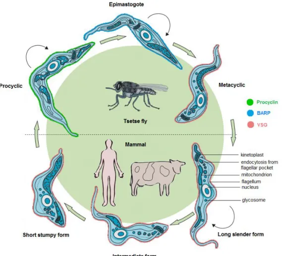
As for the other trypanosomes, Trypanosoma brucei is a heteroxenous parasite i.e. requires more than one obligatory host to complete its life cycle: a mammalian host and the blood-feeding tsetse fly vector. Infection in the mammal starts with a bite of an infected tsetse that inoculates the infective cell cycle-arrested metacyclic stage into the mammal bloodstream and draining lymphatics. T. brucei then differentiates to a long slender bloodstream form (BSF) that divides asexually by binary fission. In the mammal, parasites can live extracellularly in the blood or in the extravascular fluids and interstitial spaces of organs and tissues. At high parasite density, a yet unidentified parasite-released factor induces differentiation of long slender forms to short stumpy BSFs, passing through an intermediate form (Vassella et al., 1997, Reuner et al., 1997). Stumpy forms are non-dividing cells and the only life cycle stage transmissible to the tsetse, which can occur during a blood meal of the fly.
Once in the fly midgut, the stumpy forms differentiate into procyclic forms (PFs), which re-enter the cell cycle, actively multiplying and colonizing the fly gut. PFs undergo a complex differentiation process while migrating to the fly proventriculus. During this process each PF gives rise by an asymmetric cell division to one long and one short epimastigote; the long epimastigotes are not able to proceed in development while the short epimastigotes migrate to the tsetse salivary glands. Here, they attach to epithelial cells and start to multiply. Attached epimastigotes are the only life cycle stage known to perform meiosis (Peacock et al., 2011). Finally, in the salivary glands, epimastigotes differentiate to metacyclic forms, ready to be transmitted back to the mammalian host by a tsetse bite (Sharma et al., 2009) (Fig. 1).
Throughout its life cycle, T. brucei exhibits a series of specific adaptations that allow it to cope with the completely different environments it encounters in the mammal and the fly. The several life cycle stages reflect the alterations undergone by the parasite to be best adapted to the host’s environment while balancing survival,
6
Figure 1. Life cycle of African trypanosomes. In the bloodstream of the mammalian host,
parasites exist as a polymorphic population of bloodstream forms (BSFs) consisting of dividing (black arrows) slender forms and cell cycle arrested intermediate and stumpy forms. In the tsetse fly vector, after entering the midgut, stumpy forms differentiate to the procyclic forms (PFs), which further develop to the migrating epimastigotes. In the salivary glands, the latter transform into the infective metacyclic forms, which are injected during the next blood meal of the fly into the mammalian host (adapted from Brun et al., 2010).
proliferation and transmissibility. The process of differentiating from one life cycle stage to the other is complex and implicates deep changes in cell morphology and ultrastructure, metabolism, cell cycle and cell surface proteins, accompanied by a strong reprogramming in gene expression (Kramer, 2012).
So far, the best-studied differentiation pathway in T. brucei, and in kinetoplastids in general, is the differentiation of BSFs to PFs. Five recent studies brought to light the genome-wide changes in gene expression between these two developmental stages identifying a significant percentage of genes that are differentially expressed between them (6–40 % of all genes analyzed, depending on
7 the technology used) (Jensen et al., 2009, Kabani et al., 2009, Nilsson et al., 2010, Siegel et al., 2010, Veitch et al., 2010, Queiroz et al., 2009). This reflects the extraordinary parasite capacity of rapidly remodeling its gene expression pattern upon sensing the environmental changes that take place when switching hosts.
One of the fundamental changes occurring when T. brucei alternates between the mammal and the insect is the dramatic change of its surface protein composition. Inside the fly, PFs are coated with highly acidic and repetitive proteins, the procyclins (GPEET and EP procyclins), (Roditi et al., 1989, Clayton & Mowatt, 1989, Vassella et al., 2001) which are later exchanged for a coat of brucei alanine-rich protein (BARP) coat in epimastigotes (Urwyler et al., 2007). Further differentiation into the metacyclic stage replaces BARPs for a dense coat of metacyclic variant surface glycoproteins (MVSGs). After transmission to the mammal, a similar type of VSG (Cross, 1975) is maintained as the major surface protein in the BSF slender and stumpy forms of T. brucei (Vickerman, 1985). When stumpy forms differentiate to PFs, the VSG coat is shed and replaced by procyclins (Roditi et al., 1989) (Fig. 1).
As extracellular parasites, one of the major challenges that T. brucei is confronted with are the host’s natural defenses. The cell surface of the parasite lies at the host-parasite interface, serving as a first line of defense against the attacks mounted by the host. In PFs, the procyclin coat is crucial for parasite survival in the tsetse midgut (Ruepp et al., 1997, Acosta-Serrano et al., 2001). On the other hand, in BSFs the very dense coat of VSGs is crucial for protecting the parasite against the mammalian immune system. VSGs are molecules of approximately 55-65 kDa (Cross, 1975), which are present at the parasite cell surface as homodimers and are attached to the membrane by a GPI moiety (Ferguson et al., 1988). A stunning number of VSG homodimers are tightly and orderly packed at the parasite surface: ~107 VSG molecules per parasite (Jackson et al., 1985), which account for about 90% of its surface proteins (Grunfelder et al., 2002). Although the VSG family displays extensive sequence variation, the secondary and tertiary structure of these molecules is highly conserved (Blum et al., 1993, Carrington & Boothroyd, 1996), maybe to ensure that a dense VSG monolayer is always formed.
8
The VSG coat is a remarkable protective barrier against the innate and the adaptive immune system of the mammal. VSG protects parasites against recognition and lysis by the alternative complement pathway (Ferrante & Allison, 1983) and from anti-VSG antibody-mediated phagocytosis (Guirnalda et al., 2007, Pan et al., 2006). At low antibody titres, protection against antibody-mediated killing results in part from clearance of antibody-VSG complexes from the parasite surface, facilitated by the high rates of endocytosis and surface recycling existent in T. brucei (Barry, 1979, Natesan et
al., 2007) and the hydrodynamic forces generated by its flagellar movement (Engstler et al., 2007). Additionally, the dense VSG coat is probably shielding many other
cell-surface molecules from immune recognition such as invariant or less variable protein domains such as the VSG C-terminal domain (Schwede et al., 2011). There is also evidence that VSG shed from the membrane contributes to immune suppression of both B- and T-cell activation (Mansfield & Paulnock, 2005).
Even though all these phenomena cooperate to defend the parasite against the host immune response, T. brucei accomplishes immune evasion primarily through another exceptional strategy: antigenic variation of the VSG. As it will be discussed next, antigenic variation is as far as we know the leading process that allows the T. brucei parasite to thrive against the immune attack and persist in the bloodstream of the mammal for prolonged periods.
1.1.4 ANTIGENIC VARIATION - THE POWER OF CHANGING
Paradoxically, although the VSG coat is essential for immune evasion, it is extremely immunogenic. Most VSGs possess an N-terminal ‘variable’ domain, with high sequence divergence and which contains the epitopes exposed at the surface (Miller et al., 1984, Cross, 1984). The VSG C-terminal domain is generally more conserved and encodes the subsurface GPI-anchor signal (Cross, 1984, Carrington et
al., 1991). The VSG epitopes strongly elicit an antibody response in the mammal
(Morrison et al., 1982, Black et al., 2010). The parasite has the capacity to evade antibody-mediated killing mostly via antigenic variation of VSGs.
9
Figure 2. Antigenic variation during T. brucei infection. During infection the overall
parasitaemia, i.e. ‘total parasite burden’ in the blood varies in a wave-like manner (upper black line). Peaks correspond to maximal parasite density and valleys represent massive parasite clearance primarily by the action of VSG-specific antibodies produced by the host. A small number of VSG variants (‘major’) dominate the infections peaks, but the entire number of variants expressed at any time is greater and there may be many VSGs present in low abundance (‘minor’). Except for the ‘total parasite burden’, each line corresponds to different VSGs and peaks only once. The first peak is frequently dominated by the VSG most expressed in the initial population (day 0) (adapted from McCulloch & Field, 2015).
Typically during a T. brucei infection, VSG epitopes are recognized and activate host B-cells to produce VSG-specific antibodies against the ‘major’, predominantly expressed VSG in the population. The anti-VSG antibodies produced are largely responsible for clearing most of the parasites, leading to a drop in parasitaemia, i.e. in parasite density in the blood (Black et al., 2010). A few parasites in the population have switched their VSG and represent ‘minor’ VSG variants that have not been recognized yet by the immune system; as a result, these parasites will successfully replicate and substitute the previous parasite population. As prior VSG variants are recognized by the immune system and cleared, newly switched variants emerge, giving rise to the characteristic periodic waves of parasitaemia, with several infection peaks which reflect the episodes of parasite clearance and relapse, described for the first time in humans more than a century ago (Ross & Thomson, 1910) (Fig. 2).
Antigenic variation relies on two key features: VSG monoallelic expression and VSG switching. Monoallelic expression consists in the expression of a single VSG gene
10
out of a genomic repertoire of ~2,000 VSG genes encoding different variants of the protein (Cross et al., 2014). Therefore, each parasite expresses only one VSG variant at the surface at a time. The active VSG gene is expressed from a specialized subtelomeric locus called a bloodstream expression site (BES). Of the ~15 BESs present in the genome (Hertz-Fowler et al., 2008), only one BES is transcriptionally active, while the others are silent.
VSG switching can occur via different molecular mechanisms. The most frequent mechanism is the activation of a silent VSG by homologous recombination (HR) into the active BES (Robinson et al., 1999). Recombination is the only way to access the VSG repertoire of ~2000 genes that lie mainly in subtelomeric non-BES loci The most frequent HR switching mechanism is the so-called duplicative gene conversion. This involves duplication and insertion of an inactive telomeric or array
VSG into the active BES, replacing the previously active VSG gene. A new VSG might
also be assembled by segmental gene conversion in which segments of VSG genes and pseudogenes are copied and recombine to generate novel functional ‘mosaic’ VSGs that will replace the previously active VSG. In addition, HR can also mediate a telomere exchange in which reciprocal crossover between two telomeres (no DNA sequence is lost) and their associated VSGs takes place.
VSG switching can also be recombination-independent. This less common mechanism consists of a transcriptional activation of a different BES and silencing of the previous one with no involvement of DNA rearrangements by recombination. This is termed in situ switch (McCulloch et al., 2015, Rudenko, 2011). In contrast with BSFs,
in situ transcriptional activation is considered to be the only route by which MVSGs are
activated in metacyclic parasites (Barry & McCulloch, 2001).
Curiously, trypanosome infections typically show hierarchical expression of individual VSGs in which some variants appear early in infection while others appear progressively later (Gray, 1965, Miller & Turner, 1981, Morrison et al., 2005, Marcello & Barry, 2007). This sequential expression seems to depend on the differential activation probabilities of each VSG, which are dictated in part by the type of genetic locus they occupy. During infection, the first VSGs to be switched on are intact
11 telomeric VSGs resident in BESs and minichromosomes followed by inactive VSGs from subtelomeric arrays, and subsequently by ‘mosaic’ VSGs (Robinson et al., 1999, Morrison et al., 2005, Aitcheson et al., 2005, Marcello & Barry, 2007, Lythgoe et al., 2007, Hall et al., 2013, McCulloch et al., 2015).
We know that one infection peak can actually be composed by mixtures of subpopulations expressing more than one VSG variant (Miller & Turner, 1981, Hajduk & Vickerman, 1981, Robinson et al., 1999, Barry & McCulloch, 2001) (Fig. 2). Recent studies observed that although a small number of VSG variants dominate in each peak of infection, there was a higher than expected diversity of total VSG variants being expressed at any given time (an average of 20-30 VSG variants/peak). As a result, probably most of the intact VSG repertoire is exhausted rather early in infection and recombinatorial mechanisms such as mosaic formation should be critical to expand the
VSG repertoire. Perhaps the assembly of mosaic VSGs can even be the predominant
switch mode in nature, where infections are sustained for long periods (Hall et al., 2013, Mugnier et al., 2015).
It is important to note that VSG switching does not depend on the immune response to occur because T. brucei still switches VSG in axenic cultures (Doyle et al., 1980). Nevertheless, switching rates are noticeably higher (10-2-10-5 switches/trypanosome/generation) in strains recently isolated from nature or fly-transmitted which are ‘pleomorphic’, i.e. that retain the capacity to differentiate into stumpy forms and are tsetse transmissible (Turner & Barry, 1989, Turner, 1997). By contrast, in laboratory-adapted ‘monomorphic’ strains, which have lost the ability to naturally differentiate into the stumpy stage and complete the life cycle, switching frequencies are typically low (10-4-10-7 switches/trypanosome/generation) (Lamont et
al., 1986, Aitcheson et al., 2005, Boothroyd et al., 2009). This supports the view that
VSG switching is a stochastic process in which antibodies act chiefly a selective force, rather than a trigger.
Antigenic variation is essential for parasite survival. However, it needs to be balanced in order ensure infection chronicity. While VSG switching needs to occur at a frequency that ensures infection persistence, the appearance of new VSG variants
12
should be kept under certain limits to avoid VSG repertoire exhaustion or host immune system overwhelming (Gjini et al., 2010). The interplay between antigenic variation and stumpy differentiation seems critical for this balance. Stumpy forms are the only transmissible stage to the tsetse fly and differentiate at high parasite densities apparently through a quorum-sensing-like mechanism (Vassella et al., 1997, Reuner et
al., 1997). Although slender forms establish the first BSF population within the
mammal, stumpy forms predominate in chronic infections (MacGregor et al., 2011). Since stumpy forms do not divide and do not switch VSG they contribute to prevent population overgrow and limit the frequency of emergence of new VSG variants, optimizing the balance between transmission probability, parasite virulence and chronicity of infection (Matthews et al., 2015, MacGregor et al., 2011).
1.2 CHROMATIN
1.2.1 GENOME ORGANIZATION
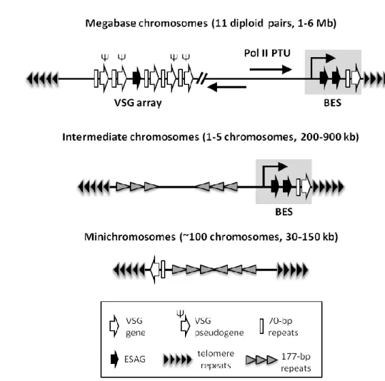
Trypanosoma brucei is a diploid organism with a nuclear genome of
approximately 35 Mb/haploid DNA content. Despite its small genome, the karyotype in
T. brucei is remarkable, consisting of more than 120 chromosomes (Berriman et al.,
2005) divided into three different classes according to their size: 11 megabase chromosomes (1-6 Mb), 1-5 intermediate chromosomes (200-900 kb) and ~100 minichromosomes (30-150 kb) (Melville et al., 2000, Wickstead et al., 2004, Cross et
al., 2014). The core of minichromosomes consists mostly of repetitive palindromes of
177-bp repeats (Wickstead et al., 2004). Intermediate chromosome structure is not entirely known but it shares with minichromosomes a large core of 177-bp repeats (Wickstead et al., 2004). Some strains of T. brucei also contain nuclear extrachromosomal circular DNAs, termed NlaIII repeat-elements (Alsford et al., 2003) of unknown function.
About 15% of the total DNA in trypanosomes corresponds to the kDNA which consists on a intertwined network of thousands of double-stranded circular DNA molecules which include large maxicircles (~23 kb) (Sloof et al., 1992) and smaller minicircles (~1 kb) (Chen & Donelson, 1980). Similarly to mitochondrial DNA in other
13 eukaryotes, maxicircles encode ribosomal RNA (rRNA) genes plus genes mostly coding for subunits of the mitochondrial membrane complexes involved in oxidative phosphorylation. The kinetoplast has a special replication and segregation mechanism, prior to nuclear DNA replication and division, respectively (Jensen & Englund, 2012, Woodward & Gull, 1990). Importantly, many of its maxicircle transcripts require a process of RNA editing in order to mature into functional and translatable messenger RNAs (mRNAs). This process is characterized by, sometimes extensive, insertions and/or deletions of uridine (U) nucleotides into the pre-mRNAs (Goringer, 2012).
Gene density in trypanosomes is extremely high, with the 26-megabase genome encoding ~9,000 putative genes encompassing all known housekeeping genes (Berriman et al., 2005). Trypanosome genome is organized in a very unusual way for eukaryotes. The majority of protein-coding genes are densely packed in large directional clusters in which genes are organized in a head-to-tail orientation. Most of these clusters are transcribed polycistronically by RNA polymerase II (Pol II). Hence, each of them is termed a polycistronic transcription unit (PTU) (Imboden et al., 1987, Muhich & Boothroyd, 1988) (see 1.3.1 Transcriptional and post-transcriptional regulation). Such type of organization resembles that of prokaryotic operons, except for the fact that in trypanosomes genes belonging to the same PTU are generally not functionally related (Berriman et al., 2005). Although polycistrons exist among other eukaryotes such as Caenorhabditis elegans, in trypanosomes they are remarkable for encompassing nearly all protein-coding genes in a genome-wide fashion: about 150 PTUs are predicted to exist in the housekeeping regions of T. brucei genome where some can harbor 100 genes (Siegel et al., 2009, Kolev et al., 2010).
1.2.2 THE LARGE VSG REPERTOIRE
Remarkably, the large family of VSGs is composed of ~2,000 genes and occupies a vast extension of the genome. Hundreds of silent VSG copies are organized in tandem arrays at proximal subtelomeric positions in megabase chromosomes or as single genes in minichromosomes (Williams et al., 1982, Berriman et al., 2005, Cross et
14
Figure 3. Distribution of VSG genes in the genome of T. brucei. Megabase chromosomes encode
all housekeeping genes, which are expressed in polycistronic transcription units (PTUs) by RNA polymerase II (Pol II) (see 1.3.1 Transcriptional and post-transcriptional regulation). Hundreds of
VSG copies are located proximal to subtelomeres in VSG arrays and include VSG genes,
pseudogenes and incomplete sequences. VSGs are transcribed in bloodstream expression sites (BESs) which reside at subtelomeres in megabase and intermediate chromosomes. A BES contains several expression site-associated genes (ESAGs) and a single functional VSG at its end.
ESAGs are interspersed through other genomic locations such as VSG arrays. Minichromosomes
can also contain VSGs at subtelomeres. Intermediate and minichromosomes contain stretches of 177-bp repeats, which in minichromosomes form a palindromic core. Typically, VSGs are flanked upstream by 70-bp repeats, important for homologous recombination-dependent VSG switching (see 1.3.2 Control of bloodstream expression sites). Arrows indicate direction of transcription. The diagram is not drawn to scale. For simplicity, genes are single-oriented but they display both orientations in the genome and can reside at both subtelomeric ends.
megabase or intermediate chromosomes (Becker et al., 2004, Hertz-Fowler et al., 2008) (Fig. 3).
In the genome of T. brucei VSGs can be grouped into four categories: ‘functional’ (encodes all recognizable features of known functional VSGs), ‘atypical’ (complete genes possibly encoding proteins with inconsistent VSG folding or post-translational modification), ‘pseudogene’ (with frameshifts and/or in-frame stop
15 codons), and ‘incomplete’ containing N-terminal or C-terminal fragments (Berriman et
al., 2005). Whereas putative functional VSGs represent a minority (13-20% in the strain
Lister 427), pseudo and incomplete VSGs predominate (Berriman et al., 2005, Marcello & Barry, 2007). Among this large family, there appears to exist small VSG subfamilies with high sequence identity at the N-terminal or C-terminal which are thought be important to provide interacting partners for mosaic VSG formation (Marcello & Barry, 2007). Part of the VSG archive is not unique (10% in T. b. brucei strain Lister 427), existing as two copies or more (Cross et al., 2014). Besides, MVSGs represent a very small part of the repertoire. In Lister 427 there are six MVSGs (Cross et al., 2014), with sequence identities consistent with those expressed in metacyclic parasites differentiated in vitro (Kolev et al., 2012).
The VSG repertoire is significantly divergent between species and strains and even between different life cycle stages or laboratory growth conditions. For examples, most VSG gene sequences in strains Lister 427 and TREU 927/4 are very distant. The closest match between both has less than 50% coding sequence (CDS) identity. An important observation was that the VSG archive also diverges considerably within the same strain (Lister 427) propagated as different life cycle forms (BSFs vs PFs) or cultured in distinct laboratories, exhibiting loss or duplication of VSG genes (Cross et
al., 2014). Besides providing us with a comprehension of the diversity of the VSG
family, such data draws attention for the importance of knowing the ‘VSGnome’ repertoire of the strain/isolate being studied.
Similarly to T. brucei, many other pathogens such as Plasmodium falciparum (malaria parasite) (Scherf et al., 2008) and Borrelia spp. (Lyme disease bacteria) (Barbour et al., 2000, Zhang et al., 1997) express their variant surface antigens at subtelomeres. This highlights the existence of preferential conditions at these genomic locations for the function of such genes. Since subtelomeres are prone to ectopic recombination it seems likely that these are privileged sites for VSG duplication and recombination, and therefore for VSG repertoire diversification; besides, it has become apparent that the characteristic reversible silencing events associated with the telomere probably sustain monoallelic VSG expression and provide transcriptional insulation of silent VSG copies, essential to antigenic variation (Barry et al., 2003). The
16
relationship between telomeric location and VSG silencing will be discussed in a subsequent section (see 1.3.2. Control of bloodstream expression sites).
1.2.3 CHROMATIN ORGANIZATION IN EUKARYOTES
Organization of DNA into chromatin is a hallmark of the eukaryotic nucleus. Chromatin is necessary for packaging the DNA molecules inside the physical limits of the nucleus and plays important roles in essential processes such as DNA replication, DNA damage repair, RNA processing and transcription. The basic unit of chromatin is the nucleosome: ~145–147 bp DNA superhelix wrapped around an octamer composed of two copies each of the histone proteins H2A, H2B, H3 and H4 (Luger et al., 1997). Interacting with the nucleosome core, there is usually a distinct histone, histone H1 (H1) or ‘linker histone’, which binds externally to the nucleosomal DNA and the ‘linker DNA’ i.e. the short DNA segments that interconnect nucleosomes, and that altogether with the nucleosome core forms the chromatosome (Simpson, 1978, Bharath et al., 2003). Histones are small proteins (~13 kDa) rich in positively charged, basic amino acids (aa) (e.g. arginine and lysine) that facilitate interaction with the negatively charged DNA molecules.
The long-held notion is that chromatin can be organized in a series of increasingly complex and compact conformations which go from ‘primary’ to higher-order ‘secondary’ and ‘tertiary’ structures. Primary structure of chromatin consists in the linear DNA molecules arranged in long nucleosomal arrays often referred as ‘beads-on-a-string’. A secondary structure is defined as that arising from the folding and condensation of individual nucleosomal arrays into chromatin fiber, such as the often mentioned ’30-nm fiber’, which is driven by short-range interactions between neighboring nucleosomes where histone H1 seems to be a key player (Robinson & Rhodes, 2006). Succeeding interactions between fiber leads to large-scale chromatin assemblages, designated as tertiary structure, which ultimately culminates in the fully condensed chromosomes typically observed during metaphase (Luger et al., 2012). Despite the well-established importance of higher-order structures for chromatin function, the conformation of such structures, particularly that of the 30-nm fiber, still
17 remains unsolved and, more recently, its existence in vivo has even been questioned (Maeshima et al., 2010, Nishino et al., 2012).
Chromatin can be organized and regulate short-range DNA domains, for example the local structure of an active promoter, or larger DNA domains (up to Mb of length) generating local structures at the micro-scale. For instance, chromatin can organize into nuclear bodies, chromatin domains or territories, which might involve several chromatin modifications and binding proteins and anchoring to nuclear structures like the nuclear lamina (Pombo & Dillon, 2015). The nucleolus is a hallmark example of a nuclear body of eukaryotes: it is the subnuclear compartment where transcription of ribosomal DNA (rDNA) by RNA polymerase I (Pol I), rRNA processing and ribosome assembly takes place (Lam & Trinkle-Mulcahy, 2015).
Chromatin domains can be subdivided in heterochromatin and euchromatin. In a simplified definition, heterochromatin is more packed (visible as electron-dense domains), gene-poor and generally less transcriptionally active. By contrast, euchromatin typically consists of open chromatin regions that are gene-rich and transcriptionally active. Transposable elements and repetitive DNA regions such as the centromere and the telomere are usually organized in constitutive heterochromatin, which suppresses transcription and/or DNA recombination at these loci, maintaining genome integrity. On the other hand, facultative heterochromatin has the potential to interchange with euchromatin, i.e. to reversible convert to more decondensed chromatin and transcriptionally active state, for instance in the context of monoallelic gene expression (Trojer & Reinberg, 2007). In most cell types repressive heterochromatin is organized around the nucleolus or tethered to the nuclear envelop, whereas euchromatin is found at more central positions in the nucleus (Misteli, 2005, Padeken & Heun, 2014, Pombo & Dillon, 2015).
Different structural and functional states of chromatin can be defined by DNA modifications and variations in nucleosome composition specified by different histone variants or histone covalent post-translational modifications (PTMs) (e.g. methylation, acetylation, phosphorylation), especially present at their N-terminal tails (Luger & Richmond, 1998). Given the ever-increasing number of histone PTMs identified, the
18
theoretical possible combinations of nucleosome components are astonishing and have the capacity to ‘code’ for a large number of chromatin states. PTMs are added or removed from histones by several histone-modifying enzymes, which ‘write’ or “erase” the code. Other chromatin-binding proteins can “read” specific histone PTMs and recruit several other factors such chromatin remodelers and chromatin architectural proteins which trigger a set of downstream events that include changes in nucleosome positioning, DNA accessibility and recruitment of other nuclear machinery e.g. transcription factors (Strahl & Allis, 2000, Lee et al., 2010, Yun et al., 2011).
Together with histone PTMs, histone variants contribute to alter nucleosome stability, chromosome structure and gene expression. Histone variants are non-allelic variants with a distinct amino acid sequence from their canonical counterparts and their deposition onto the DNA is not replication-dependent. Variants might substitute missing histones or be specifically recruited to defined genomic locations to serve specific functions (Luger et al., 2012).
The action of all these players results in a pallet of dynamic chromatin states which can influence for instance, DNA replication, RNA processing and transcription, for instance, working as epigenetic on/off transcriptional switches (Preuss & Pikaard, 2007, Venkatesh & Workman, 2015).
1.2.4 CHROMATIN ORGANIZATION IN T. BRUCEI
Alike other lower eukaryotes, T. brucei mitosis is ‘closed’, which means that nuclear envelope and nucleolus do not disassemble (Vickerman & Preston, 1970, Ogbadoyi et al., 2000). Interestingly, even though all chromosome classes segregate via a mitotic spindle (Ersfeld et al., 1998), it was recently found that T. brucei uses unconventional kinetochores which appear to have emerged exclusively in kinetoplastids early in evolution (Akiyoshi & Gull, 2014).
The main nuclear architectural features typical of eukaryotes are also observed in T. brucei such as a double bilayered nuclear envelope, nuclear pore complexes with a conserved composition (DeGrasse et al., 2009), a nucleolus and domains of
19 euchromatin and heterochromatin, the latter predominant at the nuclear periphery (Ersfeld, 2011). While the typical nuclear lamina found in metazoans seems to be absent from the parasite (Field et al., 2012), one protein with lamin-like functions, NUP-1, has been identified in T. brucei (Rout & Field, 2001, DuBois et al., 2012).and shown to be important for nucleus structural integrity and telomere positioning at the nuclear periphery (DuBois et al., 2012).
Contrary to what generally occurs, in trypanosomatids chromosomes do not visibly condense during metaphase and a 30-nm fiber has never been observed (Hecker & Gander, 1985, Burri et al., 1995). Overall trypanosome DNA is less compacted within the nucleus when compared with higher eukaryotes (Hecker & Gander, 1985) and even between two stages of the parasite life cycle, with chromatin being more compact in BSFs than in PFs (Schlimme et al., 1993). This has been suggested as an important adaptation to accommodate the rapid changes in gene expression required throughout life cycle development of the parasite (Hecker et al., 1994, Belli, 2000). Curiously, the less compacted state of trypanosome chromatin has been attributed the presence of divergent histones and differential expression of histone variants between BSFs and PFs, namely that of H1 proteins (Schlimme et al., 1993, Burri et al., 1994).
A paradigmatic example of nuclear subcompartmentalization in T. brucei is the so-called expression site body (ESB). This is an extranucleolar Pol I body, functionally distinct from the nucleolus and that appears to be solely dedicated to the transcription of the active BES in BSFs (Navarro & Gull, 2001, Chaves et al., 1998). Telomeres also occupy different nuclear positions throughout development of T. brucei. While in the bloodstream slenders, at interphase, telomeres preferentially localize at inner regions of the nucleus in the transmissible, non-dividing stumpy and in the insect PF stage, telomeres cluster close to the nuclear periphery and appear to be associated with heterochromatin (Ogbadoyi et al., 2000, Perez-Morga et al., 2001). Thus, telomeres are closer to the peripheral heterochromatic regions in those life cycle stages in which active BES is almost (Amiguet-Vercher et al., 2004) or completely inactive (stumpy and PF, respectively). While telomeric repeats are probably packed into constitutive heterochromatin, the BESs at the subtelomeres probably correspond to facultative