1
Title: Is differential diagnosis attainable in disarticulated pathological bone remains? A
case-study from a late 19th/ early 20th century necropolis from Juncal (Porto de Mós, Portugal)
Author names and affiliations:
Sandra Assis, LABOH – Laboratório de Antropologia Biológica e Osteologia Humana, CRIA/FCSH, Universidade Nova de Lisboa, Portugal; CRIA – Centre for Research in Anthropology, Faculdade de Ciências Sociais e Humanas, FCSH, Universidade Nova de Lisboa, Portugal 1200-069. Email: [email protected]
Charlotte Yvette Henderson, CIAS – Research Centre for Anthropology and Health, Department of Life Sciences, University of Coimbra, Portugal 3001-401. Email: [email protected]
Sílvia Casimiro, LABOH – Laboratório de Antropologia Biológica e Osteologia Humana, CRIA/FCSH, Universidade Nova de Lisboa, Portugal; CRIA – Centre for Research in Anthropology, Faculdade de Ciências Sociais e Humanas, FCSH, Universidade Nova de Lisboa, Portugal. Email: [email protected]
Francisca Alves Cardoso, LABOH – Laboratório de Antropologia Biológica e Osteologia Humana, CRIA/FCSH, Universidade Nova de Lisboa, Portugal; CRIA – Centre for Research in Anthropology, Faculdade de Ciências Sociais e Humanas, FCSH, Universidade Nova de Lisboa, Portugal 1200-069. Email: [email protected]
Corresponding author:
Sandra Assis, CRIA – Centre for Research in Anthropology, Faculdade de Ciências Sociais e Humanas, Universidade Nova de Lisboa, Portugal 1200-069. Email: [email protected]
Present/permanent address:
CRIA – Centre for Research in Anthropology, Faculdade de Ciências Sociais e Humanas, Universidade Nova de Lisboa, Portugal 1200-069.
2
Is differential diagnosis attainable in disarticulated pathological bone remains? A case-study from a late 19th/ early 20th century necropolis from Juncal (Porto de Mós,
Portugal)
Abstract
Differential diagnosis is a fundamental step in every palaeopathological study. It is a challenging exercise since many intrinsic and extrinsic factors may negatively impact the accurate interpretation of bone changes in human skeletal remains. Among these, the completeness and preservation of skeletal elements plays a significant role. This study aims to explore the limits of differential diagnosis in the analysis of disarticulated, fragmented bones. The sample consists of eleven adult long-bone fragments with noticeable changes. The remains were identified in a dis-articulated skeletal assemblage from the former necropolis of Juncal (Porto de Mós, Portugal), which probably closed in the late 19th century/early 20th century. They were analysed visually and with X-radiography, and the changes carefully described prior to differential diagnosis. Five bones presented signs of healed bone trauma and one showed features compatible with leg amputation. Periosteal reactions were observed in several bones, one of them resembling changes consistent with an overlying skin ulcer. Two bone specimens were identified as belonging to the same individual due to the matching bone changes. Despite the incomplete remains, a broader diagnosis was possible for most cases, which facilitated a discussion of health, medical and social care among the inhabitants of the region.
Keywords: palaeopathology, trauma, periosteal reactions, joint changes, amputation,
3
1. Introduction
Differential diagnosis occupies a central role in the challenging task of understanding disease in past human populations (Larsen, 2006). In palaeopathology, differential diagnosis is a process of ongoing improvement, as new imaging, genetic and biochemical techniques are slowly unveiling the complex co-evolutionary interactions of host-pathogens, as well as the impact of environmental changes in past disease expression (Grauer, 2012). More often, diagnostic criteria for dry bone rely on the detailed observation and description of the bone changes “coupled with a logic-driven problem-solving framework” (Klaus, 2015: 13), and using clinical imaging data from living patients as comparative sources (Mays, 2012). Even so, a considerable number of factors are described in the palaeopathological literature as impacting our ability to assess the aetiology of bone changes. These factors may be associated with the object of analysis, i.e., skeletonised human remains, the limited bone tissue response to disease and injury, and the fragmentary and/or commingled nature of some skeletal assemblages.
Commingling of human skeletal remains can occur at any stage of burial, in association with certain funerary practices, as a result of excavation and storage of skeletal assemblages, and even after it (Fox and Marklein, 2014). The study of commingled remains, defined by Osterholtz and co-authors (2014) as human and/or faunal remains that have become de-individualized due to the mixing of elements, either intentionally or unintentionally, is not a straightforward exercise especially if it aims to reconstruct past population’s health, behaviour and cultural practices. The presence of fragmentary remains, often occurring in association with commingled assemblages also increases the interpretative difficulties (Osterholtz et al., 2014). Regarding these problems, several publications focussing on theoretical and new methodological approaches in the study and interpretation of commingled, disarticulated, or disturbed skeletal remains in bioarchaeological and forensic contexts have been published (for a review see Adams and Byrd, 2008 and Osterholtz et al. 2014, and authors herein. See also Adams and Byrd, 2006; Tuller and Ðuric, 2006; Silva et al., 2009; Cabo et al. 2012; Varas and Leiva, 2012; Gregoricka, 2014; Finlayson et al., 2017; Geber et al., 2017; Mahfouz et al., 2017; Verdugo et al., 2017). Despite these problems, the difficulty in establishing a direct correlation between an isolated pathological bone and systemic conditions remains a barrier in the paleopathological study of commingled samples (Fox and Marklein, 2014). The presence of post-mortem changes (including the so-called pseudopathologies) mistakenly identified as ante-or peri-mortem signs of trauma and disease also interfere in the differential diagnosis (Pinhasi and Bourbou, 2008; Turner-Walker, 2008). In fact, poor preservation or under-representation of specific skeletal parts, both at the macroscopic and histological levels, constitutes a serious impediment to diagnostic accuracy in both undisturbed and commingled remains (Pinhasi and Bourbou, 2008; Assis et al., 2015). Pathological analysis should remain generalized, when fragments of the same individual cannot be identified (Fox and Marklein, 2014) and the temptation to over interpret the lesions avoided (Sheridan, 2017). However, the careful palaeopathological study of commingled remains is possible, as supported by several studies (e.g., Willmon et al., 2013; Brickley and Buckberry, 2015; Ellis, 2016; Williams and Polet, 2017).
This paper aims to explore the limits of differential diagnosis in the palaeopathological analysis of disarticulated and fragmentary bone pieces recovered after the accidental destruction of part of the former necropolis (dated approximately to between 1780 and the late 19th or early 20th century) of the village of Juncal (Portugal).
4
2. Material and Methods
During 2006 construction work in an abandoned courtyard in the village of Juncal (Porto de Mós, Centre of Portugal) (Fig. 1), human skeletons and bones were discovered, randomly collected and stored in wooden boxes by construction workers. This discovery occurred in the vicinity of São Miguel church, founded in 1780, and confirmed as the location of the former necropolis of Juncal. There is no available information with regard to the founding date for this necropolis, but it is likely that it ceased functioning in the late 19th century or early 20th century, following the construction of a new cemetery on the outskirts of the village.
With the aim of safeguarding further evidence, an archaeological and anthropological emergency excavation was conducted. The archaeological and anthropological surveys excavated the remains of three nonadult skeletons and numerous disarticulated and fragmentary bone pieces. Only a field assessment of the remains was possible because of the mandated for expedient reburial. Due to time and logistical constraints (prior to reburial), it was only possible to determine the minimum number of individuals, which was estimated at 169 individuals: 154 adults and 15 nonadults, based on the complete skulls and frontal bones. The estimate of the minimum number of individuals followed the recommendations of Herrmann (1990). In general, the disarticulated assemblage contained bones from almost all parts of the skeleton, each of which exhibited different levels of preservation and/or completeness (time constraints mean that this could only be assessed). Among the disarticulated bone remains, eleven had noticeable alterations; they were retained for subsequent palaeopathological analysis and diagnosis. These elements were visually inspected with the aid of a magnifying lens, and some x-rayed. All changes were carefully described according to the degree of preservation, the location of the lesion(s), and the type of bone response. A differential diagnosis followed.
3. Results and discussion
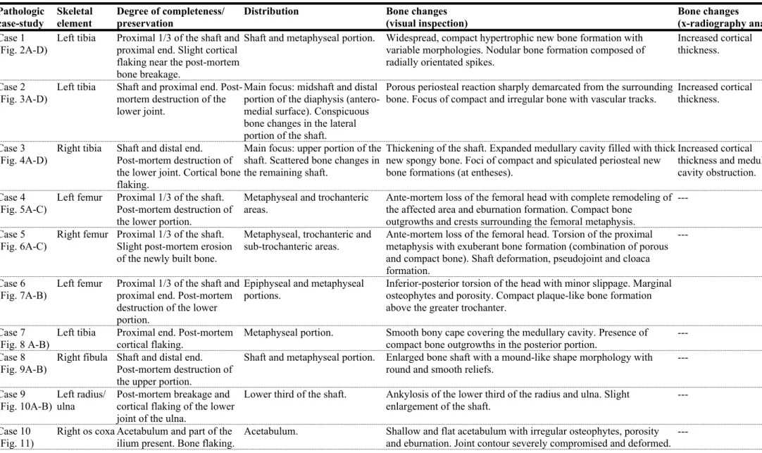
A descriptive summary of the eleven pathological cases is presented in Table 1 (to access the detailed description and differential diagnosis of each case, please see the Supplementary Materials).
Of the eleven bone fragments analysed, ten were long bones and one a fragment of a right os coxa. Nine long bones belonged to the lower limb and two to the upper limb. Apart from the post-mortem bone, no major taphonomic alterations were noticed. Six bone pieces exhibited slight cortical bone flaking. All the disarticulated bones were from adult individuals of unknown age-at-death and sex. Although sex estimation in commingled assemblages is possible, for instance, using regional or population specific long bone dimensions (e.g., the femoral head, Wasterlain, 2000), the presence of severe pathological changes made this approach unfeasible.
The presence of new bone formation was a common feature in several pathological cases, corroborating the multifactorial aetiology of periosteal reactions and their apparent lack of specificity (for a review see Weston, 2008, 2009, and 2012). This inference was particularly notorious in case 1 (Fig. 2 A-D), case 2 (Fig. 3 A-D) and case 3 (Fig. 4 A-D), in where exuberant deposits of periosteal new bone were observed along the tibial shaft. Although a positive diagnosis of chronic skin ulcer was established in case 2, in the tibial fragment of
5 case 1 it was impossible to reach a more conclusive diagnosis. In this particular case, a range of potential disorders were considered, and not fully discarded from the differential diagnosis, among them melorheostosis, hypertrophic osteoarthropathy or secondary HOA, inflammatory or infection processes, and fluorosis. Skeletal fluorosis, for instance, requires high levels of fluroide intake, normally through water consumption and eating leafy plants grown in areas with high water fluoride levels (Littleton, 1999). In Portugal, naturally occurring water with high fluoride levels only occurs in the northern part of the country, where the fluoride typically binds to calcium forming low soluble CaF2 (Eggenkamp et al., 2015), but fluorosis is not reported in the clinical literature in mainland Portugal. Thus skeletal fluorosis is an unlikely cause of the changes observed in case 1 due to the local geology, although without the dentition it cannot be completely excluded.
Despite the lack of cloaca formation (in the preserved fragment of case 3), the presence of poorly organized and dense trabeculae in the marrow, as well as of slight periosteal reactions makes a diagnosis of osteomyelitis possible. Also possible is a case of Garré’s sclerosing osteomyelitis. In a recent paper, Giuffra and co-authors (2015) described a set of bone changes observed in the right tibia of an adult male skeleton from medieval Tuscany, which resemble this at both the macroscopic and radiographic levels. The diagnosis advanced by Giuffra and colleagues (2015) was a possible case of chronic sclerosing osteomyelitis of Garré. Acquired syphilis could also cause similar changes and cannot be excluded as a differential diagnosis, albeit an improbable one.
In this assemblage, it was possible to identify five cases compatible with healed fractures (three hip fractures, one fracture of the fibula and one fracture of the radius/ulna), and one amputation. Of the three hip fractures, two were classified as extra-capsular fractures (case 4, Fig. 5 A-C and case 5, Fig. 6 A-C), and one as an intra-capsular fracture of subcapital type (case 6, Fig. 7 A-B) (Galloway, 2014). Complete displacement and non-union of the femoral head was also observed in cases 4 and 5. Although the femoral head was not recovered, one cannot exclude a case of necrosis of the femoral head, as a secondary complication, since this condition may develop as the result of a subcapital or transcervical fracture of the femur neck (Ortner, 2003). Similarly, a case of stable slipped femoral capital epiphysis (SFCE), in which only a subtle posterior slip of the epiphysis on the metaphysis occurs (Loder, 1998), cannot be completely disregarded from the differential diagnosis of case 6.
Mild to severe changes in the bone shape and size, secondary osteoarthritis, as well as a case of infection, i.e., pyogenic osteomyelitis (case 5) were diagnosed as secondary manifestations of trauma. In fact, the presence of osteoarthritis and/or extensive signs of bone remodelling led us to reject a possible relationship between those changes of cases 4 and 7 and congenital abnormalities, respectively femoral aplasia and lower leg maromelia (Barnes, 2012). In the case diagnosed as a tibial amputation (case 7, Fig. 8 A-B), the presence of a bony cap covering the medullary cavity suggests that the individual has recovered and survived the amputation. On the other hand, the absence of a pseudojoint associated with extensive callus formation disregards a case of pseudoarthrosis following, for example, a non-consolidated fracture (Aufderheide and Rodríguez-Martín, 1998; Adler, 2000). The changes observed in the right fibula of case 8 indicates a rotational ankle fracture (Fig. 9 A-B) whereas those of case 9 are compatible with a radioulnar synostosis of post-traumatic origin (Fig. 10 A-B). The congenital form of radioulnar synostosis was ruled out due to the distal location of the lesion and its association with a bony callus (Stevenson, 2006 and authors herein).
6 Finally, a severe case of osteoarthritis of the hip was diagnosed in the acetabulum of a right os coxa (case 10, Fig. 11) and in a right femoral head (case 11, Fig. 12), as justified by the presence of its most distinguishing features, namely eburnation and combined irregular marginal osteophytes, porosity, and destruction of the joint contours (Waldron, 2009). Despite the mushroom appearance of the femoral head, the presence of the fovea capitis made a diagnosis for Legg-Calvé-Perthes disease improbable. The severity of the lesions observed and their anatomical location appears to suggest that both bone fragments belonged to the same individual. This hypothesis was tested and confirmed through re-articulation of the two bone fragments.
Despite the inherent limitations of a fragmentary and commingled assemblage, the results clearly show that positive diagnosis is possible. It should be mentioned, however, that the type and exuberance of the lesions aided the differential diagnosis considerably. Nevertheless, it was impossible to determine if some of the conditions were caused or potentiated by hidden pathologies. For instance, one cannot disregard an association between hip fractures and osteoporosis. In senile or postmenopausal individuals (primary osteoporosis), a decrease in the bone tissue mass may occur (osteopenia) due to an imbalanced remodelling process (Brickley and Ives, 2008). This fact diminishes the ability of bone to repair, as well as to resist fatigue, which increases the risk of fracture (Brickley and Ives, 2008). Dry bone evidence of osteoporosis may be inferred through bone mass and microstructural analysis (Mays, 2008), as well as through the presence of fractures in the distal radius (Colles' fracture), vertebrae and at the proximal femur (hip fractures) (Agarwal, 2008; Brickley and Ives, 2008). The possible relationship between age and the presence of fractures, namely those of the hip, has been widely discussed in the clinical and palaeopathological literature (e.g., Koval and Zuckerman, 2000; Agarwal and Stout, 2003 and authors herein; Agarwal, 2008; Brickley and Ives, 2008; Mays, 2008). For example, Curate and co-authors (2013) studying a Portuguese identified skeletal assemblage found an increasing frequency of “osteoporotic fractures” among older individuals of both sexes. The frequency of fractures was also higher in females diagnosed with osteoporosis (Curate et al., 2013). The inability to infer the age and sex of those individuals displaying hip fractures made it impossible to establish any relationship between this type of lesion and osteoporosis in these disarticulated cases.
The underlying mechanism that caused the skin ulcer of case 2 is also unknown. Skin ulceration normally results from a deficient blood circulation in the extremities. In addition to chronic venous insufficiency, other conditions of vascular (e.g., atherosclerosis, arteriovenous malformation, rheumatoid arthritis, systemic lupus erythematosus, scleroderma, polyarteritis nodosa), neuropathic (e.g., diabetes, peripheral neuropathy), haematological (e.g., sickle cell anaemia), traumatic (e.g., burns, cold injury), neoplastic (e.g., skin tumour, basal or squamous cell carcinoma) and infectious origins (e.g., chronic osteomyelitis, leishmaniasis, leprosy, herpes, Madura foot, tropical ulcer, amongst others) may lead to the development of skin ulcers (Mekkes et al., 2003; Simon and McCollum, 2004; Abbade and Lastória, 2005). An identical inference can be made for the limb amputation, since this type of surgical procedure is carried out in cases of trauma, venous diseases, frostbite and burns, ergot (and other toxin) poisoning, wound infections (e.g., gas gangrene and related sepsis), sensory neuropathies and diabetes mellitus, and massive benign and malignant tumours (Kirkup, 2007). For example, prior to modern chemotherapy, improved imaging and biopsy diagnoses, and advanced reconstructive surgery, amputation was the mainstay of treatment in cases of malignant bone tumours, such as osteosarcoma (Bacci et al., 2002; DiCaprio and Friedlaender, 2003). Surgical amputation may also be carried out when co-morbidities are present. For example, it
7 is frequent in cases of chronic venous insufficiency associated with diabetes (e.g., Johannesson et al., 2009; Bruck, 1992).
The lack of sufficient information regarding the exact chronology of the remains hampered the reconstruction of the environmental and living conditions of the individuals, and their possible association with the bone conditions observed. It is known from historical sources that the village of Juncal always had a strong economic dependence on agriculture, being located in a fertile rural area (Gil, 2011). In 1770 an earthenware factory specialising in the production of decorative earthenware, dishware and tiles was built in Juncal (Martins, 2016). This factory, then called the Royal Factory of Juncal a title conferred by Queen Mary I due to the quality and prestige of the material produced, was severely destroyed during the Napoleonic wars. It was re-built in 1811 and worked until its definitive closure in 1876 (Martins, 2016). Juncal is also located in a region in which the extraction of clays, lime and stones has occurred at least since the Middle Ages (Gil, 2011). Accordingly, one cannot exclude that some of the cases described, especially those of traumatic origin, may be work-related or associated with the hazards of daily life. A close relationship with some pathologies common in Portugal at that time is also possible. In the 19th century, for instance, several diseases such as cholera, tuberculosis, yellow fever, and typhoid fever were responsible for high rates of mortality in Portugal (Silva, 2004; David de Morais, 2008; Almeida, 2011; Longo, 2015). Leprosy, syphilis, tetanus, malaria, flu, gastritis, enteritis and dysentery, scabies, gonorrhoea, leishmaniasis, brucellosis, and carbuncle (anthrax) were also common among the Portuguese adult population (Almeida, 2014). Tumours, hydropsy, apoplexy, fulminant haemorrhages, and acute or chronic inflammations were frequent, especially among older individuals (Veiga, 2004). Some of these conditions are considered comorbidities, for example, of chronic ulceration of the lower leg, or may have led to limb amputation.
Inferring access to medical care is very difficult to establish from this analysis of pathological cases. In the case of tibial amputation, the bony scar seems to suggest an effective and straight bone cut, probably performed by someone with anatomical knowledge. In contrast, the presence of fracture complications caused by ineffective immobilization and infection seems to suggest a lack of appropriate medical care. This difference in care may be due to temporal changes or differential socio-economic access to treatment, but without dating evidence for these bones this cannot be fully confirmed. Although medical knowledge in Portugal in the 19th century was as advanced as in other European countries, not all Portuguese citizens received the same medical care. While the best hospitals, physicians and surgeons were located in the main cities of Lisbon, Porto and Coimbra, the rural areas struggled with insufficient medical and sanitary conditions, as well as, human and material resources (Almeida, 2014).
The different pathological outcomes may also reflect the type of lesion and treatment available. Although the term “amputation” was only introduced in the surgical treatises of the 17th century, it is a procedure with a long history (Kirkup, 2007). Through time, the introduction of more effective surgical instruments and the development of new techniques of vessel ligature and skin suture did not completely eliminate the risk of complications, but contributed to increasing rates of survival and recovery (Kirkup, 2007). The same cannot be said, for instance, in cases of comminuted subtrochanteric femoral fractures. Nowadays, these fractures are described as one of the most difficult to manage in orthopaedic surgery (Rohilla et al., 2008; Hak et al., 2015; Wang et al., 2016), requiring complex surgical interventions and implants for fixing the broken pieces (Wang et al., 2016). None of which was available in the
8 past. Even today, numerous complications may arise during and after surgery, including blood loss, avascularity of the fragments, infection, delayed union or non-union (Koval and Zuckerman, 2000; Rohilla et al., 2008). Despite the severity of the pathological cases, all elements showed evidence of healing, meaning that the individuals have survived (at least to some extent) the traumatic event. In the most disabling cases, such as cases 4 and 5, it also indicates that individuals must have had strong social support to allow long-term survival. In summary, this study revealed that: (1) differential diagnosis is possible in fragmented remains; (2) the type, location and severity of lesions can help to reunite bones from the same individual, improving the accuracy of diagnosis; (3) trauma, infection and joint changes were some of the conditions that affected the inhabitants of Juncal in the past; (4) medical treatment was available for some conditions; and finally (5) familial and social support were probably strong in this village, which is corroborated by the survival and recovery of individuals.
4. Concluding Remarks
This paper has presented and discussed the differential diagnosis of eleven bone fragments with exuberant bone changes. Of the different categories of disease, trauma, was the most represented skeletal pathology. Signs of probable inflammation (and/or infection) of the periosteum, cortical tissue and medullary cavity, and primary and secondary joint changes (often associated with entheseal changes) were also frequent. Determining if the fractures were purely accidental or the result of underlying bone fragility is almost impossible. Even so, the presence of signs of bone healing, as well as of surgical procedures (i.e., amputation) seem to suggest that the former inhabitants of Juncal had access to some degree of care and social support during recovery, contributing to their survival. It is highly probable that some of the fractures observed negatively impacted the individual’s daily routine, affecting their mobility and the ability to self-care. For other pathological cases (i.e., case 1) it was more difficult to establish a more conclusive diagnosis.
The recovery of single bones rather than complete skeletons was a major impediment to evaluating the skeletal distribution of the bone changes, as well as to assessing the biological profile of the individuals. The former is pivotal for every differential diagnosis; the latter is important in the diagnosis of conditions that may show different distributions by age group or sex. These limiting factors hampered the access to possible hidden conditions and made the differential diagnosis in terms of the aetiology of the changes unattainable. Despite the incomplete remains, a broader diagnosis or range of diagnoses was possible for most cases, aided by the type and exuberance of the lesions. Broader diagnoses, as well as considerations as to how these changes may have impacted a person's life and the need for medical and social care still provide valuable insights into health, disease, medical and social care in past populations. Thus case studies at both an individual level and in disarticulated remains can provide palaeopathological insights for populations without complete skeletons.
Acknowledgements
S. Assis is a post-doctoral fellow (grant number: IF/00127/2014/POSDOC) of the Exploratory Project Bone Matters / Matérias Ósseas (IF/00127/2014/CP1233/CT0003) funded by
9 Fundação para a Ciência e a Tecnologia. C Henderson is funded by Fundação para a Ciência e a Tecnologia (grant number: SFRH/BPD/82559/2011). S. Casimiro is CRIA’s research grantee and funded by CRIA’s Strategic Development Plan (reference: CRIA - UID/ANT/04038). F Alves Cardoso is funded by Fundação para a Ciência e a Tecnologia FCT Investigator Award Programme (IF/00127/2014). The European Commission ESF and POPH support Fundação para a Ciência e a Tecnologia. The authors would like to thank the Clínica Universitária de Imagiologia dos Hospitais da Universidade de Coimbra (HUC), CIAS – Centro de Investigaçãoe em Antropologia da Universidade de Coimbra, Lucínia Oliveira and ArqueoHoje Lda.
This work was first presented as a poster at the 20th Congress of the European Anthropological Association, European Anthropology in a Changing World: From Culture to Global Biology, held between the 24th and 28th of August of 2016. We would like to acknowledge the participation in the poster of the students Andreia Mendes and Sónia Soares (Department of Anthropology, FCSH, Universidade Nova de Lisboa).
References
Abbade, L., Lastória, S., 2005. Venous ulcer: epidemiology, physiopathology, diagnosis and treatment. Int. J. Dermatol. 44, 449-456.
Adams, B.J., Byrd, J.E., 2006. Resolution of small-scale commingling: a case report from the Vietnam War. Forensic Sci. Int. 156, 63–69.
Adams, B.J., Byrd, J.E., 2008. Recovery, Analysis, and Identification of Commingled Human Remains. NJ, Humana Press, Totowa.
Adler, C.P., 2000. Bone Diseases: Macroscopic, Histological and Radiological Diagnosis of Structural Changes in the Skeleton. Springer, Berlin.
Agarwal, S., 2008. Light and broken bones: examining and interpreting bone loss and osteoporosis in past populations, in: Katzenberg, A., Saunders, S., (Eds.). Biological Anthropology of the Human Skeleton. 2nd edition. John Wiley & Sons, New Jersey, pp. 387-410.
Agarwal, S., Stout, S., 2003. Bone loss and osteoporosis: an anthropological perspective. Springer Science & Business Media, New York.
Almeida, M.A.P., 2011. A epidemia de cólera de 1853-1856 na imprensa portuguesa. História, Ciências, Saúde – Manguinhos, Rio de Janeiro 18, 1057-1071.
Almeida, M.A.P., 2014. As epidemias nas notícias em Portugal: cólera, peste, tifo, gripe e varíola, 1854-1918. História, Ciências, Saúde –Manguinhos, Rio de Janeiro 21, 687-708.
10 Assis, S., Keenleyside, A., Santos, A.L., Alves Cardoso, F., 2015. Bone Diagenesis and its implication for disease diagnosis: the relevance of bone microstructure analysis for the study of past human remains. Microsc. Microanal. 21, 805-825.
Assis, S., Santos, A.L., Roberts, C., 2011. Evidence of hypertrophic osteoarthropathy in individuals from the Coimbra Skeletal Identified Collection (Portugal). Int. J. Paleopathol. 1, 127-206.
Aufderheide, A., Rodríguez-Martín, C., 1998. The Cambridge encyclopedia of human paleopathology. Cambridge University Press, Cambridge.
Bacci, G., Ferrari, S., Lari, S., Mercuri, M., Donati, D., 2002. Osteosarcoma of the limb amputation or limb salvage in patients treated by neoadjuvant chemotherapy. Bone Joint Surg. [Br] 84-B, 88-92.
Balaji, R., Ramachandran, K., Krishnakumar, A., Venugopal, M., 2006. Radiological quiz – musculoskeletal. Indian J. Radiol. Imag. 16, 931‐932.
Barnes, E., 2008. Congenital anomalies, in: Pinhasi, R., Mays, S. (Eds.). Advances in human paleopathology. John Wiley & Sons, Ltd., Chichester, pp. 329-362.
Barnes, E., 2012. Atlas of developmental field anomalies of the human skeleton. Wiley-Blackwell, New Jersey.
Bedi, A., Toan Le, T., 2004. Subtrochanteric femur fractures. Orthop. Clin. North. Am. 35, 473-483.
Boel, L., Ortner, D., 2013. Skeletal manifestations of skin ulcer in the lower leg. Int. J. Osteoarchaeol. 23, 303-309.
Brickley, M., Buckberry, J., 2015. Picking up the pieces: Utilizing the diagnostic potential of poorly preserved remains. Int. J. Paleopathol. 8, 51-54.
Brickley, M., Ives, R., 2008. The bioarchaeology of metabolic bone diseases. Academic Press, Oxford.
Bruck, L., 1992. A standardized trans-tibial amputation method following chronic occlusive arterial disease. Prosthet. Orthot. Int. 16, 157-162.
Bugler, K., White, T., Thordarson, D., 2012. Focus on ankle fractures. Bone & Joint.
http://www.boneandjoint.org.uk/content/focus/ankle-fractures (accessed 14.09.2016). Bullough PG., 2010. Orthopaedic pathology. Mosby/Elsevier, Maryland Heights, Mo.
Burgener, F., Kormano, M., Pudas, T., 2008. Bone and joint disorders: differential diagnosis in conventional radiology. 3rd edition. Georg Thieme Verlag, Stuttgart.
11 Cabo, L.L., Dirkmaat, D.C., Adovasio, J.M., Rozas, V.C., 2012. Archaeology, mass graves, and resolving commingling issues through spatial analysis, in: Dirkmaat, D.C. (Ed.). Companion to Forensic Anthropology. John Wiley & Sons Ltd, Chichester, pp. 175-195. Castriota-Scanderbeg, A., Dellapiccola. B., 2005. Abnormal skeletal phenotypes: from simple signs to complex diagnoses. Springer-Verlag, Berlin.
Cavanaugh, J., Holman, G., 1965. Hypertrophic osteoarthropathy in childhood. J Pediatr 66, 27-40.
Curate, F., Albuquerque, A., Correia, J., Ferreira, I., Lima, P.L., Cunha, E., 2013. A glimpse from the past: osteoporosis and osteoporotic fractures in a Portuguese identified skeletal sample. Acta Reumatol. Port. 38, 20-27.
David de Morais, J.A., 2008. Tifo epidémico em Portugal: um contributo para o seu conhecimento histórico e epidemiológico. História da Medicina 16, 214-230.
DiCaprio, M., Friedlaender, G., 2003. Malignant bone tumors: limb sparing versus amputation. J. Am. Acad. Orthop. Surg. 11, 25-37.
Eggenkamp, H.G.M., Marques, J.M., Neves, O., 2015. A geochemical atlas of the Portuguese waters. Onderzoek und Belevung, Bessum.
Ellis, M.A.B., 2016. Presence and absence: an exploration of scurvy in the commingled subadults in the spring street Presbyterian Church collection, Lower Manhattan. Int. J. Osteoarchaeol. 26, 759–766.
Finlayson, J.E., Bartelink, E.J., Perrone, A.; Dalton, K., 2017. Multimethod resolution of a small-scale case of commingling. J. Forensic Sci. 62, 493-497.
Fox, S.C., Marklein, K., 2014. Primary and secondary burials with commingled remains from archaeological contexts in Cyprus, Greece, and Turkey, in: Osterholtz, A.J., Baustian, K.M., Martin, D.L., (Eds.). Commingled and disarticulated human remains: working toward improved theory, method, and data. Springer, New York, pp. 193-211.
Galloway, A., 2014. The lower extremity, in: Wedel, V., Galloway, A., (Eds.). Broken bones: anthropological analysis of blunt force trauma. Charles C. Thomas, Springfield, pp. 245-313. Geber, J., Hensey, R., Meehan, P., Moored, S., Kadore, T., 2017. Reassessing the age, sex and metric variation of commingled human remains from a 1911 excavation of a Neolithic passage tomb complex in North-West Ireland. Int. J. Osteoarchaeol. 27, 131–142.
Gil, L.C.S., 2011. O castelo de Porto de Mós: da arqueologia à arquitectura uma visão de complementaridade. O castelo do século XII a XV. Dissertação de Mestrado em Arqueologia. Universidade Nova de Lisboa, Lisboa.
12 Giuffra, V., Vitiello, A, Giusiani, S., Caramella, D., Fornaciari, G., 2015. A possible case of Garré’s sclerosing osteomyelitis from Medieval Tuscany (11th–12th centuries). Int. J. Paleopathol. 11, 51–55.
Grauer, A., 2012. Introduction: The Scope of Paleopathology, in: Grauer, A., (Ed.). A Companion to Paleopathology. John Wiley and Sons, Chichester, pp. 1-14.
Gregoricka, L.A., 2014. Assessing life history from commingled assemblages: the biogeochemistry of inter-tooth variability in Bronze Age Arabia. J. Archaeol. Sci. 47, 10-21. Hak, D., Wu, H., Dou, C., Mauffrey, C., Stahel, P., 2015. Challenges in subtrochanteric femur fracture management. Orthopedics, 38, 498-502.
Herrmann, B., Grupe, G., Hummel, S., Piepenbrink, H., Schutkowski, H., 1990. Praehistorische Anthropologie. Springer Verlag, Berlin.
Ihde, L., Forrester, D., Gottsegen, C., Masih, S., Patel, D. B., Vachon, L. A., White, E. A., Matcuk, G. R., 2011. Sclerosing bone dysplasias: review and differentiation from other causes of osteosclerosis. Radiographics 31, 1865–1882.
Jaffer, Z., Nelson, M., Beighton, P., 1981. Bone fusion in the foetal alcohol syndrome. J. Bone Joint Surg. Br. 63B, 569–71
Johannesson, A., Larsson, G.-U., Ramstrand, N., Turkiewicz, A., Wiréhn, A.-B., Atroshi, I., 2009. Incidence of Lower-Limb Amputation in the Diabetic and Nondiabetic General Population. Diabetes Care 32, 275-280.
Kirkup, J., 2007. A history of limb amputation. Springer-Verlag, London.
Klaus, H.D., 2015. Paleopathological rigor and differential diagnosis: Case studies involving terminology, description, and diagnostic frameworks for scurvy in skeletal remains. Int. J. Paleopathol. DOI: 10.1016/j.ijpp.2015.10.002.
Koval, K., Zuckerman, J., 2000. Hip fractures: a practical guide to management. Springer Science & Business Media, New York.
Larsen, C., 2006. The changing face of bioarchaeology: an interdisciplinary science, in: Buikstra, J., Beck, L., (Eds.). Bioarchaeology: the contextual analysis of human remains. Academic Press, Burlington, pp. 359-374.
Littleton, J., 1999. Paleopathology of Skeletal Fluorosis. Am. J. Phys. Anthropol. 109, 465-483.
13 Longo, C., 2015. Epidemias: perspectiva de Portugal com principal enfoque em Lisboa e na peste branca (tuberculose).Cadernos de Cultura Medicina na Beira Interior da Pré-história ao século XXI 29, 109-120.
Lovell, N., 1997. Trauma analysis in paleopathology. Yearb. Phys. Anthropol. 40, 139–170. Mahfouz, M.R., Mustafa, A., Fatah, E.E.A., Herrmann, N.P., Langley, N.R., 2017. Computerized reconstruction of fragmentary skeletal remains. Forensic Sci. Int. 275, 212– 223.
Martins, M.F., 2016. Os administradores da fábrica do Juncal e a real casa da Nazaré. O Ideário Patrimonial 7, 57-65.
Mays, S., 2008. Metabolic bone disease, in: Pinhasi, R., Mays, S., (Eds.). Advances in human paleopathology. John Wiley & Sons, Ltd., Chichester, pp. 215-251.
Mays, S., 2012. The relationship between Paleopathology and the Clinical Sciences, in: Grauer, A., (Ed.). A Companion to Paleopathology. John Wiley and Sons Ltd., Chichester, pp. 285-309.
Mekkes, J., Loots, M., Van Der Wal, A., Bos, J., 2003. Causes, investigation and treatment of leg ulceration. Brit. J. Dermatol. 148, 388-401.
Ortner, D., 2003. Identification of pathological conditions in human skeletal remains. Academic Press, Amsterdam.
Ortner, D., 2008. Differential diagnosis of skeletal lesions in infectious disease, in: Pinhasi, R., Mays, S., (Eds.). Advances in human paleopathology. John Wiley and Sons Ltd., Chichester, pp. 191‐214.
Osterholtz, A.J., Baustian, K.M., Martin, D.L., 2014. Commingled and disarticulated human remains: working toward improved theory, method, and data. Springer, New York.
Pinhasi, R., Bourbou, C., 2008. How representative are human skeletal assemblages for population analysis, in: Pinhasi, R., Mays, S., (Eds.). Advances in human paleopathology. John Wiley & Sons, Ltd., Chichester, pp. 31-44.
Resnick, M., Kransdorf, M., 2005. Bone and Joint Imaging. Elsevier Saunders, Philadelphia. Roberts C., 2000. Trauma in biocultural perspective: past, present and future work in Britain, in: Cox M., Mays S., (Eds.). Human Osteology in Archaeology and Forensic Science. Greenwich Medical Media, London, pp. 337–356.
Rohilla, R., Singh, R., Magu, N.K., Siwach, R.C., Sangwan, S.S., 2008. Mini-incision dynamic condylar screw fixation for comminuted subtrochanteric hip fractures. J. Orthop. Surg. 16, 150-155.
14 Shapiro, F., 2001. Pediatric orthopaedic deformities: basic science, diagnosis and treatment. Academic Press, San Diego.
Sheridan, S.G., 2017. Bioarchaeology in the ancient Near East: Challenges and future directions for the southern Levant. Am. J. Phys. Anthropol. 162, 110–152.
Silva, A.M., Crubézy, E., Cunha, E., 2009. Bone weight: new reference values based on a modern Portuguese Identified Skeletal Collection. Int. J. Osteoarchaeol. 19, 628–641.
Silva, J., 2004. O imaginário social das epidemias em Portugal no Século XIX. Lusíada 2, 95-125.
Simon, D., McCollum, C.N., 2004. Management of venous leg ulcers. Br. Med. J. 328, 1358-1362.
Singh, R., Kamal, T., Roulohamin, N., Maoharan, G., Ahmed, B. and Theobald, P., 2014. Ankle Fractures: A Literature Review of Current Treatment Methods. Open J. Orthop. 4, 292-303.
Steinbock, R.T., 1976. Paleopathological diagnosis and interpretation. CC Thomas, Springfield.
Stevenson, R., 2006. Limbs, in: Stevenson, R., Hall J., (Eds.). Human malformations and related anomalies. Oxford University Press, Oxford, pp. 835-934.
Szőke, G., Kiss, S., Terebessy, T., Holnapy, G., 2009. Pediatric Orthopedics, in: Miklós, S.M., Sim, F., (Eds.). Color Atlas of Clinical Orthopedics. Springer Verlag, New York, pp. 242-384.
Tuller, H., Ðurić, M., 2006. Keeping the pieces together: Comparison of mass grave excavation methodology. Forensic Sci. Int. 156, 192–200.
Turner-Walker, G., 2008. The chemical and microbial degradation of bones and teeth, in: Pinhasi, R., Mays, S., (Eds.). Advances in human paleopathology. John Wiley & Sons, Ltd., Chichester, pp. 3-29.
Varas, C.G., Leiva, M.I., 2012. Managing commingled remains from mass graves: considerations, implications and recommendations from a human rights case in Chile. Forensic Sci. Int. 219, e19–e24.
Veiga, T.R., 2004. A População Portuguesa no Século XIX. CEPESE e Edições Afrontamento Lda., Porto.
Verdugo, C, Kassadjikova, K,Washburn, E., Harkins, K.M., Fehren-Schmitz, L., 2017. Ancient DNA clarifies osteological analyses of commingled remains from midnight Terror Cave, Belize. Int. J. Osteoarchaeol. 27, 495–499.
15 Waldron, T., 2009. Paleopathology. Cambridge University Press, Cambridge.
Wang, J., Ma, J.-x., Jia, H.-b., Chen, Y., Yang, Y., Ma, X.-l., 2016. Biomechanical evaluation of four methods for internal fixation of comminuted subtrochanteric fractures. Medicine 95, 1-6.
Wasterlain, R.S. 2000. Morphé: análise das proporções entre os membros, dimorfismo sexual e estatura de uma amostra da colecção de esqueletos identificados do Museu de Antropologia da Universidade de Coimbra. Dissertação de mestrado em Evolução Humana. Universidade de Coimbra, Coimbra.
Weston, D., 2008. Investigating the specificity of periosteal reactions in pathology museum specimens. Am. J. Phys. Anthropol. 137, 48-59.
Weston, D., 2009. Brief communication: paleohistopathological analysis of pathology museum specimens: can periosteal reaction microstructure explain lesion etiology? Am. J. Phys. Anthropol. 140, 186-193.
Weston, D., 2012. Nonspecific infection in paleopathology: interpreting periosteal reactions, in: Grauer, A., (Ed.). A Companion to Paleopathology. John Wiley and Sons Ltd., Chichester, pp. 492-512.
Williams, F. L’E, Polet, C., 2017. A secondary mandibular condylar articulation and collateral effects on a Late Neolithic mandible from Bois Madame rockshelter in Arbre, Belgium. Int. J. Paleopathol. 16, 44-49.
Willmon, R., Coqueugniot, H., Holowkad, S., Dutour, O., Pfeiffer, S., 2013. Fibrous dysplasia of the temporal bone: A case from the Glen Williams Ossuary, Ontario, Canada. Int. J. Paleopathol. 3, 269-273.
Zimmerman, M., Kelley, M., 1982. Atlas of human palaeopathology. Praeger Publisher, New York.
Figure 1. Location of the village of Juncal in relation to Lisbon, the capital of Portugal.
Figure 2. A and B: extensive hypertrophic new bone formation with dense and variable morphologies (arrowhead). Medial and posterior views, respectively. C: note the presence of a large and smooth area of new bone formation (arrowhead) with a spiculated bony nodule above (arrowhead). Antero-medial view. D: increased cortical thickness of the shaft (arrowheads). (Scale: 2cm).
Figure 3. A: porous periosteal reaction sharply demarcated from the surrounding bone (arrowhead). B: detail of the porous bone lesion (arrowhead). Medial views. C: foci of spiky and reticulated bone formations and vascular tracks on the lateral and posterior portions of the shaft (arrowhead). Lateral view. D: increased cortical thickness of the shaft (arrowheads). (Scale: 2cm).
Figure 4. A: abnormal enlargement of the upper third of the diaphysis (arrowhead). B: note the expanded medullary cavity, in cross-section, filled with thick trabecular bone. Posterior views. C: dense foci of periosteal new bone formations entangled by vascular tracks. Lateral view. D: increased cortical thickness and medullary cavity obstruction of the proximal third of the tibia shaft (arrowhead). (Scale: 2cm).
Figure 5. A and B: ante-mortem loss of the femoral head with remodeling of the affected area (arrowheads). Anterior and posterior views, respectively. C: detail of the affected area, showing the presence of new bone outgrowths and crests (mainly at entheses) surrounding the femoral metaphysis (arrowheads). Proximal views. (Scale: 2cm).
Figure 6. A and B: details of the bony cape covering the medullary cavity, as well as of the bone outgrowths (arrowhead). Anterior and posterior views, respectively. (Scale: 2cm).
Figure 7. A: torsion of the proximal metaphysis with exuberant bone formation and shaft deformation (arrowhead). Posterior view. B and C: details of the lesion showing cloacae and pseudo-joints, respectively (arrowheads). Proximal views. (Scale: 2cm).
Figure 8. A: inferior-posterior torsion of the head with minor slippage. Presence of a plaque-like bone formation above the greater trochanter, and affecting the gluteus medius muscle insertion (arrowhead). Anterior view. B: femoral head showing marginal osteophytes and porosity (arrowhead). Posterior view. (Scale: 2cm).
Figure 9. A: lower portion of the fibula showing a bone callus and a slight oblique malalignment of the shaft (arrowhead). Lateral view. B: detail of the bone callus and of the irregular bone surface in the space of attachment of the tibiofibular and posterior talofibular ligaments (arrowheads). Medial view. (Scale: 2cm).
Figure 10. Shallow and flat acetabulum with irregular marginal osteophytes, porosity and eburnation. Note that the joint contour is severely compromised and deformed (arrowhead). Lateral view. (Scale: 2cm).
Figure 11. Flattened femoral head with a mushroom appearance. Presence of severe joint changes: porosity, eburnation and marginal osteophytes. Anterior view. (Scale: 2cm).
Figure 12. A: synostosis of lower third of the radius and ulna. Anterior view. B: anterior surface showing a slight elevation with smooth relief (arrowhead). Note the post-mortem damage of the ulna and the widespread flaking of the cortical bone surface. (Scale: 2cm).
1
SUPPLEMENTARY MATERIAL
Is differential diagnosis attainable in disarticulated pathological bone remains? A case-study from a late 19th/ early 20th century necropolis from Juncal (Porto de Mós, Portugal)
Sandra Assis1,*, Charlotte Yvette Henderson2, Sílvia Casimiro1, Francisca Alves Cardoso1 1LABOH – Laboratório de Antropologia Biológica e Osteologia Humana, CRIA/FCSH, Universidade Nova de Lisboa, Portugal; CRIA – Centre for Research in Anthropology, Faculdade de Ciências Sociais e Humanas, FCSH, Universidade Nova de Lisboa, Portugal 1200-069.
2CIAS – Research Centre for Anthropology and Health, Department of Life Sciences, University of Coimbra, Portugal 3001-401.
*Corresponding author: Sandra Assis, CRIA – Centre for Research in Anthropology, Faculdade de Ciências Sociais e Humanas, Universidade Nova de Lisboa, Portugal 1200-069. Email: [email protected]
I. Description and differential diagnosis of the pathological cases
The mosaic of bone changes observed may be attributed to a broader range of disorders that include circulatory, traumatic, metabolic, and infectious diseases, among others. In the forthcoming pages, the description and differential diagnosis of each pathological case are presented, highlighting the most probable aetiologies.
Case 1: portion of a left tibia, shaft and proximal end (Fig. 2A-D)
Description: Exuberant superimposed periosteal bone formation was observed along the tibial
diaphysis. The periosteal bone outgrowths were characterized by dense and uneven structure, whose morphology ranged from sharp ridges, more pronounced in the lateral portion of the diaphysis, to irregular bony plaques slightly detached from the outer-surface of the tibia and more numerous on the medial portion. Bone spicules were seen near the upper joint contours. Also, irregular bone excrescences were present at the site of attachment of the posterior ligaments. A well-defined nodular bone elevation (30 x 26 mm) composed of radially orientated spikes was observed on the anterior surface of the midshaft, and above a compact, smooth bone deposit that appears to expand distally. Radiological study revealed an increased cortical thickness, more exuberant in the anterior surface. No sign of osteoclastic activity was identified.
Differential diagnosis (circulatory, infectious/inflammatory, metabolic and neoplastic conditions and skeletal dysplasia): The bone changes observed resemble those of
hypertrophic osteoarthropathy (or secondary HOA – for a review see Assis et al., 2011). Hypertrophic osteoarthropathy is a circulatory condition that manifests through a generalized and symmetrical production of periosteal new bone (Resnick and Kransdorf, 2005), more conspicuous at both the ends and shafts of long and short tubular bones. The periosteal new bone deposition presents a solid thin or thick (often undulating and cloaking) appearance
2 (Burgener et al., 2008) with a multi layered laminated structure (Balaji et al., 2006). In this case, the absence of the remaining skeleton constitutes a limiting factor for investigating the full distribution of the bone lesions, namely its symmetry. Nevertheless, the apparent involvement of the cortical tissue, which appears unaffected in HOA (Cavanaugh and Holman, 1965) makes a diagnosis for this condition less probable.
Extensive periosteal reactions similar to those described for case 1 may also develop in cases of nonspecific and specific infections, such as osteomyelitis, treponematosis and leprosy. Osteomyelitis is a non-specific condition of the bone that affects not only the medullary cavity, but also the periosteum (periosteal reactions) and the cortex (osteitis) (Steinbock, 1976; Aufderheide and Rodríguez-Martín, 1998). In severe cases, inflammation reaches the outer surface of the cortex and penetrates the periosteal membrane. As a result, considerable changes may develop such as necrosis of the bone tissue (sequestrum), formation of exuberant periosteal reactions, with single or multiple layers, and eventually forming an involucrum (Steinbock, 1976; Ortner, 2003; Waldron, 2009); and the formation of cloacae or draining sinuses (Steinbock, 1976; Ortner, 2003; Resnick and Kransdorf, 2005). These changes are pathognomonic for the differential diagnosis of pyogenic osteomyelitis (Ortner, 2008; Waldron, 2009). Bilateral periostitis, osteitis and osteoperiostitis and gummatous osteomyelitis are the most common findings in the long bones of those affected by acquired syphilis, a treponematosis (Steinbock, 1976). In tibiae, the bone-forming lesions may produce an anterior non-structural “bowing” called “sabre shin”. Leprosy, especially of the lepromatous type, may cause extensive, diffuse, and bilaterally symmetrical musculoskeletal abnormalities (Steinbock, 1976). The long bones, namely the tibia and fibula may exhibit nonspecific periosteal thickening, mainly in the lower portion (Steinbock, 1976). Apart from the periosteal new bone formation and thick cortical bone, the non-involvement of the medullary cavity and lack of cloaca formation (in the preserved fragment) seems incompatible with a case of osteomyelitis. The morphology and distribution of lesions on the bone surface is also distinct from those caused by syphilis and leprosy. Nevertheless, one cannot completely discount a possible relationship between the changes observed and an unknown inflammatory or infectious condition (or conditions).
Fluorosis, a metabolic disturbance caused by a long-standing intake of high levels of fluoride may induce severe and generalized periosteal hyperostosis, with marked mineralization of the interosseous ligaments of the forearm and legs, new bone deposition at periosteal and endosteal levels, and cortical bone resorption (Zimmerman and Kelley, 1982; Ortner, 2003). This case shows a set of bone changes compatible with a diagnosis of fluorosis, namely: diffuse periosteal hyperostosis and irregular bony excrescences formation at the site of attachment of ligaments. Despite that, no signs of cortical resorption was observed. Skeletal fluorosis requires high levels of fluoride intake, normally through water consumption and eating leafy plants grown in areas with high water fluoride levels (Littleton, 1999). In Portugal, naturally occurring water with high fluoride levels only occurs in the northern part of the country, where the fluoride typically binds to calcium forming low soluble CaF2 (Eggenkamp et al., 2015), but fluorosis is not reported in the clinical literature in mainland Portugal. Thus skeletal fluorosis is an unlikely cause of these changes in this individual due to the local geology, although without the dentition it cannot be completely excluded.
The extension of the lesion seems incompatible with some benign and malign bone tumours, such as osteochondroma and osteosarcoma, respectively, since these conditions tend to produce bony excrescences or masses in areas of rapid bone growth, i.e., metaphyses
3 (Waldron, 2009). In contrast, the superimposed layering of thick new bone observed mainly in the anterior surface of the tibia, seems to mirror some of the changes described for melorheostosis or Leri’s disease, a rare sclerosing bone dysplasia. In this condition, dense compact bone is laid down on the periosteal surface in successive layers that expand distally, leading to cortical and medullary hyperostosis that resembles flowing dripping candle-wax (Aufderheide and Rodríguez-Martín, 1998; Ortner, 2003; Resnick and Kransdorf, 2005; Ihde et al., 2011). Osteoma-like bone protrusions and/or soft tissue ossifications may also be present (Burgener et al., 2008). Despite the absence of the characteristic nodular, dripping candlewax pattern, a case of melorheostosis cannot be completely excluded from the differential diagnosis.
Case 2: portion of a left tibia, shaft and proximal end (Fig. 3A-D)
Description: Thick periosteal new bone formation (12 x 5 cm) was observed on the medial
portion of the tibia. The periosteal lesion presented a plaque-like morphology, with well-defined contours and a porous appearance. This was slightly stretched above and beyond the bone surface causing an abnormal enlargement of the affected area. On the outer surface of the periosteal lesion, cortical venous imprints were visible. Scattered foci of new bone formation with a spiculated appearance were recorded in the lateral portion of the diaphysis. A combination of compact and porous new bone formation with a reticulated organization was also observed in the lateral portion of the tibia shaft. The use of conventional X-radiography revealed an increased cortical density of the midshaft with partial obstruction of the medullary cavity.
Differential diagnosis (infectious/inflammatory conditions): In the clinical literature, an association between skin ulcers and periosteal new bone formation has often been reported (Resnick and Kransdorf, 2005). The radiographic signature is normally characterized by a considerable amount of new bone on the outer surface of the cortex, often producing undulating bony contours. These bone changes are almost invariably located on the diaphyseal and metaphyseal segments of the lower limb tubular bones (Resnick and Kransdorf, 2005). On dry bone remains, large chronic ulcers, especially those of venous stasis manifest through “… plaquelike deposition of periosteal bone of considerable thickness, roughly copying the outline of the ulcer” (Ortner, 2003: 207). According to Boel and Ortner (2013), four skeletal features should be considered in the differential diagnosis of ulcers, namely, the location (on the anterior and medial surface of the tibia shaft), morphology (lesions sharply demarcated from the surrounding bone), type of bone response (predominantly proliferative, however, lytic foci may also develop), and the arrangement of the newly built bone (predominantly porous indicating a chronic condition). Other changes, including cortical thickening accompanied by periosteal reaction with thickened bark-like bone formation, striation and vascular tracks may also be observed (Boel and Ortner, 2013). Apart from lytic lesions, all of the above-mentioned features were observed, suggesting a positive diagnosis of periosteal reactions secondary to an overlying skin ulcer of unknown origin (see discussion).
Case 3: portion of a right tibia, shaft and distal end (Fig. 4A-D)
Description: Abnormal thickening of the midshaft. The post-mortem breakage of the proximal
end exposed the bone in cross-section revealing a thin cortical outer-shell and an expanded medullary cavity filled with dense spongy bone. Periosteal new bone formation with a
4 compact and spiculated morphology was observed in the lateral portion of the shaft and at the enthesis of the insertion of the popliteus muscle. The radiological study revealed an increased thickness of the cortical tissue and a slight constriction of the medullary cavity, more noticeable above the enlarged area.
Differential diagnosis (infectious conditions): Despite the absence of the most distinguishing
features of pyogenic osteomyelitis (for details see description of Case 1), the presence of poorly organized and dense trabeculae in the marrow, as well as of slight periosteal reactions makes a diagnosis of osteomyelitis possible. Also possible is a case of Garré’s sclerosing osteomyelitis. Albeit rare, this chronic, non-purulent osteomyelitis is more frequent in children and young adults and preferentially affects the shafts of long bones (e.g., tibial metaphyses) and the jaw (Adler, 2000; Ortner, 2003). It manifests through cortical thickening, considerable periosteal and endosteal new bone formation, new spongy bone filling the marrow cavity, and extensive and very dense osteoclerosis (under radiography analysis) (Adler, 2000; Ortner, 2003). Cloaca formation is unlikely (Ortner, 2003). In a recent paper, Giuffra and co-authors (2015) described a set of bone changes observed in the right tibia of an adult male skeleton from medieval Tuscany, which resemble this at both the macroscopic and radiographic levels. The diagnosis advanced by Giuffra and colleagues (2015) was a possible case of chronic sclerosing osteomyelitis of Garré. Acquired syphilis could also cause similar changes and cannot be excluded as a differential diagnosis, albeit an improbable one.
Case 4: portion of the shaft of a left femur (Fig. 5A-C)
Description: Ante-mortem loss of the femoral head and neck, as well as of the great
trochanter. A close inspection revealed the complete remodelling of the metaphysis, which appeared as a flat and smooth surface, interspersed by porous areas, and showing a small area of eburnation. Bone projections and crests of different sizes and shapes were seen surrounding the femoral metaphysis. A major bone formation circa 3 cm in size was seen in the upper portion of the gluteal tuberosity. The femoral head was not recovered.
Differential diagnosis (trauma, circulatory and congenital conditions, osteoarthritis): The loss of the femoral head and the complete remodelling of the metaphysis is compatible with a complete extra-capsular fracture and associated displacement of the head (not recovered). Due to the extensive remodelling of the affected area, two types of extra-capsular fracture are possible: basicervical and intertrochanteric. The former are uncommon except in children or adults with an underlying pathological condition (Galloway, 2014). The latter may develop as a result of high-energy trauma (in young individuals) or associated with simple falls, especially in cases of osteoporosis, muscle atrophy and in those with a greater propensity for falling (e.g., elderly individuals) (Galloway, 2014). Despite the loss of the femoral head, one cannot exclude a case of necrosis of the femoral head, as a secondary complication. Necrosis of the femoral head may develop as the result of a subcapital or transcervical fracture of the femur neck (Ortner, 2003). Other contributing causes include hip dislocation, femoral head epiphysiolysis, haemolytic anaemia, and limb immobilization (Aufderheide and Rodríguez-Martín, 1998). The presence of extensive signs of bone remodelling and eburnation (secondary osteoarthritis – see differential diagnosis of case 10 and 11) in the affected area allowed us to exclude a case of femoral aplasia; that is, a proximal focal deficiency that affects the development and ossification of the femoral neck and head. In severe cases, the neck appears as a fibrocartilaginous band that connects the shaft with the femoral head at the acetabulum (Barnes, 2012).
5
Case 5: upper portion of the shaft of a right femur (Fig. 6A-C)
Description: Ante-mortem loss of the femoral head. Severe torsion of the femoral shaft
associated with a massive bone formation. In fact, few anatomical landmarks (i.e., the greater trochanter) can be identified due to the bone mass formed. The new bone formation was characterized by an irregular morphology and combined compact and porous bone. Some cloacae penetrating the body of the new bone formation and into the core of the femur were seen. In the superior portion of the lesion, a widened and flat area with a porous surface, which resembled an articular surface was observed.
Differential diagnosis (trauma and infection conditions): These changes suggest an
extra-capsular fracture (subtrochanteric type) with complete displacement of the femoral head and significant damage to the trochanters and contiguous areas of attachment of muscles and ligaments (Galloway, 2014). Although the femoral head was not recovered, one cannot exclude the occurrence of necrosis as a complication. The exuberant amount of callus produced may mirror the degree of instability of the fracture and the vascularity of the affected area. Unstable fractures tend to produce an increasing amount of callus, whereas in the poorly vascularised areas of the skeleton (e.g., the midshaft of the tibia) the production of a callus is scarce (Bullough, 2010). Signs of complications, such as misalignment of the shaft (malunion), angulation of the upper portion, infection (i.e., probably pyogenic osteomyelitis, due to the presence of cloacae), and a possible pseudo-joint were also observed in this case. These changes suggest that the fracture was not reduced or that the reduction was not maintained during healing (Lovell, 1997). Furthermore, it also indicates some degree of bone comminution; that is, the fragmentation of the affected area into two or more bone pieces (Galloway et al., 2014), and their possible penetration through the skin. In subtrochanteric fractures, the degree of comminution may be related to the type of energy applied or the degree of fragility of the bone tissue. High-energy trauma is associated with extensive fragmentation that may affect larger portions of the femur. Similar effects may occur in older individuals due to bone fragility, even in lower-energy injuries (Koval and Zuckerman, 2000; Galloway et al., 2014; Hak et al., 2015). These post-traumatic complications are in agreement with those encountered in the clinical practice. They generally heal slower than intertrochanteric fractures and impose challenges on reduction due to the deforming muscles forces (Hak et al., 2015). High biomechanical stress coupled with decreased vascularity is also reported as causing malunion, delayed union, nonunion or implant failure (Bedi and Toan, 2004; Hak et al., 2015).
Case 6: portion of a left femur, shaft and proximal end (Fig. 7A-B)
Description: Atypical head morphology characterized by an inferior-posterior torsion (medial
plane), with head-neck fold formation, and subsequent reduction of the femoral neck angle. A slight bone callus was observed above the trochanteric fossa. Joint changes, such as marginal osteophytes and porosity, were seen on the femoral head. Porous bone formations were also seen in the fovea of the femoral head. A compact, plaque-like new bone formation with a smooth relief and sharp contours was identified above the greater trochanter, affecting the insertion site of the gluteus medius muscle.
Differential diagnosis (circulatory conditions, trauma and osteoarthritis): To a certain extent,
the bone changes observed resemble those of a slipped femoral capital epiphysis (SFCE) and Legg-Calvé-Perthes disease. The former is an adolescent hip disorder characterized by a
6 postero-inferior displacement of the femoral head in relation to the neck (Szöke et al., 2009; Bullough, 2010), sometimes following trauma of the physeal plate – a type I Salter-Harris epiphyseal fracture (Castriota-Scanderbeg and Dellapiccola 2005). The most common features are head-neck fusion in a slipped position, head dislocation, and irregularities of the neck, such as abrasion and resorption (Ortner, 2003). The latter is an avascular necrosis of the femoral epiphyses (Shapiro, 2001; Ortner, 2003; Szöke et al., 2009) that typically shows a “mushroom-like” appearance, absence of the depression for the ligamentum teres, porosity and exuberant bony growth (Aufderheide and Rodríguez-Martín, 1998; Ortner, 2003). Whereas Legg-Calvé-Perthes disease is improbable due to the absence of the most distinguishing features. A case of stable SFCE, in which only a subtle posterior slip of the epiphysis on the metaphysis occurs (Loder, 1998), cannot be completely disregarded. Independent from the timing of their occurrence (in infancy, leading to SFCE, or in adulthood) the changes observed are consistent with a healed intra-capsular fracture of the femoral neck (subcapital type) that caused slippage of the head and secondary osteoarthritis (see differential diagnosis of case 10 and 11). Subcapital fractures are more common and result from abduction forces that provoke a break beneath the joint surface along the line of the epiphyseal plate (Galloway, 2014).
Case 7: proximal end of a left tibia (Fig. 8A-B)
Description: The most striking feature of this case was the presence of a bony layer or “cap”
that has completely covered the medullary cavity. The bony layer presented a smooth surface in the inferior portion of the metaphysis, and exhibited sharp bone outgrowths with an upward development in the posterior portion. No evidence of joint changes were seen in the proximal articular surface.
Differential diagnosis (trauma and congenital conditions): This case is consistent with a tibial
amputation. Before the assistance of effective anaesthesia and antibiotics, many people died undergoing amputation (Roberts, 2000). Extreme pain and shock, blood loss, followed by an increasing risk of infection and failed healing were major complications that might have impacted their survival (Kirkup, 2007). The presence of a bony cap covering the medullary cavity clearly indicates that the individual has recovered and survived the amputation. This evidence also excludes a case of lower leg maromelia (or congenital amputation), a rare transverse limb deficiency that occurs due to a failure in the limb mesenchyme. As a result of which the limb development ceases below a certain level, normally below the knee, and the limb will not reach its full extent (Barnes, 2012). The absence of a pseudojoint associated with extensive callus formation disregards a case of pseudoarthrosis. This is caused by the nonunion of fractured extremities, which can occur due to a lack of immobilization leading to continuous mobility (Aufderheide and Rodríguez-Martín, 1998; Adler, 2000). Other causes of nonunion include deleterious conditions, such as infection or inadequate blood supply, and it is clear evidence of inadequate bone healing (see Lovell, 1997 and authors herein).
Case 8: portion of a right fibula, shaft and distal end (Fig. 9A-B)
Description: Enlarged bone elevation in the lateral portion of the metaphysis. The bone lesion
exhibited a round shape with smooth reliefs. Smooth and irregular bone ridges were observed in the surface for the interosseous membrane. An oblique bone platform starting at the tip of the distal end of the fibula and comprising the lateral malleolus and the triangular subcutaneous area, was observed slightly above the bone shaft and on the lateral surface. The