Ana Filipa Patrício Amendoeira
Licenciada em Bioquímica
Steroid-BODIPY theranostics:
Receptor-mediated cell uptake and applications
in cancer Diagnostics and therapy
Dissertação para obtenção do Grau de Mestre em
Bioquímica para a Saúde
Orientador: Maria Alexandra Núncio de Carvalho Ramos
Fernandes, Professora Doutora, FCT/UNL
Co-orientador: Fernanda Marujo Marques, Doutora,
CTN/IST
Março, 2020
Ana Filipa Patrício Amendoeira
Licenciada em Bioquímica
Steroid-BODIPY theranostics:
Receptor-mediated cell uptake and applications
in cancer Diagnostics and therapy
Dissertação para obtenção do Grau de Mestre em
Bioquímica para a Saúde
Orientador: Maria Alexandra Núncio de Carvalho Ramos
Fernandes, Professora Doutora, FCT/UNL
Co-orientador: Fernanda Marujo Marques, Doutora,
CTN/IST
Júri:
Presidente: Professora Doutora Maria Teresa Nunes Mangas Catarino Arguente: Doutora Maria Cristina das Neves Oliveira
Vogal: Professora Doutora Maria Alexandra Núncio de Carvalho Ramos Fernandes
Faculdade de Ciências e Tecnologia, Universidade Nova de Lisboa
Steroid-BODIPY theranostics: Receptor-mediated cell uptake and applications in cancer Diagnostics and therapy
Copyright © Ana Filipa Patrício Amendoeira, Faculdade de Ciências e Tecnologia, Universidade Nova de Lisboa.
A Faculdade de Ciências e Tecnologia e a Universidade Nova de Lisboa têm o direito, perpétuo e sem limites geográficos, de arquivar e publicar esta dissertação através de exemplares impressos reproduzidos em papel ou de forma digital, ou por qualquer outro meio conhecido ou que venha a ser inventado, e de a divulgar através de repositórios científicos e de admitir a sua cópia e distribuição com objetivos educacionais ou de investigação, não comerciais, desde que seja dado crédito ao autor e editor.
Acknolowdgements
Firstly, I would like to specially thank to my supervisor Professor Alexandra Fernandes which was a patient mentor, contributing to my development in the world of science and helping me whenever possible. To Professor Pedro Baptista which always contributes with his ideas and calmed his students. Both have given me the opportunity to develop as a scientist and as human being. Thank you both for all your support and opportunities. To Dr. Fernanda Marques, my co-supervisor, for all support and availability. I would also like to thank my professor Teresa Catarino for her patience and support during these years of master degree.
All members of 319 and 315 laboratories, specially to my master student colleagues Inês Pombo, Diogo Sequeira and Beatriz Oliveira, and Daniela Ferreira. Without them it would have been impossible to do this work. Thank you for your help, inspiration and motivation.
My friends, for always being there, for the support, advices and motivation that gave me. They are too many and those who are special know it. However, there is a small group of special friends who have supported me recently in difficulty times. They know who that and they will always be in my heart.
To my parents, for trusting me and understanding for the missing moments. To my sister for your patience and for listening to my good and bad emotions. To my little nieces for giving me such joy.
Thank you all!
Ana Filipa Amendoeira (Pipa)
Resumo
O cancro continua a ser uma das principais causas de morte a nível mundial, apesar da investigação intensiva dos mecanismos da patologia e o desenvolvimento de novas abordagens terapêuticas. Por conseguinte, é urgente incrementar o desenvolvimento de novas plataformas de diagnóstico e tratamento, que sejam mais seletivas, poupando as células saudáveis, e que suplantem a resistência das células cancerígenas. Reporta este a um estudo de derivados de estradiol – testosterona -, e de nortestosterona conjugados ao corante BODIPY como potenciais agentes de teranóstico multimodal (tomografia por emissão de positrões (PET) / fluorescência e terapia fotodinâmica (PDT).
Foram realizados ensaios de captação celular em células relevantes do cancro da mama e da próstata, assim como em fibroblastos normais, utilizando microscopia de fluorescência de forma a avaliar as respetivas vias de tráfego celular. Os resultados mostraram uma internalização inespecífica dos conjugados em ambas as células normais e cancerígenas, sugerindo uma captação celular dependente de energia, através de endocitose mediada por cavéolas. Co-culturas 2D indicaram ainda que os conjugados são mais específicos para as células cancerígenas.
Os ensaios de viabilidade celular mostraram que os conjugados BODIPY não são tóxicos para ambas as células normais e cancerígenas. Em contraste, e em consequência da irradiação de luz visível, foi observado um efeito intenso de morte celular. Os resultados demonstraram que os conjugados de esteroides-BODIPY (EE2-C8 e HA-4198) são potenciais fotossensibilizadores para PDT contra as células do cancro da mama e da próstata. Será necessária investigação adicional para auferir mais pistas sobre o mecanismo de ação induzido por estas plataformas após a PDT.
Palavras-Chave: Cancro, Conjugados esteroides-BODIPY, Captação celular, Terapia
Abstract
Cancer is still one of the leading cause of death worldwide despite the intensive investigation of the disease mechanisms and the development of new therapeutic approaches. Therefore, it is urgent to develop novel diagnostic and treatment platforms more selective and sparing healthy cells and overcoming resistance of cancer cells. Here, we report the study of estradiol-, testosterone- and nortestosterone derivates conjugated to BODIPY dye as a potential multi-modality theranostic agents (positron emission tomography (PET)/ fluorescence and photodynamic therapy (PDT)).
Cellular uptake assays were performed in relevant breast and prostate cancer cells, as well as in normal fibroblasts, using fluorescence microscopy, in order to evaluate the trafficking pathways. Results showed a non-specific internalization of conjugates in cancer and normal cells, suggesting an energy-dependent cellular uptake through caveolae-mediated endocytosis. 2D co-cultures demonstrated that the conjugates are more specific for cancer cells.
Cell viability assays showed that BODIPY conjugates are non-toxic for cancer and normal cells. In contrast, upon visible light irradiation a severe cell death effect was observed. Results demonstrated that EE2-C8 and HA-4198 steroid-BODIPY conjugates are potential photosensitizers for PDT against breast and prostate cancer cells. Future work are needed to gain more clues into the mechanism of action induced by these platforms after PDT.
Table of contents
Acknolowdgements ... vii
Resumo ... ix
Abstract ... xi
Figure Index ... xvii
Table Index ... xxi
Acronyms and List of Abbreviations ... xxiii
1. Introduction ... 1
1.1. Cancer: an overview ... 1
1.1.1 Incidence and Mortality ... 1
1.1.2 Breast and Prostate Cancers ... 2
1.2. Steroid Hormones... 3
1.2.1. Estrogens ... 4
1.2.2. Androgens ... 5
1.2.3. Hormone Receptors Signaling Pathways ... 6
1.2.4. Hormone Receptors and Cancer ... 7
1.3. Cellular uptake mechanisms ... 7
1.3.1. Phagocytosis ... 8
1.3.2. Pinocytosis ... 9
1.3.2.1. Clathrin-mediated Endocytosis (CME) ... 9
1.3.2.2. Clathrin-independent Endocytosis ... 9 1.4. Cancer Theranostics ... 11 1.4.1. Photodynamic Therapy (PDT) ... 12 1.4.1.1. BODIPY-based photosensitizers ... 13 1.4.1.2. Steroid-BODIPY conjugates ... 14 1.5. Objectives ... 15
2. Methods and Materials... 17
2.1. Compounds ... 17
2.2. Human Cell Lines ... 18
2.2.1. Maintenance of Cell Cultures ... 19
2.3.1. Sample preparation ... 21
2.3.2. SDS-PAGE and transfer to PVDF membrane ... 21
2.3.3. Primary and secondary antibodies incubation ... 22
2.3.4. Film Exposition ... 23
2.4. Intracellular tracking of steroid-BODIPY conjugates in cancer and normal cells ... 23
2.5. Internalization of Steroid-BODIPY with specific inhibitors ... 24
2.6. Assessment of cell uptake and trafficking of steroid-BODIPY conjugates ... 24
2.6.1. Active vs. passive transport ... 24
2.6.2. Inhibition of endocytosis of steroid-BODIPY conjugates in the triple negative breast cell line (MDA-MB-231) ... 25
2.6.3. Intracellular localization of Steroid-BODIPY in Breast and Prostate cancer cell lines 25 2.6.3.1. Hoechst 33258 and LAMP-2 Staining ... 25
2.7. Internalization of steroid-BODIPY conjugates in a 2D Co-culture ... 26
2.8. Cell Viability Assays ... 26
2.9. Visible light irradiation ... 27
2.10. Statiscal Analysis ... 27
3. Results and Discussion ... 29
3.1. Expression of steroid receptors by Western-Blot ... 29
3.1.1. Estrogen Receptor α expression ... 29
3.1.2. . Androgen Receptor (AR) expression ... 30
3.2. Intracellular tracking of steroid-BODIPY conjugates in cancer and normal cells ... 30
3.2.1. Steroid-BODIPY conjugates in breast cancer cells ... 31
3.2.2. Steroid-BODIPY conjugates in prostate cancer cells ... 33
3.2.3. Steroid-BODIPY conjugates in primary normal Fibroblasts ... 36
3.3. Internalization of Steroid-BODIPY with specific inhibitors ... 37
3.4. Assessment of cell uptake and trafficking of steroid-BODIPY conjugates ... 41
3.4.1. Active vs. passive transport ... 41
3.4.2. Inhibition of endocytosis of steroid-BODIPY conjugates in the triple-negative breast cell line (MDA-MB-231) ... 42 3.4.3. Intracellular localization of Steroid-BODIPY in Breast and Prostate cancer cell lines
3.5. Internalization of steroid-BODIPY conjugates in a 2D Co-culture ... 47
3.6. Cell Viability Assays ... 49
3.7. Visible Light Irradiation ... 50
4. Conclusions and Future Perspectives ... 53
5. References ... 55 6. Annexes ... 63 6.1. Annex A ... 63 6.3. Annex C ... 72 6.4. Annex D ... 73 6.5. Annex E ... 74
Figure Index
Figure 1.1 - Distribution of estimated number of new cases and mortality by Europe in 2018 of all
cancer types in both sexes, all ages. ... 2
Figure 1.2 - Structures of representative examples of steroid hormones. ... 4
Figure 1.3 - Schematic illustration of the primary structure of steroid receptors and its functional domains.. ... 5
Figure 1.4 - General schematic illustration of steroid hormones signalling at cellular level.. ... 6
Figure 1.5 - Classification of endocytosis pathways based on the proteins that are involved in the internalization of particles and solutes. ... 8
Figure 1.6 - Schematic representation of different endocytosis pathways. ... 10
Figure 1.7 - Schematic illustration of general clinical procedure in a clinical of PDT. ... 12
Figure 1.8 - Structure of BODIPY dye core. ... 14
Figure 2.1 - Structures of estradiol-BODIPY conjugates. ... 17
Figure 2.2 - Structures of androgen-BODIPY conjugates. ... 17
Figure 2.3 - Semi-dry transfer system.. ... 22
Figure 3.1 - Estrogen α Receptor (ERα) expression in MCF-7, MDA-MB-231, LNCaP, PC-3 cell lines and Fibroblasts.. ... 29
Figure 3.2 - Androgen Receptor (AR) expression in MCF-7, MDA-MB-231, LNCaP, PC-3 cell lines and Fibroblasts. ... 30
Figure 3.3 - Brighfield and Fluorescence images of steroid-BODIPY conjugates in MCF-7 breast cancer cell line at 6h. ... 31
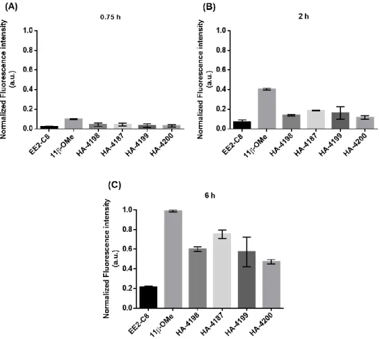
Figure 3.4 - Normalized Fluorescence intensity of MCF-7 cells incubated for (A) 0.75 h, (B) 2 h and (C) 6 h with steroid-BODIPY conjugates.. ... 32
Figure 3.5 - Normalized fluorescence intensity of MDA-MB-231 cells incubated for (A) 0.75 h, (B) 2 h and (C) 6 h with steroid-BODIPY conjugates.. ... 33
Figure 3.6 - Brighfield and Fluorescence images of estradiol-BODIPY conjugates in PC-3 cells at 6h. ... 34
Figure 3.7 - Normalized fluorescence intensity of PC-3 cells incubated for (A) 0.75 h, (B) 2 h and (C) 6 h with steroid-BODIPY conjugates. ... 35
Figure 3.8 - Normalized fluorescence intensity of LNCaP cells incubated for (A) 0.75 h, (B) 2 h and (C) 6 h with steroid-BODIPY conjugates.. ... 36
Figure 3.9 - Brightfield and fluorescence of steroid-BODIPY conjugates in Fibroblasts (normal cells) at 6h. ... 37
Figure 3.10 - Normalized fluorescence intensity of Fibroblasts incubated for 6 h with steroid-BODIPY conjugates. ... 37
Figure 3.11 - Internalization of estradiol-BODIPY derivates (EE2-C8 and 11β-OMe) in MCF-7 breast cancer cell line with or without E2. ... 39
Figure 3.12 - Internalization of estradiol-BODIPY derivates (EE2-C8 and 11β-OMe) in MDA-MB-231 breast cancer cell line with or without E2. ... 39
Figure 3.13 - Internalization of androgen-BODIPY derivates (HA-4198 and HA-4187) in PC-3
prostate cancer cell line with and without T.. ... 40
Figure 3.14 - Internalization of androgen-BODIPY derivates (HA-4198 and HA-4197) in LNCaP
prostate cancer cell line with or without T. ... 40
Figure 3.15 - Internalization of steroid-BODIPY conjugates in PC-3 cells at 4 °C or 37 °C during
6 h. ... 42
Figure 3.16 - Inhibition of endocytosis of EE2-C8 estradiol-BODIPY conjugates in MDA-MB-231
cell line. ... 43
Figure 3.17 - Internalization and localization of estradiol-BODIPY conjugates (EE2-C8 and 11β-OMe) in MCF-7 breast cancer cell line at 6h. ... 45
Figure 3.18 - Internalization and localization of androgen-BODIPY conjugates (4198 and
HA-4187) in PC-3 prostate cancer cell line at 6h.. ... 46
Figure 3.19 - Internalization of EE2-C8 estradiol-BODIPY conjugate in a co-culture of
MDA-MB-231 cells and fibroblasts.. ... 48
Figure 3.20 - Internalization of HA-4198 androgen-BODIPY conjugate in a co-culture of PC-3
cells and Fibroblasts. ... 49
Figure 3.21- Cell viability of estradiol-BODIPY conjugates determined by means of the MTS
assay. ... 50
Figure 3.22 - Breast cancer cell and normal cell (Fibroblasts) death following PDT induced by
visible light and (A) EE2-C8 or (B) 11β-OMe estradiol-BODIPY conjugates as photosensitizers.. ... 51
Figure 3.23 - Prostate cancer cell and normal (Fibroblasts) death following PDT induced by visible
light and (A) HA-4198 or (B) HA-4187 androgen-BODIPY conjugates as photosensitizers. ... 52
Figure 6.1 - Brighfield and fluorescence images of androgen-BODIPY conjugates in MCF-7
breast cancer cell line at 6h.. ... 63
Figure 6.2 - Brighfield and fluorescence images of steroid-BODIPY conjugates in MDA-MB-231
breast cancer cell line at 6h.. ... 64
Figure 6.3 - Brightfield and fluorescence images of androgen-BODIPY conjugates in PC-3
prostate cancer cell line at 6h.. ... 65
Figure 6.4 - Brightfield and fluorescence images of steroid-BODIPY conjugates in LNCaP
prostate cancer cell line at 6h. ... 66
Figure 6.5 - Normalized fluorescence intensity of the (A) MCF-7, (B) MDA-MB-231, (C) PC-3 and
(D) LNCaP cells incubated for 0, 0.75, 2 and 6 h with steroid-BODIPY conjugates.. ... 67
Figure 6.6 - Brightfield and fluorescence images of steroid-BODIPY conjugates in Fibroblasts at
6h.. ... 68
Figure 6.7 - Brightfield and fluorescence images of steroid-BODIPY conjugates in PC-3 cells at
6h.. ... 69
Figure 6.8 - Internalization and localization of estradiol-BODIPY conjugates (EE2-C8 and 11
Figure 6.9 - Internalization and localization of androgen-BODIPY conjugates (4198 and
HA-4187) in LNCaP prostate cancer cell line.. ... 71
Figure 6.10 - Cell viability of androgen-BODIPY conjugates determined by means of the MTS
assay. ... 72
Figure 6.11 - Cell viability of steroid-BODIPY conjugates determined by means of the MTS
assay.. ... 72
Figure 6.12 - Prostate cancer cell death following PDT induced by visible light and 50 μM of
Table Index
Table 2.1 - Characteristics of the steroid-BODIPY conjugates studied. ... 18 Table 2.2 - Human cancer cell lines used in this work and its general characteristics. ... 19 Table 2.3 - Three buffer system of semi-dry transfer... 22
Acronyms and List of Abbreviations
AF1 Transactivation function 1 domain
AF2 Transactivation function 2 domain
AR Androgen Receptor
ATCC American Type Culture Collection
ATP Adenosine triphosphate
BODIPY 4,4-difluoro-4-bora-3a,4a-diaza-s-indacene
BRCA1 Breast cancer 1 susceptibility gene
BRCA2 Breast cancer 2 susceptibility gene
BSA Bovine Serum Albumin
CAV1 Caveolin-1
CCP Clathrin coated pits
CCV Clathrin coated vesicles
CIE Clathrin-independent endocytosis
CME Clathrin-mediated endocytosis
CTCF Corrected Total Cell Fluorescence
DHEA dehydroepiandrosterone
DHEAS Dehydroepiandrosterone sulfate
DHT dihydrotestosterone
DMEM Dulbecco’s Modified Eagle Medium
DMEM/F-12 Dulbecco’s Modified Eagle Medium/ Nutrient Mixture F-12) DMSO Dimethyl sulfoxide
DNA Deoxyribonucleic acid
DTT 1,4-Dithiothreitol
E2 Estradiol
ECL Enhanced chemiluminescence
EDTA Ethylenediamine tetraacetic acid
EE2 Ethynylestradiol
EIPA 5-(N-ethyl-N-isopropyl)-Amiloride
ER Estrogen Receptor
ESR1 Estrogen Receptor 1 gene
ESR2 Estrogen Receptor 2 gene
FBS Fetal bovine serum
FITC Fluorescein isothiocyanate
Hoechst Phenol,4-[5-(4-methyl-1-piperazinyl)[2,5'-bi-1H-benzimidazol]-2'-yl]- trihydrochloride
HPR Horseradish peroxidase
LAMP-2 Lysosome-associated membrane glycoprotein 2
LAMP2A Lysosome-associated membrane glycoprotein 2 A
LDI Laser diode intensity
LNCaP Human metastatic prostate carcinoma cell line
MCF-7 Human metastatic breast adenocarcinoma cell line
MDA-MB-231 Human metastatic breast adenocarcinoma cell line
MEM Non-essential amino acid
MTS 3-(4,5-dimethylthiazol-2- yl)-5-(3 carboxymethoxyphenyl)-2-(4-sulfophenyl)-2H-tetrazolium
NADH Nicotinamide adenine dinucleotide
NADPH Nicotinamide adenine dinucleotide phosphate
PBS Phosphate Buffered Saline
PC-3 Human metastatic prostate adenocarcinoma cell line
PCR Polymerase Chain Reaction
PDT Phorodynamic therapy
Pen/Strep Penicillin/Streptomycin
PET Positron emission tomography
PMS Phenylmethylsulfonyl fluoride
PR Progesterone receptor
PS Photosensitizer
PVDF Polyvinylidene fluoride
ROS Reactive Oxygen species
RPMI Roswell Park Memorial Institute medium
RT Room Temperature
SDS Sodium dodecyl sulfate
SDS-PAGE Sodium dodecyl sulfate Polyacrylamide gel electrophoresis
SPECT single photon emission computed tomography
T Testosterone
TBST Tris Buffered Saline with Tween
TRICT Tetramethylrhodamine
UV Ultraviolet
1.
Introduction
1.1. Cancer: an overview
The human body has approximately 3x10¹³ cells and the average human lifespan includes 1016
cell divisions (1). A precise control of cellular division and differentiation through a network of complementary mechanisms that regulate cell proliferation and death are of most importance. Thus, the uncontrolled multiplication of cells in a specific location it would be clinically described as neoplasia (2,3).
Cancer is a large group of diseases of higher multicellular organisms. It is characterized by alterations in the expression of a variety of genes resulting in the uncontrolled growth and division of abnormal cells. Usually, can invade surrounding tissue and can metastasize to other organs. Metastases are the leading cause of death from cancer (4). Cancer can be caused by a variety of changes in gene expression resulting in dysregulated balance of cell proliferation and cell death (5). These alterations are the consequence of the interaction between genetic factors, lifestyle and three types of external agents, such as: chemical carcinogens (tobacco-smoking, food and drinking water contamination); physical carcinogen (UV and ionizing radiation); and biological carcinogens (infections caused by viruses, bacteria or parasites) (1,5).
Some types of cancers can be cured by conventional therapies such as surgery, radiotherapy, or chemotherapy if they are detected early. Additionally, avoiding the exposure to common risk factors, a significant proportion of cancers could be prevented (4).
1.1.1 Incidence and Mortality
Cancer is a major public health problem worldwide and the second leading cause of mortality. According to the World Health Organization (WHO), cancer was responsible for an estimated 9.6 million deaths in 2018. Globally, about 1 in 6 deaths is due to cancer (4). Approximately 70% of cancer deaths occur in low- and middle-income countries. Cancer incidence and mortality are rapidly growing worldwide due to the growth and aging of the population, particularly in less developed countries (6). The incidence of cancer is also due to an increase of established risk factors such as overweight, smoking, absence of physical activity, and altered reproductive patterns related to economic development and urbanization.
A significant proportion of cancer deaths is caused by lung, colorectal, stomach, liver and breast cancers. The most recurrent types of cancer vary between different global regions and among men and woman (6).
In Europe, the total number of cancer cases and cancer deaths in 2018 are approximately 23% and 20%, respectively. These estimated numbers represent only 9% of the global population.
Figure 1.1 presents the distribution of all new cancer cases and deaths in Europe in 2018 for
both sexes in all ages. For both sexes combined, female breast cancer is the most commonly diagnosed cancer (12.4% of the total cancer cases) in Europe followed by colorectal cancer (11.8%), lung cancer (11.1%) and prostate cancer (10.6%). Lung cancer is the leading cause of cancer death (20% of the total deaths) in Europe following by colorectal cancer (12.5%), breast cancer (7.1%), pancreas (6.6%) and prostate cancer (5.5%). In females, breast cancer is the most commonly diagnosed cancer and the leading cause of death. In contrast, the most frequently diagnosed cancer in males is prostate cancer and lung cancer for mortality (6).
In Portugal, cancer incidence is similar to Europe. This disease was the cause of death of 27,900 patients in 2016, 3% more than the previous year (7).
In the last decade, cancer has been extensively investigated at various levels, such as physiological, cellular and molecular levels promoting the remarkable development of new approaches for cancer treatment (8).
1.1.2 Breast and Prostate Cancers
Breast and Prostate cancers are two malignant diseases that have some remarkable similarities namely they are tumours of female and male accessory sex organs, respectively and are both characterized by hormone-sensitive cancers, which could respond to hormone therapy (9). These tumours constitute one of the most commonly diagnosed cancers in Europe as well as the leading causes of cancer-related mortality for women and men, respectively (9).
Breast cancer is a complex disease and the second cause of cancer-associated death among various women (10). Worldwide, there are about 2.1 million new breast cancer cases diagnosed
Figure 1.1 - Distribution of estimated number of new cases and mortality by Europe in 2018 of all cancer types in both sexes, all ages. Adapted from GLOBOCAN 2018.
in 2018. In the majority of the countries (154 of 185), this condition is the most diagnosed cancer and is also the leading cause of cancer death in over 100 countries (6).
In addition, almost 1.3 million new cases and 359,000 related deaths of prostate cancer were estimated worldwide in 2018. This type of cancer is considered the second most frequent cancer and the fifth leading cause of cancer death in men (6).
Several factors such as genetical susceptibility and environmental factors, could be related with initiation and development of breast and prostate cancers (10). In breast cancer, family and thus genetic predisposition are the most important risk factors (9). The majority of inherited breast cancer cases are due to mutations in tumour suppressor genes BRCA1 and BRCA2. Additionally, prolonged and unopposed exposure to estrogen is another significant risk factor for breast cancer. Other risk factors associated to this malignancy, include age, race, radiation exposure, weight, exercise, alcohol consumption, smoking and previous breast disease. As with breast cancer, a genetic predisposition is also an important risk factor in prostate cancer. In fact, prostate cancer has many risk factors in common with breast cancer. Prolonged exposure to steroid hormones, like breast cancer but in this case androgens, is also an important risk factor for prostate cancer. This hormone-dependent fact is supported by the elimination of prostate cancer risk in men that have genetic anomalies blocking androgen production or suffer early castration. Prostate cancer has other risk factors similar to breast cancer as age, obesity, low-fiber and high-fat diet, and prostate inflammation (9).
A better understanding of the cellular and molecular mechanisms associated to breast and prostate cancers, and the identification of new biomarkers for early diagnostics and prognosis could contribute to design effective cancer therapeutic approaches (10). An example of these biomarkers in hormone-dependent cancers are steroid hormones receptors.
1.2. Steroid Hormones
Steroids are a class of organic compounds with a four-ring skeleton composed by 17 carbon atoms. They can differ significantly by modifications in functional groups attached to the core, the oxidation state of the individual rings, alterations to the ring structures and degree of unsaturation. Steroids include, for example, some anti-inflammatory drugs, dietary lipids (cholesterol) and, especially sex hormones such as estradiol and testosterone. These molecules have two fundamental biological functions: (1) several steroids are signalling molecules that activate steroid hormone receptors, and (2) are important components of cell membrane structures, such as cholesterol, controlling membrane fluidity (11).
Steroid hormones are a class of hormones synthetized from a common precursor molecule, cholesterol. These hormones act as chemical messengers in the body and control several
physiologic mechanisms such as the development and function of the reproductive system (12). They are grouped depending on their biological function: estrogens (female sex steroids), androgens (male sex steroids), progestins, glucocorticoids and others (Figure 1.2) (11). These molecules are small lipophilic ligands that bind with high affinity to their respective receptors, intracellular proteins which are included in the nuclear family of transcription factors (12). Signal transduction is mediated by these receptors through genomic and nongenomic actions (12). Steroid hormone receptors are present in many cells but that levels are elevated in premalignant and malignant cells (11). The development and growth of numerous human cancers are influenced by these molecules (13).
Steroids and steroid receptors played an important role in the occurrence of cancer as well as target for this condition therapeutics. Thus, currently, this is a promising area of research (13).
1.2.1. Estrogens
Estrogens, known as female hormones, are organic compounds with a core structure composed of 17 carbon-carbon bonds arranged as four fused rings. All estrogens have 18 carbons being known as C18 steroids with identical chemical structures and function (14). Estrogens are small lipophilic molecules essential to the function of the female reproductive system (13). These molecules are mainly produced by the ovary and transported via blood stream to specific target tissues. The most important and potent form of natural estrogen is 17β-estradiol (E2) followed by less effective estrogens, estrone and estriol (15). E2 is an important regulator of growth, differentiation and function in a variety of target tissues, including the female and male reproductive systems, mammary gland, and cardiovascular and skeletal systems (11,16). The principal biological effects of E2 are triggered through the estrogen receptors (ER) (16).
The ER is classified as two distinct forms, ERα and ERβ, which are encoded by different genes (ESR1 and ESR2, respectively). However, these proteins are members of the nuclear hormone receptors superfamily of transcription factors sharing high degree of homology (14,16). The major difference between the two ER forms is the amino terminal domain, ERβ has a shorter than ERα – see Figure 1. 3. The full length size of human ERα has 595 amino acids and 66 kDa while human ERβ protein has 530 amino acids and 54 kDa. Besides full-length ERα isoform (66 kDa), two shorter isoforms (36 kDa and 46 kDa) have been described as consequence of products of
alternative splicing, or the presence of alternate start codons. Additionally, ERβ has five well-known isoforms (14,17).
1.2.2. Androgens
Androgens are a class of steroid hormones that are synthesized by the testis and adrenal glands consisting of cholesterol 19-carbon derivates. Androgens play a key role in the development and survival of male reproductive tissues, as prostate, by influencing gene expression levels. They are also precursors for estrogens, female sex hormones. The most abundant androgen in men is testosterone (16). However, there are other forms of androgens, including dihydrotestosterone (DHT), androstenedione, and dehydroepiandrosterone (DHEA) and its sulfonylated derivate (DHEAS). Testosterone or DHT are more potent than Androstenedione and DHEAS. In addition, weakly activating androgen receptors can also be metabolized into the more potent androgens (18). These steroid hormones primarily perform their functions through their respective receptor (androgen receptor) (19) .
Androgen receptor (AR) is a ligand-dependent transcription factor which is activated in the presence of androgens (20,21). This receptor is a member of the superfamily of nuclear hormone receptors sharing several structural and functional characteristics with other receptors (22–24). The AR protein consists of 910-919 amino acids and has 110 kDa (Figure 1.3) (19,25) . In the absence of androgens, the AR is stabilized in an inactive state, in the cytoplasm, by a complex of chaperone proteins (heat-shock proteins) (18,19). Upon androgen binding AR is translocated into the nucleus and subsequently can initiate expression of genes by binding to a specific DNA sequence which are recognized by the AR (21,26).
Figure 1.3 - Schematic illustration of the primary structure of steroid receptors and its functional domains. The primary structure of human steroid receptors: ER – estrogen receptor; AR – androgen receptor. Region A/B contains transactivation function 1 domain (AF1); Region C the DNA-binding domain; Region D is the hinge region; Region E consist of ligand-binding domain; and Region F the transactivation function 2 (AF2) domain. Adapted from (12).
1.2.3. Hormone Receptors Signaling Pathways
There are two distinct types of hormone receptors signaling, usually named as genomic and non-genomic pathways (12,14). Generally, in the non-genomic pathway, upon hormones binding to the respective hormone receptor in the cytoplasm, a conformational change occurs and the ligand-receptor complex translocates into the nucleus. In the nucleus, the ligand-receptor binds to the chromatin at specific response elements sequences or transcription initiation complexes on the DNA, activating or repressing transcription (Figure 1.4) (14,25,27–29). Despite hormone-dependent transcription, the principal mechanism of action for hormone receptors, hormones can also signal without direct binding of the hormone receptors to the DNA (14,18,27). These non-genomic pathway occurs rapidly (in seconds to minutes) and includes the activation of second messenger signaling cascades that involve kinases, calcium flux, phospholipases, among others secondary messengers (18). This rapid signaling results from binding to specific extranuclear receptors, which is localized mostly in the cell membrane (28).
Figure 1.4 - General schematic illustration of steroid hormones signalling at cellular level. Steroid Hormones can bind to cytoplasmatic receptors or membrane-associated receptors. When the hormones bind to cytoplasmatic recepors, the complex hormone-receptor is translocated to the nucleus activating transcription (genomic pathway). On the other hand, when the hormones bind to membrane receptors activates second messengers signalling cascades. Adapted from (29).
1.2.4. Hormone Receptors and Cancer
There is evidence that steroid hormones, as estrogens and androgens, influence many human hormone-dependent cancers, such as breast and prostate cancers, through diverse mechanisms mediated by respective steroid hormone receptors (30). Breast and prostate cancers shares a diversity of molecular similarities.
For breast cancer, estrogens represents a role in tumour development and progression. Approximately 80% of all breast cancers express estrogen receptors (ER) (16,31). Thus, these receptors are a potential biological target for diagnosis and therapy in hormone-dependent breast cancers (30). Usually, ER are overexpressed on the membrane and nucleus of breast cancer cells providing a possible mechanism for targeted drug delivery (11). Furthermore, the case is similar for prostate cancer diagnostic and therapy. High levels of testosterone have been linked with a diversity of diseases, namely prostate cancer (16). The progression of this cancer, however, have been associated to elevated AR expression in malignant tissue indicating that AR plays an important therapeutic target for prostate cancer treatment (16,19,21).
Therefore, the study of steroid hormones, as well as, their respective steroid receptors have a great potential as diagnostic or prognostic biomarkers during tumour development and progression (30).
1.3. Cellular uptake mechanisms
Cellular trafficking of substances and signaling are the most important mechanisms for biological activity, which is regulated by cellular membrane. Cells may internalize steroid via various mechanisms, including passive diffusion (metabolic energy-independent) and transporter-mediated mechanisms as facilitated diffusion and active transport (metabolic energy-dependent) (32–34). Conventionally, steroid hormones cross the cell membrane bi-phospholipid layer through passive diffusion, due to their hydrophobicity. Alternatively, are known that sex hormones, namely estrogens and androgens, binds with cell membrane-bound G-protein coupled receptors to perform their genomic or non-genomic functions (28,33).
Endocytosis mediated by membrane receptor also may be involved in cellular uptake of steroid hormones. However, the purpose of the steroids endocytosis in nuclear receptor-mediated functions it is still unclear (32,33). In all mammalian cells, endocytosis is a fundamental used to communicate with the extracellular environment. This is an energy-dependent process whereby cells internalize portions of cell membrane, cell-surface receptors, and a diversity of soluble molecules and ions, such as nutrients, from the extracellular environment (34,35). However, small cells can also be internalized forming vesicles that carry its contents into the cells.
Cells have numerous mechanisms for endocytosis. The traditional classification of endocytosis mechanisms is divided by size into two categories: phagocytosis, the uptake of large particulates (“cell eating”), and pinocytosis, the uptake of fluids and solutes (“cell drinking”) (36–38) . The latest category is sub-categorized in other mechanisms based on the proteins and lipids involved in endocytic processes. Accordingly, pinocytosis is divided into clathrin-mediated endocytosis (CME) and clathrin-independent endocytosis (CIE) (36,37). This last category is still sub-divided in caveolae- mediated endocytosis, clathrin- and caveolae-independent endocytosis and macropinocytosis (37). Figure 1.5 illustrated a schematic representation of the different endocytosis pathways.
Nonetheless, these mentioned endocytic pathways are focused in four essential steps: (1) specific binding at the cell surface; (2) plasma membrane pinching and budding off; (3) roping of the trafficking vesicle obtained and (4) trafficking of the vesicle to cytoplasm (36,37).
1.3.1. Phagocytosis
Phagocytosis is an endocytosis pathway and consists in the ingestion of large particles by an actin-dependent mechanism. This process is typical of specialized cells, including immune cells, as dendritic cells, macrophages, monocytes, neutrophils, and mast cells. Nevertheless, it is also associated to the nutrient, pathogens, dead cells and cell debris uptake (37,39). Primarily, the phagocytic pathway requires a specific recognition of the particles through receptors at surface of cell membrane. This recognition leads to a membrane distortion that surrounds the particle, engulfs it, and ends with the formation of a phagosome (36,37,40). Thereafter, the phagosome undergo maturation, fuses with lysosomes for degradation (36).
Figure 1.5 - Classification of endocytosis pathways based on the proteins that are involved in the internalization of particles and solutes. Adapted from (36).
1.3.2. Pinocytosis
1.3.2.1. Clathrin-mediated Endocytosis (CME)
CME is one of the most studied and important pathways of cellular entry in all mammalian cells. This route is responsible for uptake of essential nutrients, efficient receptors signalling and synaptic vesicle recycle in neurons (36,37). CME is initiated by the attachment of particles at the cell membrane surface, forming clathrin coated pits (CCP). After invagination, CCP are pinched off from the plasma membrane by dynamin (a small GTPase), triggered the formation of clathrin coated vesicles (CCV). Then, clathrin coat is discarded and the vesicles fuse with endosomes and subsequently with lysosomes initiating a degradation process. Alternatively, endosomes containing particles can be transported back to cell membrane for endosomes recycling – see
Figure 1.6. Clathrin, a cytosolic protein, together with adaptor proteins (APs), constitute the
coated pits (36,37,41).
1.3.2.2. Clathrin-independent Endocytosis
The most studied clathrin-independent internalization mechanisms is the caveolae-mediated endocytosis. This endocytic pathway is responsible for diverse biological functions, including lipid regulation, vesicular transport, and cell signalling (37,42). Caveolae are cholesterol-rich cell membrane microdomains composed mainly of caveolin proteins (caveolin-1 and caveolin-2). Endocytosis via caveolae is initiated by the formation of plasma membrane flask-shaped invagination that contains a striated coat of caveolin. After internalization, the vesicles are transformed into caveossomes and finally the particles can be released to the cytoplasm or fused with lysosomes for degradation – see Figure 1.6 (36,37,41–43). Caveolae have been associated to several disease conditions, namely cancer, making their mechanisms investigation of potential biomedical relevance (42).
Figure 1.6 - Schematic representation of different endocytosis pathways. (1) Clatherin-mediated endocytosis; (2) Phagocytosis; (3) Macropinocytosis; (4) Claveolae-mediated endocytosis; and (5) Clathrin- and caveolae-independent endocytosis. Adapted from: What is membrane trafficking . 2018. Available from: https://www.mechanobio.info/what-is-the-plasma-membrane/what-is-membrane-trafficking/#ITEM-1574-0). Acecessed February 2020.
1.4. Cancer Theranostics
There are several therapies for cancer treatment. Currently, the most common conventional therapies used for cancer treatment are surgery, chemotherapy and radiotherapy. The method of treatment is chosen depending mainly on the type and stage of cancer. There is no general methodology to treat all types of cancer due to the heterogeneity and the complexity between different patients with the same cancer and different types of cancer. Conventional therapies for cancer treatment have some significant restrictions because of side effects, incomplete tumour eradication, lack of selectivity, and the variable tumour response to treatment and clinical behaviour. Another problem of these conventional therapies is the increased resistance to treatment. Although these disadvantages they are still used for cancer treatment (44–46). Despite the recent biomedical advancements of these conventional therapies for cancer treatment, it is important the investigation and development of new diagnostic and treatment approaches. Recently, has been developed new technologies which could clinically advantageous for cancer diagnostic and treatment (47).
Cancer theranostics is a new technology that combines diagnosis and therapy. This new approach has a great potential for personalized cancer treatment. Therefore, personalized medicine has the capacity to optimize targeted delivery and dosing of treatments having purpose to decrease delays in treatment and facilitate patient care (46,48).
Molecular imaging is a technique diagnostic system which allowed to visualize, measure and characterize biological mechanisms at the cellular and molecular levels in humans. Molecular imaging of cancer is a growing area and it can be executed with several imaging tools providing sensitive non-invasive information of tumour properties. Ligands used can be labelled with a fluorescent dye for optical imaging, a positron emitting radionucleotide for positron emission tomography (PET), a contrast agent for magnetic resonance imaging (MRI) or a gamma emitting radionucleotide for single photon emission computed tomography (SPECT) imaging (46,49).
There are many tools that can combine imaging and therapy. The use of light as an activation mechanism had a marked development in cancer treatment. It can be used directly, triggering physiological changes in cells and tissues, or indirectly, inducing the formation of by-products (effector molecules). These types of light-induced therapies include particularly photodynamic therapy (PDT) (46,50).
A theranostic approach requires agents that simultaneously have the capacity of targeting, imaging, and treatment (46). An example are the receptor based fluorescence ligands, specifically a series of estrogen-, testosterone- and 19-nortestosterone conjugates linked to a BODIPY dye (16).
1.4.1. Photodynamic Therapy (PDT)
PDT is widely used in the treatment of several types of tumours, such as breast and prostate cancers (51,52). PDT is an alternative method for cancer treatment and combines three principal components, a light-activated chemical known as a photosensitizer (PS), light irradiation at an appropriate wavelength, and molecular oxygen from tissues to induce cell death through oxidative damage (53–55). Individually, each of these components do not develop a biological response. However, a mixture of the three factors promotes the formation of reactive oxygen species (ROS), as singlet oxygen (1O
2), hydrogen peroxide (H2O2), superoxide anions (O2•-) and hydroxyl radicals
(OH•) (52). These ROS produced intracellularly have short lifetimes and can provoke irreversible damage to crucial cellular macromolecules such as DNA, amino acids, proteins, phospholipids, leading to ablation of the tumour cells(56).
Clinically, the treatment is dependent on the uptake of the PS into malignant tissue. PSs are usually administered by systemic or topical application but normally it is executed by intravenous injection (57). Once the optimal tissue concentration is optimized, the malignant tissue can then be exposed to light with a convenient wavelength for a pre-determined time triggering the selective activation of the PS. Subsequently, this PS activation produces highly reactive ROS and can induce cell apoptosis and/or necrosis, immune response and microvasculature shutdown in the tumour – see Figure 1.7 (52,55). This alternative approach is a clinically approved therapy and has several of advantages over other cancer therapies. These advantages include low toxic, good selectivity, side effects, minimal invasiveness, and it can be combined with other cancer therapies (51,58). Despite these advantages, PDT has various drawbacks such as lack of tumour selectivity; photobleaching of PS; photosensitization; the need of an oxygen-rich environment and absence of accepted light dosimetry; short tissue penetration depth; as a local treatment, cannot be used to treat metastasized cancers (50,59,60). Therefore, these limitations make the development of PDT a challenge by increasing the interest of theranostics approaches.
1.4.1.1. BODIPY-based photosensitizers
Photosensitizers are compounds which have been used in the treatment of various medical conditions. These class of compounds is a key factor in PDT. A PS is considered to be an ideal drug for cancer PDT if it has the subsequent characteristics: (1) selectivity to tumor cells and rapidly clear from the normal tissues; (2) rapid body clearance; (3) capacity to be delivered via several administration routes; (4) good water solubility; (5) chemical and physical stability; (6) dark biological stability with no cytotoxicity; (7) high molar extinction coefficients; (8) photostability; (9) activation at visible/near-infrared wavelengths for good tissue penetration (53,56,57).
The PS classification is based on its functional capabilities and the structural characteristics have an important position. These type of compounds are divided into three groups: first, second and third generation. First generation PS are porphyrin-based drugs which are aromatic tetrapyrrolic structures. Due to the disadvantages of the first generation PSs, the clinical use of these photosensitizing drugs is limited and an extensive investigation was required in order to improve the efficiency of PSs (45,55,56,61,62). The second generation PSs, such as chlorin, phthalocyanines, and porphyrins, were designed to reduce side effects, increase tumour selectivity, and to increase depth penetration of light with longer wavelength (600 nm - 800 nm) (61,63). These type of PSs include a broad group of compounds that can be applied in numerous therapeutic applications. Because of solubility problems of a multiple PS in aqueous medium, the development of delivery models could be advantageous for PS transportation to the tumour site improving the cellular uptake. Second generation PSs have been bound to drug carrier molecules, such as steroid hormones, originating third generation PSs. Thus, with the rapid advancements in this study area, a variety of new and more specific PSs has revealed potential for cancer therapy (62,64).
Among a wide range of chromophores, Boro-dipyrromethene (BODIPY) is one of the most commonly used fluorescent dye (31). BODIPY is a fluorescent dyes and structural analogs of porphyrins (Figure 1.8). These molecules have several attractive properties, including facile synthesis and structural flexibility, good solubility, peculiar spectroscopic properties (narrow absorption and emission bands), strong absorption coefficients in the visible range, high fluorescence quantum yields, good photostability, and others (31,65). In addition to these greater chemical and physical properties, BODIPY is cell membrane-permeable, and thus used in a variety of biological applications, such as helping to monitor mechanisms of action in living systems as well as to guide surgery (11,31). The potential use of BODIPY dye in PDT has been suggested because of its capacity to generate singlet oxygen. However, in recent times, techniques to introduce 18F radioisotopes into BODIPY has been recently described, in order to provide a potential development of dual fluorescence/PET imaging probes (16). BODIPY dyes have also been used to label several ligands including DNA, proteins, peptides, carbohydrates lipids, and in particular, steroid hormones (65,66).
1.4.1.2. Steroid-BODIPY conjugates
Nowadays, a major problem of chemotherapeutic agents clinically is the lack of selectivity. Therefore, the development of selective and potent therapeutic agents is under continuous investigation in order to target tumour tissue without affecting cell viability of normal cells (16).
Around 80% of all breast cancers express estrogen receptors (ER-positive). Thus, the first line of treatment for hormone-dependent breast cancer is hormone therapy and drugs that target and block ER. Hormone steroid receptors have also become attractive targets for molecular imaging due to their implication in the growth hormone-dependent cancer types, such as breast and prostate cancers. Testosterone is the most hormone in men. In case of prostate cancer, low levels of testosterone led to this condition. As well as in breast cancer, hormone receptors, as androgen receptor, also provide significant prognostic information for the diagnosis and treatment of hormone-dependent tumours. Therefore, hormone receptor-imaging has a great potential for breast and prostate cancers screening, staging, response assessment and guiding therapies (11,16,31).
Recently, has been reported the development of multimodality theranostics agents as BODIPY dyes conjugated to estradiol-, testosterone- and 19-nortestosterone. 17α-ethynylestradiol (EE2)-BODIPY conjugate, with a linear eight carbon spacer chain, presents the highest relative binding affinity (RBA) for the ERα. However, the 11β-methoxy derivate of EE2-C8-BODIPY conjugate improve in vivo localization. Additionally, four androgen-BODIPY conjugates shown significant RBA for the AR and thus, cellular uptake studies and continuous investigation of these conjugates are important for the improvement of cancer diagnostic and therapy strategies (16,67,68).
In summary, these conjugates provide a potential platform as a receptor-based fluorescence probes for imaging breast and prostate cancers, PSs for PDT, and also as a PET imaging agents upon 18F radioisotope substitution (16).
1.5. Objectives
Steroid receptors studies have shown their potential use as biological targets for hormone responsive cancers. Receptors overexpression in human cancer cells and their binding properties provides an important role for malignant tumours localization.
The main goal of this work was to evaluate the cellular uptake of estradiol-, testosterone- and nortestosterone-derivates conjugated to BODIPY as potential multi-modality theranostic agents (PET/fluorescence and PDT) in relevant breast and prostate cancer cells by fluorescence microscopy. To assess conjugates potential for photodynamic therapy (PDT), irradiation studies were also done.
In order to reach this main goal, various tasks were set:
i. Effects of conjugates in tumour and normal cells uptake; ii. Steroid receptor blocking with specific inhibitor;
iii. Understand the mechanism of cellular uptake of conjugates; iv. Localization of conjugates in tumour cells;
v. Interaction of conjugates with tumor and normal cells in co-culture;
vi. Irradiation studies in tumour and normal cells in order to assess conjugates potential for PDT.
2.
Methods and Materials
2.1. Compounds
Steroid-BODIPY conjugates studied were synthesized in the Faculty of Medicine and Health Sciences (Université de Sherbrooke, Canada). These compounds are estradiol-, testosterone- and nortestosterone derivates conjugated to BODIPY dye (as fluorescent probe) (Figures 2.1
and 2.2). The characteristics of the conjugates are described in Table 2.1. Solid compounds were
stored at 4°C protected from light.
Figure 2.1 - Structures of estradiol-BODIPY conjugates: EE2-C8 (17 α-[1-heptyne-7-(4-ethynylphenyl)-(4,4-difluoro-8-(1,3,5,7-tetramethyl-4-bora-3a,4a-diaza-s-indacen)]-3,17β-estradiol); and OMe - 11β-methoxy-3,17β-estradiol).
Figure 2.2 - Structures of androgen-BODIPY conjugates: HA-4198 (17α-[4,4-difluoro-8-(4/
-ethynylphenyl)-1,3,5,7-tetramethyl-4-bora-3a,4a-diaza-s-indacene]-19-nortestosterone); HÁ-4187
Table 2.1 -Characteristics of the steroid-BODIPY conjugates studied.
Due to the low solubility of the conjugates in water, dimethyl sulfoxide (DMSO) (Sigma, St. Louis, USA) was used to solubilize the steroid-BODIPY conjugates. DMSO is an organic, polar and aprotic solvent commonly used to solubilize several nonpolar or poorly soluble drugs due to their amphipathic properties (69). The stock solutions of each conjugate were prepared at a concentration of 10 mM in DMSO and stored at -20°C protected from light. The concentrations of compounds used to perform the biological assays were prepared by diluting each stock solution in complete medium.
2.2. Human Cell Lines
In this work, four types of adherent human cancer cell lines were used to evaluate the effect of the steroid-BODIPY conjugates after green light irradiation, two from breast adenocarcinoma cells (MCF-7 and MDA-MB-231) and two from prostate cancer cells (PC-3 and LNCaP). Table 2.2 summarizes details and cancer cell lines general characteristics, such as origin, morphology, steroid receptors (estrogen receptor and androgen receptor) and culture medium.
Name/Code Molecular Formula
Molecular Weight (g.mol-1) EE2-C8 (-3,17β-estradiol) C44H46BF2N2O2 699.0 11β-OMe (-11β-methoxy-3,17β-estradiol) C45H49BF2N2O3 714.7 HA-4198 (-19-nortestosterone) C39H43BF2N2O2 620.6 HA-4187 (-7α-methyl-19-nortestosterone) C40H45BF2N2O2 634.6 HA-4199 (-7α-methyl-nortestosterone) C41H47BF2N2O2 648.6 HA-4200 (-testosterone) C40H45BF2N2O2 634.6
Table 2.2 - Human cancer cell lines used in this work and its general characteristics such as origin, morphology, steroid receptors and culture medium used.
Abbreviations: (ER – Estrogen Receptor; AR – Androgen Receptor; (+) – positive; (-) – negative; RPMI 1640 - Roswell Park Memorial Institute 1640 Medium (GibcoTM by Life Technologies, Invitrogen, California,
USA); DMEM – Dulbecco’s Modified Eagle Medium (GibcoTM by Life Technologies, Invitrogen, California,
USA); FBS – Fetal Bovine Serum (GibcoTM by Life Technologies, Invitrogen, California, USA); Pen/Strep –
Penicillin/Streptomycin solution (GibcoTM by Life Technologies, Invitrogen, California, USA); MEM - non-essential amino acid (Sigma, St. Louis, USA).
Additionally, as a normal cell line, primary dermal fibroblasts from foreskin of a neonatal African American was also used in this work. Fibroblasts were cultured in DMEM culture medium supplemented with 10% of FBS and 1% Pen/Strep. Fibroblasts morphology is spindle-shaped, and the cells are bipolar and refractile.
All cell types used in this work were obtained from American Type Culture Collection (ATCC®, Virginia, USA).
2.2.1. Maintenance of Cell Cultures
Cell lines were cultured in RPMI 1640 medium or Complete Culture Medium constituted by DMEM medium (see Table 2.2) supplemented with 10% (v/v) FBS, 1% (v/v) Pen/Strep solution (solution with 10000 U/mL penicillin, 10000 μg/mL streptomycin) in culture flasks of 25 and 75 cm2 (SPL
Cancer
Cell Line Origin Morphology
Steroid Receptors Culture Medium % (v/v) PC-3 Metastatic prostate adenocarcinoma Epithelial ER(+); AR(-) RPMI 1640; 10% FBS; 1% Pen/StreP LNCaP Metastatic
prostate carcinoma Epithelial
ER(-); AR(+) RPMI 1640; 10% FBS; 1% Pen/StreP MCF-7 Metastatic breast adenocarcinoma Epithelial ER(+); AR(+) DMEM; 10% FBS; 1% Pen/Strep; 1x MEM MDA-MB-231 Metastatic breast adenocarcinoma (triple negative breast
cancer cell model)
Epithelial ER(-); AR(+) DMEM; 10% FBS; 1% Pen/Strep; 1x MEM
Life Sciences, South Korea). Cell cultures were maintained in a CO2 incubator (Sanyo, Osaka,
Japan) at 37°C in a humidified atmosphere (99%) and 5% (v/v) of CO2. For breast cancer cell
lines, MCF-7 and MDA-MB-231, 1% (v/v) of MEM were added to the DMEM medium.
Cell cultures were subcultured routinely to maintain exponential growth. Upon reaching approximately 80% of confluence (Nikon TMS, Nikon Instruments, Tokyo, Japan), cells were subcultured to avoid loss of growth due to contact inhibition and lack of nutrients. To this end, the culture medium was removed and discarded and 2 or 3 mL of TrypLETM Express (GibcoTM by Life
Technologies, Invitrogen, California, USA) was added to help cell detachment. After approximately 5 min of incubation in the CO2 incubator, 1 or 1.5 mL of fresh culture medium was
added to block trypsin activity. Then, the cell suspension was transferred to 15 mL falcon tubes (SPL Life Sciences, South Korea) and centrifuged for 5 min at 300 x g and 15°C (Sigma 3-16K 10280, Osterode am Harz, Germany). The supernatant was discarded and the pellet was resuspended in 1 mL of fresh medium.
Cells were counted by the Trypan blue exclusion method in a hemocytometer (Hirschmann, Eberstadt, Germany). For that, 350 μL of culture medium were mixed with 50 μL of cellular suspension obtained during the subculturing procedure and 100 μL of 0.4% (v/v) Trypan blue solution (GibcoTM by Life Technologies, Invitrogen, California, USA). Viable cells were
immediately visualized through an optical inverted microscope (Nikon TMS, Nikon Instruments, Tokyo, Japan) and cell density (cells/mL) determined by multiplying the number of total cells, the volume of the hemocytometer chamber (104 mL-1) and the dilution factor (10) divided by the
number of squares counted - Equation 1.
Equation 1.
𝐶𝑒𝑙𝑙 𝑑𝑒𝑛𝑠𝑖𝑡𝑦 (𝑐𝑒𝑙𝑙𝑠/𝑚𝐿) = 𝑇𝑜𝑡𝑎𝑙 𝑐𝑒𝑙𝑙𝑠 𝑐𝑜𝑢𝑛𝑡𝑒𝑑 𝑁𝑢𝑚𝑏𝑒𝑟 𝑜𝑓 𝑠𝑞𝑢𝑎𝑟𝑒𝑠× 10
4 (𝑐ℎ𝑎𝑚𝑏𝑒𝑟 𝑣𝑜𝑙𝑢𝑚𝑒) × 10 (𝑑𝑖𝑙𝑢𝑡𝑖𝑜𝑛 𝑓𝑎𝑐𝑡𝑜𝑟)
For each 25 and 75 cm2 culture flask, the volume of cell suspension (estimated by equation 1)
was added to fresh culture medium to perform a final volume of 5 and 15 mL, respectively. Subsequently, cell cultures were incubated at 37°C in a humidified atmosphere of 99% and 5% (v/v) of CO2.
Cell cultures were periodically analyzed for possible mycoplasma contamination. This quality control assessment was performed by Polymerase Chain Reaction (PCR) detection on total genomic DNA extracted from the studied cultures (quality control is routinely performed by lab members).
2.3. Expression of steroid receptors by Western-Blot
Expression of estrogen α and androgen receptors was quantified by Western-Blot and performed for every cell line mentioned in section 2.2.
2.3.1. Sample preparation
For protein extraction, cell lines were seeded as described in section 2.2.1.At 80% of confluence, cells were washed three times with PBS 1x and harvested with a scrapper in 1 mL of PBS 1x for microfuge tube on ice. Samples were centrifuged for 5 min at 700 x g at 4°C (Sigma 3-16K 10280) and the supernatant was discarded. A final spin was done to remove all supernatant. The pellet was resuspended in 30 μL of cell lysis solution 4x NaCl-Tris-EDTA buffer (150 mM NaCl; 50 mM Tris, pH=8; 5 mM EDTA), 1x protease inhibitor (complete ULTRA Tablets, Mini, EASYpack, Roche, Switzerland), 1x phosphatase inhibitor (PhosStop, Roche), 0.1% (w/v) 1,4-Dithiothreitol (DTT; AMRESCO, USA), 2% (w/v) NP-40 (Thermo Scientific, MA, USA) and 1 mM phenylmethylsulfonyl fluoride (PMSF; Sigma, St. Louis, USA) and samples stored at -80 °C, until further processing.
All samples were thawed and submitted to 2 min 30 s of continue pulses at ultrasonic bath in ice and preserved on ice for 1 min to avoid sample heating and the denaturation of proteins. Cell lysates were centrifuged for 5 min at 700 x g at 4°C and supernatants were used for protein quantification.
Protein concentrations were determined using the Pierce Protein Assay (Thermo Scientific, MA, USA). A calibration curve was performed with standard BSA solutions with different concentrations (0-2000 μg/mL) and protein samples were diluted 1:10 in ultrapure water (18.2 MΩ.cm-1 at 25 °C). Thereafter, 150 μL of Pierce reagent was added and samples incubated for 5
min at room temperature (RT). Absorbances were measured at 660 nm with a Tecan Infinite F200 microplate reader (Tecan, Männedorf, Switzerland). For western blot analysis, 20 or 50 μg of total protein extracts were transferred to another Eppendorf tube with Sodium Dodecyl Sulfate (SDS) Loading Buffer 4x and 3% (w/v) DTT and incubated overnight at RT.
2.3.2. SDS-PAGE and transfer to PVDF membrane
A 10% acrylamide-bisacrylamide gel (SDS-PAGE) was prepared and samples were loaded in the gel (120 V for approximately 1 h). The transfer of proteins from the gel to the PVDF membrane (AmershamTM HybondTM 0.45 PVDF, GE Healthcare, Life Sciences, Little Chalfont, UK) was
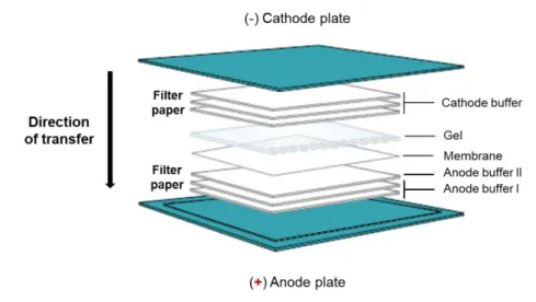
performed using a semi-dry system transfer to provide a faster and more efficient transfer. For that, a specific semi-dry transfer device with the anode plate as the base and three-buffer system to achieve efficient transfer of proteins (see Table 2.3) were used. The semi-dry transfer system
is represented in Figure 2.3, in which the membrane is in contact with the SDS-PAGE gel. The transfer process was run at 130 mV for 45 min.
Table 2.3 -Three buffer system of semi-dry transfer.
The efficiency of the protein transfer process was analysed by staining the membrane with a Ponceau S solution for 5 min. Thereafter, the membrane was washed with ultrapure water (18.2 MΩ.cm-1 at 25 °C) in order to observe the protein bands.
2.3.3. Primary and secondary antibodies incubation
To quantify estrogen and androgen receptors, specific antibodies against these proteins were used. Following transfer process, membrane was blocked with 5% (w/v) non-fat milk solution in TBST (50 mM Tris, 150 mM NaCl and 0.1% (v/v) Tween 20, pH 7.5) during 1 h at RT with agitation. This blocking step was done for possible non-specific binding because of the high protein affinity of membrane. Then, each PVDF membrane was incubated with a primary antibody in 5% non-fat milk in TBST, anti-androgen receptor (1:5000) (Abcam, Cambridge, UK) or anti-estrogen receptor α (1:500) (Sigma, St. Louis, USA), for 1h at RT with agitation or overnight at 4 °C, respectively. After primary antibody incubation, the membrane was washed three times with TBST 1x during 5
Buffer Composition
Anode I 0.3 M Tris, pH 10.4, 10 % (v/v) methanol
Anode II 25 mM Tris, pH 10.4, 10 % (v/v) methanol
Cathode 25 mM Tris, 40 mM 6-amino-n-caproic-acid, 10 % (v/v) metanol, pH 9.4
min with agitation and incubated with the appropriate secondary antibody, anti-rabbit IgG HPR-linked antibody (1:2000) (Cell Signalling Technology, Massachusetts, USA) for 1 h with agitation. Protein bands in the membrane were detected as described in section 2.3.4.
Actin levels were used as a control to normalize the results. Consequently, the membrane was incubated with stripping buffer (0.1 M glycine, 20 mM magnesium acetate and 50 mM potassium chloride) three times for 10 min with agitation to remove specific antibody binding. The same procedure of blocking and antibodies incubation described previously was done with anti-β-actin (1:5000) (Sigma, St. Louis, USA) primary antibody, and anti-mouse IgG HPR-linked antibody (1:3000) (Sigma, St. Louis, USA) as secondary antibody.
2.3.4. Film Exposition
In order to detect protein bands in the membrane, a WesternBright ECL substrate (Advansta, Menlo Park, California, USA) was applied to the membrane and incubated for 5 min. ECL is a chemiluminescent HRP substrate for western blots imaging. After ECL incubation, the film was exposed to the membrane in light absence. Densitometric analysis was performed by using Image J software to determined protein band quantification.
2.4. Intracellular tracking of steroid-BODIPY conjugates in cancer and
normal cells
Taking advantage of BODIPY fluorescence, the internalization of steroid-BODIPY conjugates in breast and prostate tumor cell lines and in normal cells was investigated by fluorescence microscopy.
All cell lines mentioned in section 1.2 were seeded on a 24-well plate (SPL Life Sciences, South Korea) with a cell density of 1x10⁵ cells/well and incubated for 24 h at 37 °C in a 99% humidified
atmosphere and 5% (v/v) CO2. After this time, the medium was replaced by fresh media (without
phenol red) supplemented with 50 μM of conjugate or 0.5% (v/v) DMSO (vector control). Cells were visualized at 0 h, 45 min, 2 h and 6 h using a Ti-U Eclipse inverted microscope (Nikon, Tokyo, Japan). Images of BODIPY-conjugates fluorescence were acquired using a FITC filter (excitation at 480/30 nm in the blue region and emission at 535/45 nm). Before visualizing the fluorescence, the culture medium was replaced by fresh medium (without phenol red). The images were treated and analyzed with Image J software allowing the quantification of the Corrected Total Cell Fluorescence (CTCF) for each cell according with Equation 2. CTCF was quantified for five cells analyzed per image in three random microscopic fields in duplicate for each cell line.


![Figure 2.2 - Structures of androgen-BODIPY conjugates: HA-4198 (17α-[4,4-difluoro-8-(4 / - -ethynylphenyl)-1,3,5,7-tetramethyl-4-bora-3a,4a-diaza-s-indacene]-19-nortestosterone); HÁ-4187 -7α-methyl-19-nortestosterone); HÁ-4200 -testosterone); an](https://thumb-eu.123doks.com/thumbv2/123dok_br/15783511.1077251/43.892.129.761.610.986/structures-androgen-conjugates-ethynylphenyl-tetramethyl-nortestosterone-nortestosterone-testosterone.webp)