Luís Leónidas Cardoso
Dissertation presented to obtain the Ph.D degree in
Integrative Biology and Biomedicine
Instituto de Tecnologia Química e Biológica António Xavier | Universidade Nova de Lisboa
The role of the gut microbiota in the
subsistence of antibiotic resistance
Oeiras,
March, 2020
The role of the gut microbiota in the
subsistence of antibiotic resistance
Luís Leónidas Cardoso
Dissertation presented to obtain the Ph.D degree in
Integrative Biology and Biomedicine
Instituto de Tecnologia Química e Biológica António Xavier | Universidade
Nova de Lisboa
Research work coordinated by:
ii
Cover image:
A depiction of Escherichia coli in the mammalian gut environment, in which it has to co-exist and compete with several microbial species. Credits go to Joana Carvalho for the original illustration.
iii The work described in this PhD thesis received financial support from Fundação para a Ciência e Tecnologia, through the grant PD/BD/106003/2014, awarded to Luís Leónidas Cardoso.
This work was conducted at Instituto Gulbenkian de Ciência (IGC), Oeiras, Portugal, under the supervision of Dr. Isabel Gordo.
v
Acknowledgements
This thesis is a product of a long journey- 5 years of scientific life, full of exciting and joyful moments, and also some hardships. If I made it out, it is thanks to the people who cherished and supported me throughout this quest, and whom I would like to acknowledge for our shared deed.
I would first like to thank my family, and in particular my parents, for their constant and unconditional support. They have been exceedingly comprehensive in these 5 years and their wisdom was essential in helping me handle adversity. I would like to thank my paternal grandparents, who have been present throughout my life have been role-models for me in many aspects. To my cousins, which have been true brothers and sisters to me in many occasions. To my maternal grandparents, to uncle Casimiro, to Têtê and other loved ones which are not with us anymore, but have had a great impact in shaping the person that I am today.
I would like to thank Isabel for betting on me and giving me the opportunity to perform this work, for the supervision and for all that she taught me along the thesis. Also, for letting me be a part of a group full of wonderful people. I learned a lot with everyone, from bacterial genetics with Roberto, to mice handling and gavages with Nelson and Catarina, statistics and sequencing tips by Ricardo and Hugo, together with many helpful know-hows passed by our mommy Dani. Paulo’s judgment and experience were really important in shaping our story, and his great sense of humor helped a great deal to cope with tough moments. Massimo’s contribute was the perfect add-on, the theoretical component that allowed us to go one step forward. It has been a pleasure working with both of you, and I am looking forward to share with the world our endeavors. I would also like to extend my thanks to the former and current members of the BE and BAS groups, for their helpful nature and strong companionship.
I would like to thank the IGC Community. Everyone was extremely helpful and good natured, from the ladies at the cantine to the admin office, making the life at the
vi
Institute more enjoyable and cozier. Of course, this work was only possible thanks to the support and resourcefulness of the members of the Institute facilities, and I am particularly thankful to them. I would also like to acknowledge the PhD Programme personnel, namely Élio, Manuela and Ana for all the support. I would also like to acknowledge my thesis committee, Karina Xavier and Joana Sá, thank you for your insight and helpful discussions.
I would like to thank a core group of friends that I had the pleasure to meet, some since high school, others during my stay at the IGC - Gonçalo Matos, Filipe Vieira, Henrique Colaço, Yash Pandya, André Barros e André Carvalho. Thank you for the profound bro-hood and for all the moments of laughter.
I would like thank my fellow colleagues from IBB2015. In them I have found true and lasting friendship. Although we don’t meet as frequently as before, every time we do is a moment to cherish and remember.
I would also like to do a special mention to my “outside of IGC” friends. I thank João F., Ricardo C., Manuel S. and Miguel L. for the unwinding moments of board and roleplay gaming - I hope we can have more of those together one day.
I would like to thank Dibengos, who have reached for me even when I did not know I needed it. For the solidarity and for the simple, genuine, happy moments.
To those mentioned, and to those that I did not mention directly but were an important part of this journey, my thanks for making this achievement possible.
ix
Summary
Antibiotic resistance is one of the major contemporary threats to global health. Studies on evolutionary biology, molecular biology and genetics have revealed that many phenomena contribute for the subsistence of resistant bacteria. The environment has been shown to be a key factor, capable of altering fitness costs and the epistasis patterns between resistance determinants. Still, few studies have ventured to assess the costs of antibiotic resistance in natural environments, and such studies are centered on pathogens. It is now known that commensal bacteria can act as reservoirs of resistance, and that resistant commensals can evolve to express pathogenicity and share resistance genes with pathogens. Here, we explore how selection acts on resistant, commensal E. coli in the mouse gut.
We observe that the fitness effects of resistance mutations in the gut are not predictable by experiments in standard laboratory media, and that after an antibiotic perturbation, the presence of microbiota affects the outcome of competitions with the sensitive strain. The costs become host-specific and lead to situations in which the resistant strain bears no cost, suggesting a role of the microbiota in the maintenance of resistance. We then report that when resistant bacteria are evolving in this system, they acquire a multitude of mutations that do not correspond to classic compensatory mutations, and that the latter appear at a different pace in mice carrying different microbiota. For the studied mutants, adapting to the gut environment seems to prevail over compensation. We also present a study in which we look for frequency-dependent selection in two environments with different complexity. We find an association between frequency-dependent selection and secretome-related functions in a minimal medium environment, and we observe Darwinian selection in the mouse gut, with a single mutant dominating the competition against the wild-type and other mutants in genes related to the secretome.
The research presented on this thesis highlights the gut microbial community as a factor that influences the survival of resistant and multi-resistant bacteria, and as a whole, stresses the importance of studying bacteria in environments that reflect
x
their place in nature. Following studies taking into account the biotic environment in which bacteria are inserted may help to prevent and reverse resistance.
xi
Resumo
A resistência a antibióticos é uma das maiores ameaças contemporâneas à saúde a nível global. Estudos de biologia evolutiva, biologia molecular e genética revelaram vários fenómenos que contribuem para a subsistência das bactérias resistentes. O ambiente é um factor-chave, capaz de alterar os custos de fitness e os padrões de epistasia entre determinantes de resistência. Ainda assim, os estudos focados no custo da resistência em ambientes naturais são escassos e centrados em agentes patogénicos. Hoje, as bactérias comensais são reconhecidas como reservatórios de resistência, e sabe-se que comensais resistentes podem evoluir no sentido de expressar patogenicidade e partilhar genes de resistência com bactérias patogénicas. Aqui, exploramos como é que a seleção actua sobre E. coli comensais resistentes, no intestino de ratinho.
Reportamos que no intestino, os efeitos no fitness das mutações de resistência, não correspondem ao previsto através de meios de laboratório convencionais, e que após uma perturbação através de um antibiótico, a presença da microbiota afecta o desenlace de competições com a estirpe sensível. Os custos tornam-se específicos consoante o hospedeiro, levando a situações nas quais as estirpes resistentes não têm custo, o que sugere um papel da microbiota na manutenção da resistência. Reportamos de seguida que quando bactérias resistentes evoluem neste sistema, adquirem uma variedade de mutações que não correspondem a mutações compensatórias clássicas. Estas aparecem a um ritmo diferente em ratinhos com microbiotas diferentes. Para os mutantes estudados, a adaptação ao ambiente do intestino parece prevalecer em relação à compensação. Por último, apresentamos um estudo no qual procuramos por selecção dependente da frequência em dois ambientes de complexidade distinta. Encontramos uma associação entre selecção dependente da frequência e funções relacionadas ao secretoma num ambiente de meio mínimo, mas observamos selecção Darwiniana no intestino, no qual um mutante domina a competição contra o wild-type e contra outros mutantes para genes ligados ao secretoma.
xii
O trabalho de investigação presente nesta tese realça a comunidade microbiana do intestino como um factor capaz de influenciar a sobrevivência de bactérias resistentes e multirresistentes e dá ênfase à importância de estudar as bactérias em ambientes que reflictam o seu lugar na natureza. Estudos subsequentes que tenham em consideração o ambiente biótico que rodeia as bactérias poderão contribuir para a prevenção e a reversão da resistência.
xiii
Table of Contents
Acknowledgements ... V Summary ... IX Resumo ... XI Table of Contents………..………XIII Thesis Outline ... XVIIChapter I – Introduction ... 19
Main targets of antibiotics ... 20
Cell wall biosynthesis ... 21
Replication ... 21
RNA and protein synthesis ... 22
Metabolism ... 25
The acquisition of resistance ... 26
Resistance through horizontal gene transfer ... 27
Resistance acquisition through mutations ... 29
Mutation rates and effects ... 30
Environmental effects on mutation rate ... 31
Mechanisms of resistance ... 32
Decreased target access ... 33
Reduced target affinity and target protection ... 35
Target protection by modification ... 37
Antibiotic inactivation ... 38
The fitness effects of antibiotic resistance ... 39
The cost of antibiotic resistance ... 40
Selection favoring the maintenance of resistance ... 40
Costless mutations ... 41
xiv
Co-selection and cross resistance ... 44
Compensation ... 45
Environment dependence of fitness effects ... 50
Epistasis ... 51
Gut commensal bacteria ... 54
Commensal bacteria as a reservoir of resistance ... 54
The gut microbiota ... 58
Escherichia coli as a model and as a gut commensal ... 60
Aims ... 62
Chapter II – Personalized Fitness Cost of Antibiotic Resistance in the Mouse Gut ... 63
Abstract ... 64
Introduction ... 65
Methods ... 67
Escherichia coli and mice strains ... 67
In vitro competitions ... 68
In vivo competitions ... 69
Selection coefficient and epistasis calculations ... 70
Microbiota analysis ... 71
Statistical analysis ... 73
Streptomycin detection test ... 74
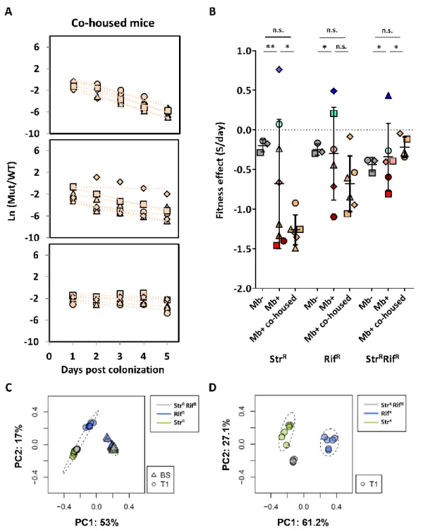
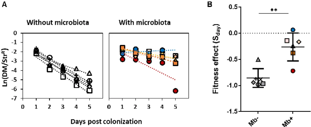
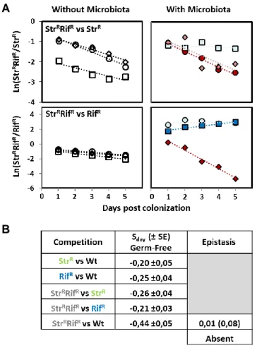
Results ... 75
Environment-dependence of resistance mutations and epistasis ... 75
Costs of antibiotic resistance in the absence of microbiota ... 76
The effect of microbiota in the cost of resistance ... 80
Antibiotic perturbation increases variation in microbiota composition .... 81
Normalization of the microbiota reduces variance of fitness effects ... 83
Extended competitions reveal a late disadvantage of multi-resistance .. 85
The effect of microbiota in the cost of resistance in a mouse adapted strain ... 86
xv
Discussion ... 88
Causes for variation, nutrition and cross-feeding ... 88
Mutation nature and pleiotropy ... 91
In vitro predictions and future approaches to measure fitness costs ... 92
Natural strains and generalizations ... 93
Supplementary Material ... 96
Chapter III – Adaptation Prevails Over Compensation in the Mouse Gut ...103
Abstract ...104
Introduction ...105
Methods ...107
Escherichia coli and mice strains ...107
In vivo evolution ...108
Reversion test...109
DNA extraction for population sequencing ...110
DNA extractions and whole-genome sequencing analysis ...110
Microbiota analysis ...112
Results ...113
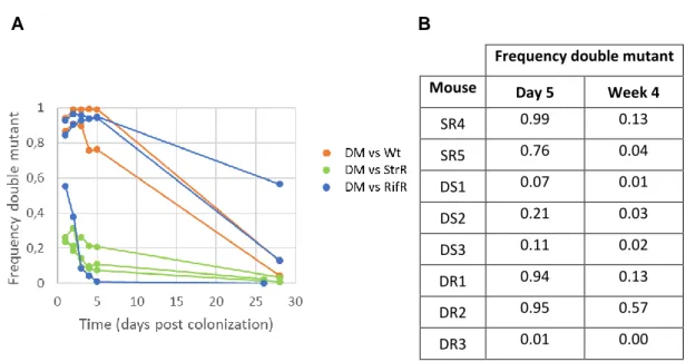
Litter-specific occurrence of compensatory mutations ...113
Nature of classic compensatory mutations ...116
Amplicon sequencing suggests clonal interference between the double mutant’s evolving lineages ...117
Within-host evolution did not lead to reversion of resistance ...118
Discussion ...119
Microbiota effects on the onset of compensatory mutations ...119
Predictability of antibiotic perturbation ...121
Nature of compensatory mutations ...121
Nature of adaptive mutations ...123
The rarity of fixation ...126
xvi
Future directions ...128
Supplementary material ...130
Chapter IV – Frequency-Dependent Selection Acting on Secretome Polymorphisms ...135
Abstract ...136
Introduction ...137
Methods ...139
Escherichia coli and mice strains ...139
In vitro competitions and selection coefficient calculation ...143
Growth curves ...144
In vivo competition ...145
Results ...147
Secretome deletion polymorphisms show magnitude frequency dependent selection ...147
Isolated growth does not fully predict competitive fitness ...150
Darwinian selection prevails over secretome polymorphism stability in the mammalian gut ...155
Discussion ...158
Frequency dependent fitness effects and secretome mutants ...158
Absence of dependency and NFDS ...160
Positive directional selection and frequency dependent effects ...161
Beneficial effects of whole gene deletions ...164
Darwinian selection acting on secretome genes ...165
Chapter V - General Discussion ...169
xvii
Thesis Outline
When a bacterium acquires a mutation, its survivability will depend on the fitness effect of the genetic modification. The fitness effect can vary across environments, reflecting different selective pressures, which may stem from the activity of other microbes.
This study was designed to evaluate the contribution of microbiota to the fitness effect of chromosomal mutations in a natural environment – the mouse gut - focusing on how the gut microbiota changes the fitness effect of mutations conferring antibiotic resistance, how it conditions the evolution of antibiotic resistant strains, and to assess the occurrence of stable polymorphisms for public traits in simple and complex environments.
Chapter I presents a general introduction on antibiotic resistance, going
through the cell targets of the main classes of antibiotics, how resistance is acquired, and the mechanisms of resistance. Afterwards, the chapter addresses the fitness cost of resistance and the known mechanisms by which bacteria reduce or circumvent this cost. The role of commensal strains as a reservoir and as a source of resistance determinants is then addressed. The major aims of this thesis are stated at the end of this chapter.
In Chapter II the fitness of resistant Escherichia coli strains was measured through in vivo competitions, showing that the cost of chromosomal resistance mutations is affected by the presence of microbiota, in a host-specific manner.
Chapter III follows the evolution of resistant E. coli in the gut environment,
identifying mutations through sequencing and revealing a prominent role of adaptation relative to compensation.
xviii
Chapter IV is a stand-alone chapter, which looks at frequency-dependent
fitness effects on single gene deletion polymorphisms in E. coli, finding frequency-dependence for secretome genes in a simple environment, and strong, positive selection in the mouse gut.
Chapter V highlights the main findings of this study, briefly contextualizes them
within the current literature and proposes approaches to be addressed in future research.
19
Chapter I – Introduction
CHAPTER I
Introduction
20
The incorporation of antibiotics into medical practice was one of the major landmarks in the history of medicine. Their utilization enhanced the combat against diseases of bacterial origin, many of which fatal by the time of their discovery 1, and
allowed the prevention of opportunistic infections in delicate medical practices 2,
resulting in an effective reduction in human morbidity and mortality caused by bacterial infections 3. However, over time, the intensive use of antibiotics has led to
the dissemination of resistant bacteria, making the treatments ineffective 1.
The prospect got worse with the early reports of multi-resistance 4,5, which
allows pathogenic bacteria to accumulate means of survival to treatments 6. These
worrisome news led to a response of the pharmaceutical industry, with the design of synthetic antibiotics, some of which directed to new cell targets. However, resistance to these new agents was developed 7, indicating that bacteria can evolve and acquire
resistance to synthetic compounds as well. As a consequence of the unrestrained, extensive and excessive use of multiple antibiotics for decades 8, antibiotic resistance
has turned into one of the major public health problems 1,2, and might lead to the
failure of quimioterapy based treatments 9 due to the spread at epidemic levels of
infections carried out by resistant bacteria 10. A post-antibiotic era, in which common
infections can once again kill, is indeed a real possibility 11.
Main targets of antibiotics
Antibiotics are grouped in several classes based on their composition, target and mechanism of action. Antibiotics target fundamental processes of the cell, such as cell wall biosynthesis, nucleic acid synthesis, both at the level of folate synthesis, DNA replication and transcription into RNA, and at the level of protein synthesis by targeting subunits of the ribosome 12. Following is a brief description of the
mechanism of action of the most commonly used antibiotics, grouped by the targeted cellular process.
21
Cell wall biosynthesis
Bacterial cell walls are made of peptidoglycans, long sugar polymers. The peptidoglycan undergoes cross-linking of glycan strands through cross links between peptides. In this process, penicillin binding proteins (PBP) are essential for the cross linking of the D-alanyl-alanine portion of the peptide chains by glycine residues. There are two main classes of antibiotics targeting cell wall synthesis, β-lactams, including extended spectrum β-lactams and large spectrum carbapenems, and glycopeptides. β-lactams compete with alanine for PBP by binding to it, preventing transpeptidation and leading to synthesis disruption 13. Glycopeptides such as vancomycin also act
on the transpeptidation step by binding to the D-alanyl D-alanine portion of the peptides, also preventing the action of PBP on these residues 14. The prevention of
the synthesis of peptidoglycan leads to lysis of the bacterial cell 13.
Replication
Within the bacterial cell cycle, the chromosomal DNA is replicated in order to pass genetic information to both daughter cells. Topoisomerases, such as DNA gyrase and topoisomerase IV are essential in this process, as they control and maintain the topological state of DNA molecules 15. Gyrase acts while DNA is
unwinded by helicase during replication and during the elongation by RNA polymerase in transcription by nicking double-stranded DNA, introducing negative supercoils and resealing the nicked ends. The functional gyrase is a topoisomerase made by joining two A subunits, responsible by the nicking and sealing, and two B subunits, that introduce negative supercoils. In Gram-negative bacteria, quinolones such as nalidixic acid and fluoroquinolones such as ciprofloxacin target DNA replication by binding to the gyrase A subunit with high affinity and blocking its strand cut and reseal ability, although certain analogues such as norafloxacin seem to inhibit gyrase activity while binding directly to DNA 16. In Gram-positive bacteria, quinolones
22
primary target is topoisomerase IV 17, which nicks and separates DNA strands after
replication without introducing negative supercoils 18. As with gyrase binding in
Gram-negative bacteria, quinolone action leads to the impairment of DNA replication. In both gram positive and gram-negative bacteria, topoisomerase inhibitors lead to the generation of single and double strand breaks and culminate in apoptosis of proliferating cells 19. While quinolones are typically bactericidal, nalidixic acid was
shown to turn bacteriostatic in very high drug concentrations, a paradox that may be related with a secondary effect of the drug blockading RNA synthesis 20.
RNA and protein synthesis
Through the action of the DNA-dependent RNA polymerase, the information coded in DNA molecules is used to synthesize messenger RNA or functional non-coding RNA 21, in a process known as transcription.
RNA polymerase is a complex enzyme composed by five subunits, two α, one β, one β’ and an ω subunit 22. Rifamycins, including rifampicin, inhibit DNA-dependent
RNA synthesis by strongly binding in a pocket of the β subunit of the RNA polymerase, deep within the DNA/RNA channel. The binding does not occur in the RNA polymerase active site but sterically blocks the extension of the nascent RNA chain after the RNA transcript becomes 2 or 3 nucleotides in length 23, effectively
23
Figure 1 - Mechanism of action of rifampicin. A) Using a DNA strand as a template, RNA
polymerase forms an elongated mRNA molecule that corresponds to the coded gene. B) By binding to the β-subunit of the DNA-dependent RNA polymerase, rifampicin, also known as rifampin, blocks the elongation of messenger RNA and impairs transcription. Adapted from
24.
The messenger RNA sequence is then processed by the multimeric structure known as ribosome to generate peptides and proteins, with each RNA triplet, named as codon, corresponding to a specific amino-acid residue. This process is called translation. The bacterial ribosome is composed of two subunits, 30S and 50S, made by RNA enveloped by proteins. 30S is composed of 16S rRNA and 21 proteins (S1– S21) whereas 50S is composed of 5S and 23S rRNAs and 36 proteins (L1–L36) 25.
As the two units work together to perform translation, different antibiotics affect protein biosynthesis by targeting either one or the other subunit.
Aminoglycoside antibiotics, such as streptomycin, kanamycin, gentamicin and kanamycin are positively charged molecules, and their uptake depends on their interaction with the negatively charged components of the outer membrane which
24
lead to an increase in permeability. This process allows the entrance to the cytoplasm through energy-dependent, electro-transport-mediated process 26. Once in the
cytoplasm, aminoglycosides interact with the 16S rRNA of the 30S ribosome subunit through hydrogen bonds in the first ribosome binding site for tRNA, the aminoacyl binding site (A site). Some aminoglycosides, including streptomycin, cause mistranslation of proteins and premature termination of translation 27,28. The
mistranslated proteins can cause damage to the cytoplasmic membrane and facilitate aminoglycoside entry, leading to an increased inhibition of protein synthesis and mistranslation, culminating in cell death 29. Other aminoglycosides, such as
kasugamycin, act by blocking peptide chain initiation 27. (Figure 2). Tetracyclines
also interfere with the binding of the t-RNA to the ribosomal A site by acting upon the conserved sequences of 16S rRNA, inhibiting the elongation phase of protein synthesis with a bacteriostatic effect 30,31.
Commonly used drugs such as chloramphenicol, macrolides and oxazolidinones act on the 50S subunit of the ribosome. Chloramphenicol interacts with the peptidyl transferase cavity of the 23S rRNA, also preventing the binding of the tRNA to the A site. Macrolides affect translocation in the early stage of protein synthesis, by targeting the peptidyl transferase center of the 23S rRNA, leading to the premature detachment of incomplete peptide chains. 12. Oxazolidinones,
including the synthetic linezolid, bind to the peptidyl tRNA binding site of the ribosome (P site). Besides affecting the initiation of protein synthesis by inhibiting the formation of the initiation complex, oxazolidinones affect the formation of 70S (the joining of the
25 two subunits to initiate protein synthesis), and the translocation of the peptide chain if the two ribosomal subunits are already performing translation 32.
Figure 2 – Mechanisms of action of aminoglycoside antibiotics. All
aminoglycosides bind to the 30S subunit of the ribosome. Depending on their structure, they may act by blocking the initiation of protein synthesis or by causing mRNA misreading, which can lead to the the block of ongoing translation and cause translation errors. Streptomycin in particular causes mRNA misreading, promoting mistranslation and translation termination. Adapted and modified from 33.
Metabolism
Some antibiotics have as targets central metabolic pathways of the cell. For instance, sulfonamides and trimethoprim target the essential folic acid pathway. Folate is a critical precursor for the synthesis of glycine, methionine, thymidine triphosphate and purines 34. Sulfonamides inhibit dihydropteroate synthase through
competition with the natural substrate, while trimethoprim acts at a later stage, inhibiting dihydrofolate reductase. Both drugs are bacteriostatic. However, their combination has a synergistic effect, killing the cell and leading to a reduced
26
probability of evolving resistance 35. Besides allowing for the expansion of the
spectrum of targets of current antibiotics, the development of these drugs is a promising approach to eliminate persistent bacteria - subpopulations of sensitive bacteria that are slow-growing but metabolically active, surviving exposure to antibiotics that act on the dividing clones 36.
Despite the use of a multitude of diverse compounds with the purpose of leading to their death or inhibition, bacteria tend to gain resistance recurrently. To understand how bacteria become able to respond to these harmful agents, it is essential to consider the biologic processes by which resistance is obtained.
The acquisition of resistance
Antibiotic resistance occurs in nature and is found in agricultural, non-clinical environments 37. Furthermore, relatively recent studies indicate that antibiotic
resistance is ancient, with targeted metagenomic studies showing the presence of diverse antibiotic resistance determinants in thousands-of-years-old permafrost samples 38,39, and with multi-resistance being reported in environments that were
isolated for millions of years 40.
As many antimicrobial compounds are produced by living organisms, bacteria in constant contact with them have evolved to survive in their presence. These bacteria are considered to be intrinsically resistant to one or more antibiotics 41,42.
However, the public health threat of antibiotic resistance does not only come from the expansion of intrinsically resistant bacteria, but also from the acquisition of resistance by previously susceptible strains, including life-threatening pathogens in clinical settings. Resistance can be acquired by spontaneous chromosomal mutations or through the acquisition of genes carrying resistance determinants, obtained from resistant organisms through horizontal gene transfer of mobile genetic elements.
27
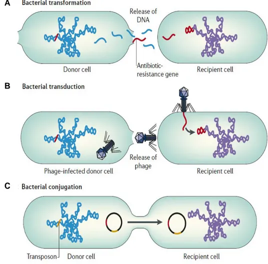
Resistance through horizontal gene transfer
Horizontal gene transfer (HGT) of antibiotic resistance genes has been shown to occur through 3 main mechanisms: transformation 43,44, which involves the
incorporation of external DNA 45; conjugation 46, a contact-dependent exchange of
genetic material 47 through the transfer of conjugative plasmids 48 or transposons 49;
transduction 50,51, in which the foreign DNA is transferred through bacteria viruses
called phages 52 (Figure 3).
HGT is now considered to be a key player in the evolution of bacteria 53. While
it typically ensues between different strains and species, the transfer of homologous regions can also occur between higher taxa 54,55. As an example, bacteria can acquire
antibiotic resistance by transformation with genes of plant origin 56, evidentiating how
“promiscuous” bacteria can be. Horizontal gene transfer can occur in various natural habitats. Gene transfer through conjugative plasmids alone has been observed in distinct environments such as soil and rhizosphere 57,58, plant surfaces 59, water 60
and in the mammalian gut 61,62. Furthermore, environmental conditions can boost the
occurrence of HGT. As an example in the context of the mammalian gut, HGT through conjugation 63 and transduction 64 is greatly increased when the intestine is
28
Figure 3 – Horizontal gene transfer between bacteria. A) Transformation occurs when
naked DNA is released on lysis of an organism and is taken up by another organism. The antibiotic-resistance gene can be integrated into the chromosome or plasmid of the recipient cell. B) In transduction, antibiotic resistance genes are transferred from one bacterium to another by means of bacteriophages and can be integrated into the chromosome of the recipient cell. C) Conjugation occurs by direct contact between two bacteria: plasmids form a mating bridge across the bacteria and DNA is exchanged, which can result in acquisition of antibiotic-resistance genes by the recipient cell. Transposons are sequences of DNA that carry their own recombination enzymes that allow for transposition from one location to another. As other mobile genetic elements, transposons can carry antibiotic-resistance genes. Adapted from 65.
B
AB
29 Additionally, horizontal gene transfer processes can lead to a very fast spread of new genes in nature. For instance, resident E. coli in the mouse gut can transfer prophage genes at an epidemic level to an invading strain in a matter of days, a process that precedes the occurrence of adaptive point mutations 66.
Antibiotic resistance genes coded on mobile genetic elements are frequently acquired in integrons 67. These DNA elements can be found in conjugative plasmids,
phages and transposons, and act as assembly platforms that incorporate exogenous open reading frames through site-specific recombination and convert them to functional genes by securing their expression 68. Integrons typically encode an
integrase, a primary recombination site and an outward-orientated promoter, providing all of the tools for the transcription of the captured gene. Multiple genes can be sequentially integrated as gene cassettes, allowing for the accumulation of functional resistance determinants 67. In fact, integron-bearing mobile genetic
elements are thought to have been major agents in the fast spread of multi-resistance in Gram-negative bacteria through horizontal gene transfer 69. Very large,
integron-like structures can also be found in the chromosome – the super-integrons. Although not mobile, these sequences can contain hundreds of accessory genes, including cassettes related to antibiotic resistance 70, and are present in many bacterial
species, being considered to have an important role in genome evolution 68.
Resistance acquisition through mutations
Despite the important role of HGT in its spread, antibiotic resistance can originate de novo in sensitive strains. Some bacteria, such as the intracellular parasite Mycobacterium tuberculosis, are mostly clonal, and typically acquire resistance in this way 71. Antibiotic resistance through de novo mutation often occurs
through single nucleotide substitutions that modify the drug target, but resistance can also be acquired through other classes of spontaneous mutations. For instance, insertions and deletions generate frameshifts and premature stop codons in genes
30
involved in antibiotic susceptibility 72, while gene duplication can lead to an increased
dosage of antibiotic hydrolytic enzymes and efflux gene pumps 73. While gene
duplications are often unstable, they can facilitate the development of a stable resistance phenotype, by allowing survival and population expansion until the occurrence of point mutations conferring higher levels of resistance 74. Point
mutations can confer resistance without modification of the primary antibiotic target. One such example regards point mutations in regulatory regions of porin coding genes that reduce porin expression 75, decreasing susceptibility. Point mutations and
deletions on such genes can lead to porin impermeability 76,77 culminating in an
increased resistance to carbapanems in Enterobacteraceae. Another example regards multi-drug (MDR) efflux pumps. Point mutations on gene repressors of MDR efflux pump genes, or in the gene regulator to which they bind can lead to overexpression of the efflux pump. On the other hand, mutations of the coding region may change the substrate binding, both at the range and affinity level 41, resulting in
an elevated level of resistance.
Mutation rates and effects
Mutations can occur through errors in DNA replication or through DNA damage. As cell division ensues, mutations with fitness effects spanning from fitness benefits to lethality can occur. In haploid microbes, the mean genome mutation rate is estimated to be of 0.003 mutations per DNA replication 78. In E. coli, the mutation rate
is estimated to be close to this value 78, and mutation accumulation studies in this
species estimate that roughly 1 in every 15 mutations is deleterious (rate of 0.0002;
79), while 1 in every 150 newly arising mutations is beneficial (rate of 2 x 10⁻⁵, 80), in
line with theoretical and molecular evolution studies suggesting that most mutations are neutral 81,82 or nearly neutral 83–85. Across haploid species, the variation in
mutation rate per base pair is high (≈16000-fold). However, the variation in mutation rate per genome is quite low (≈2.5-fold), suggesting a selective pressure towards a balance between deleterious effects of mutations and the maintenance of a minimal mutation rate in haploid organisms 78. This hypothesis is supported by data on
31 crenarchaeon Sulfolobus acidocaldarius and the bacterium Thermus thermophilus, which have evolved in extreme heat conditions, is about 5 times lower than in their mesophile counterparts, possibly reflecting an adjustment of the mutation rate to strong purifying selection 86.In bacteria, mutation frequencies are generally found to
be between 10-10 and 10-9 per replicated base pair 87. While this mutation rate per
nucleotide reflects a low probability of a specific mutation to occur, bacterial populations in nature can reach high population size. Furthermore, mutations conferring resistance can occur in multiple positions in the same target gene. As an example, a classical study mapped 17 mutational distinct alterations able to confer rifampicin resistance 88. The disposal of multiple mutational options also promotes
the recurrent appearance of resistance mutations in natural populations. Additionally, the rate of emergence of antibiotic resistance mutants is affected by cell physiology, genetics, and by aspects of the environment, such as temperature 89 and physical
structure 90.
Environmental effects on mutation rate
The mutation rate towards resistance can also change with the presence and dose of antibiotics themselves. In fact, sublethal concentrations of antibiotics can increase the rate and frequency of HGT, recombination and mutagenesis 91, while
the nature of the selected resistance mutations can vary with the dose of antibiotic
92. Furthermore, drug exposition can increase the mutation rate towards resistance
to the antibiotic - as an example, the exposition to ciprofloxacin can increase 10000-fold the rate at which ciprofloxacin resistance mutations occur 93,94, through DNA
damage and the activation of the SOS response 94. A mutagenic effect has also been
described for other fluoroquinolones 95 and for streptomycin 96.
Successive selective pressures, such as the use of different antibiotics can lead to the emergence of “mutator” bacteria. These strains originate through certain mutations in core genes related with DNA repair, which vastly increase genome mutation rate, sometimes up to 1000-fold 97. In vitro studies indicate that resistance
32
rifampicin and ciprofloxacin 99 emerge more frequently in mutator lines of E. coli.
Mutator phenotypes were reported to occur in natural populations of pathogenic 100–
102 and commensal bacteria 103,104 and allow for greater resistance levels than
non-mutator populations, particularly when full resistance requires more than one mutational step 99,105. Furthermore, some mutations conferring mutator phenotype
also increase the recombination rate and are under frequent horizontal gene transfer themselves 104, promoting the sharing of potentially beneficial traits such as virulence
factors and antibiotic resistance determinants.
Resistance through chromosomal mutations can be easily produced in a laboratory with culturable bacteria through a fluctuation test, by letting bacteria grow into very high population size while accumulating mutations, followed by a strong selection with an antibiotic - a test adapted from classic studies on the origin of resistance phenotypes to phages 106 and to antibiotics themselves 107,108. This test is
often used to estimate the mutation rate of bacteria in a given environment 109. Due
to the ease of production and the immediate acquisition of a selective phenotype, chromosomal resistance is one of the most studied types of genetic alteration. As many spontaneous resistance mutations occur in essential genes, the study of the different types of resistance has led to important findings in molecular and cell biology of bacteria, often coupling molecular and evolutionary mechanisms together.
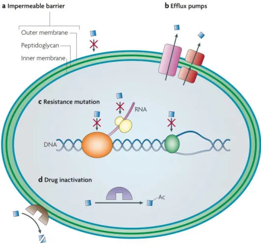
Mechanisms of resistance
There are several mechanisms by which bacteria can avoid antibiotic mediated killing or inhibition (Figure 4). These fall in 3 main categories: mechanisms that decrease target access and minimize the intracellular concentration of the antibiotic; mechanisms that modify the antibiotic target by genetic mutation or post-translational modification; and mechanisms that lead to the inactivation of the antibiotic through hydrolysis or modification 41.
33
Decreased target access
One way through which bacteria limit the access of the antibiotic target is by reducing permeability. As the outer membrane of Gram-negative bacteria forms a permeability barrier 110,111, hydrophilic antibiotics have to enter the cell through outer
membrane porin proteins, which are thought to function as non-specific channels in Proteobacteria 111. Through the down-regulation of porin expression 112, and through
the replacement of porins with selective channels 113, bacteria can block the entry of
hydrophilic antibiotics inside the cell.
Bacteria can also limit the access to intra-cellular targets through the expression or overexpression of efflux pumps. Bacterial pumps are a means of active transport and are a major contributor for the intrinsic resistance of Gram-negative bacteria 110. Some pumps have a narrow substrate specificity [e. g. tetracycline
pumps) 114], but many are able to transport a wide range of structurally dissimilar
compounds. Such transporters can confer resistance to a multitude of antibiotics, and hence are known as multidrug resistance (MDR) efflux pumps. While efflux pumps are ubiquitous in the chromosome of bacteria, some are also coded in mobile genetic elements 115–117 and can be shared through horizontal gene transfer.
Another important mechanism of reduction of target access has to do with population level phenotypes such as biofilm formation. Bacteria can encase themselves in a self-produced polymer matrix made of polysaccharide and protein and DNA. These structures can harbor a single or more bacterial species living in a socio-microbiological way 118, and frequently confer increased tolerance to antibiotics
and disinfectant chemicals 119, by physically reducing the exposition to the antibiotic
through slow penetration 120. Additionally, biofilms typically generate a gradient of
nutrients and oxygen that lead to concentration driven changes in division rate and metabolic activity, with some cells developing a level of tolerance by fine tuning of the expression of genes involved in the additional resistance mechanisms such as efflux pumps and degrading enzymes, leading to further reduced target access 119.
34
Figure 4 – Main mechanisms of resistance in gram-negative bacteria. a)
Impermeable barriers. Some bacteria are intrinsically resistant to certain antibiotics (blue squares) due to membrane impermeability, while others can limit drug entry through changes in gene expression that reduce drug permeability. b) Efflux pumps. Bacteria can also limit target access through the activity of these pumps, which secrete antibiotics to the outside of the cell. c) Resistance mutations. These mutations modify the target protein, and reduce target affinity, for example, by disabling the antibiotic-binding site but leaving the cellular functionality of the protein intact. d) Inactivation of the antibiotic. Inactivation can occur by modification covalent modification of the antibiotic, such as that catalyzed by acetylases (purple) acting on aminoglycosides, or by degradation of the antibiotic, such as that catalyzed by β-lactamases acting on β-lactams. Adapted from 37.
35 growing in a biofilm when compared to planktonic growth [e. g. 105-fold increase in mutability for Pseudomonas aeruginosa PA01, 121], and horizontal gene transfer is
also known to be increased in biofilms 122, which in turn can increase the chance of
acquiring antibiotic resistance mutations.
Reduced target affinity and target protection
Most antibiotics bind specifically to their targets with high affinity and impair the target’s function. However, certain naturally occurring mutations and gene recombination in the target structure can prevent antibiotic binding while still allowing for the target to carry its function, even if not optimally. Such alteration in the target site is a common, ubiquitous mechanism of resistance, as examples of clinical strains with this type of resistance can be found for every single class of antibiotic, regardless of the mechanism of action 123.
Perhaps the most classic examples of resistance through reduced target affinity refer to beta-lactams. The acquisition of spontaneous mutations in penicillin binding proteins conferring resistance has been reported for several genera of bacteria, including Haemophilus influenzae, Helicobacter pylori, Proteus mirabilis,
Acinetobacter baumanii, Pseudomonas aeruginosa, Streptococcus pyogenes and Listeria monocytogenes 123. Strikingly, these altered targets with reduced affinity can
be shared by horizontal gene transfer. One the best examples refer to β-lactam resistance derived in bacteria such as Streptococcus pneumoniae 124 and pathogens
such as Neisseria gonorrhoeae 125 and Neisseria meningitidis 126 through the
formation of “mosaic” penicillin binding proteins. These proteins are generated through transformation and recombination with acquired DNA coding beta-lactam insensitive variants of the proteins, usually originated from closely related species, including bacteria of commensal nature 126. Another reported situation refers to the
acquisition of the mecA gene by methicillin resistant Staphylococcus aureus. The gene encodes an alternative penicillin-binding protein 2 (PBP2a), and is carried on a
36
large genetic element, the staphylococcal cassette chromosome mec, which is presumed to have been acquired by horizontal gene transfer from other
Staphylococcus species 127.
Mutations conferring resistance to rifampicin and streptomycin are also classic examples of reduced target affinity. Rifampicin is a relevant drug in the treatment of pathogenic bacteria, being particularly important to treat tuberculosis 128. Resistance
to rifampicin typically occurs through chromosomal mutations in the rpoB gene, which codes for the target of rifampicin, the β subunit of the DNA-dependent RNA polymerase. The genome alterations include point mutations, with certain single nucleotide modifications being sufficient to grant high levels of resistance, but can also occur through small insertions and deletions 129,130, most of which occurring in a
81 base pair restricted region of the rpoB gene called cluster I 129. Additional
resistance mutations can occur in other rpoB regions, such as cluster N, cluster II and cluster III 131, but only a fraction of the resistance mutation spectrum is
responsible for the majority of clinical rifampicin resistance in Mycobacterium
tuberculosis 129. Most resistance mutations map directly in a fork domain of the RNA
polymerase, proximal to the catalytic site, or in adjacent regions 88,132. Amino-acid
substitutions in these sites are expected to affect the conformation of the binding pocket and lower its affinity for rifampicin 132, not allowing the drug to bind and block
transcription elongation.
Mutations in the ribosomal protein S12, coded by the rpsL gene 133 and in the
16S rRNA 530 loop, coded by the rrn operons 134,135 can confer high levels of
streptomycin resistance. The ribosome 30S subunit contains a conformational switch that is important for the optimization of translation. The H27 switches from an error-prone, ribosomal ambiguity form (ram), and an alternative hyperaccurate, “restrictive” form 136. Streptomycin in particular acts by stabilizing the error prone state and
increasing the binding of non-cognate tRNA. Most S12 resistance mutations and 16S mutations in the 530 loop lead to changes in ribosome accuracy. In the respective mutants, the ram state is very destabilized, and the stabilization induced by streptomycin does not trap the ribosome in such error prone state 136. Due to the
37 balancing equilibrium between ribosomal states, in a fraction of these mutants, streptomycin presence can even become essential 137,138.
Target protection by modification
The antibiotic target can be protected by modifications that do not require mutational change. Protection of targets has been found to be a clinically relevant mechanism of resistance to several antibiotics. One example is the action of erythromycin ribosome methylase on the 16S rRNA, protecting the target from being bound by macrolides and lincosamines 139. Another example is the
chloramphenicol-florfenicol resistance methyltransferase. This enzyme methylates the position A2503 of the 23SrRNA, conferring resistance to a wide range of drugs with nearby targets, such as phenicols, pleuromutilins, streptogramins, lincosamides and oxazolidonones
140. Resistance to aminoglycosides can also be granted through 16S rRNA
methylation 141,142. Protective agents can also associate with the antibiotic target.
Such examples are the quinolone resistance qnr genes, which encode pentapeptide repeat proteins. These agents bind to DNA gyrase and topoisomerase IV and protect the enzymes of inhibition by quinolones. While the mechanism of action of these resistance determinants is not fully understood, a model of the mechanism of action based on structural data of QnrN1 suggests that these agents might interact with topoisomerase-quinolone complexes, effectively rescuing the enzyme and allowing it to re-ligate DNA, thus preventing the formation of double-strand DNA breaks that typically occur with the antibiotic action 143. Some of the antibiotic resistance genes
conferring target protection can be encoded by cryptic genes, and a recent study in
Salmonella enterica shows that such genes can be activated and confer resistance
through mutations that affect their expression – a chromosomal mutation induced the expression of an aminoglycoside adenyl transferase in stringent conditions, leading to resistance to streptomycin and spectinomycin 144. On a similar note, mutations
38
of streptomycin resistance in Mycobacterium tuberculosis, Staphylococcus aureus and Escherichia coli through methylation of the 16S rRNA 530 loop 145, effectively
protecting the target from the antibiotic action.
Antibiotic inactivation
Antibiotic inactivation is a major mechanism of antibiotic resistance. Naturally occurring antibiotic resistance through antibiotic modification was first reported in 1940, with the discovery of penicillinase 146, roughly a decade after the discovery of
penicillium 147. Since then, thousands of enzymes degrading or modifying antibiotics
of different classes have been identified, including β-lactams, aminoglycosides, phenicols and macrolides 41. Some of these enzymes are able to degrade different
antibiotics of the same class 148, inclusively to modified β-lactams that were designed
to be effective against strains producing natural β-lactamases 149,150. The carriage of
these extended-spectrum β-lactamases and carbapenemases has led to a fast, world-wide spread of strains that are resistant to clinically used β-lactam antibiotics
151–153.
Many antibiotics have hydrolytically susceptible chemical bonds which are essential to their biological activity. β-lactamases, conferring resistance to β-lactams, esterases, conferring macrolide resistance and fosfomycin resistance epoxidases cleave these vulnerable bonds, destroying antibiotic activity. As hydrolytic enzymes require only water as a co-substrate, they can be excreted by bacteria and intercept antibiotics before they reach the cell 154.
Another common type of antibiotic modification is group transfer, which is carried out by the large and diverse family of group transferases. These enzymes modify antibiotics covalently, resulting in the prevention of target binding by steric hindrance. Various chemical groups can be transferred, including acyl, thiol, phosphate, nucleotidyl and ribitoyl groups 41,154. These strategies require molecular
39 with the antibiotic, and hence are active only in the cytosol 154. Aminoglycoside
antibiotics are particularly susceptible to modification, since these large molecules can be deactivated by the action of different classes of enzymes 41. Group transfer
mediated antibiotic inactivation is also known to act on other antibiotics such as chloramphenicol 155 and streptogramin 156 through acetyltransferases, and rifamycins
through a phosphotransferase enzyme 157. A bioinformatic analysis of GenBank
sequences coupled with heterologous expression experiments suggests that cryptic orthologues of the latter are present across environmental and pathogenic Gram-positive bacteria 157, further supporting that cryptic embedded genes may be a
significant fraction of the antibiotic resistome 158. Besides hydrolysis and group
transfer, antibiotics can also be inactivated by oxidation 159,160, and by the action of
lyases 161, although these routes seem to be much less common in nature 154.
The fitness effects of antibiotic resistance
A critical aspect for the maintenance of an antibiotic resistant bacteria lies in its ability to compete with antibiotic sensitive ones. In order to predict which strains are maintained in a population, evolutionary biologists estimate and compare their fitness — a quantitative measure of a genotype’s competitive ability. Fitness is derived from all phenotypes affecting the ability to survive and reproduce in a given environment. In the absence of strong random genetic drift, fitness will determine the frequency change of a population’s genotypes over time 162.
Fitness can be measured in one of two ways: as absolute fitness and as relative fitness. Absolute fitness refers to the variation in absolute numbers of a genotype, while relative fitness refers to the frequency change of the genotype in the population
163. As every environment has a limit to the number of individuals that it can carry —
a carrying capacity — relative fitness is commonly used to predict the fate of a given genotype — maintenance, fixation or extinction. In bacteria, relative fitness is typically measured through a direct competitive fitness assay, in which competing strains are
40
co-cultured in the same set of growth conditions, or estimated by measuring and comparing quantifiable growth traits, such as maximum growth rate, in single culture growth 164. When a change in the genetic information affecting one or more
phenotypes occurs, it will have a fitness effect in the individuals carrying it. This fitness effect can lead to a higher fitness by rendering a fitness benefit or lead to a lower fitness by imposing a fitness cost.
The cost of antibiotic resistance
As previously discussed, antibiotic targets are often core components of the cell. These components are typically involved in essential cellular functions, such as replication 165, transcription 166, translation 167, and cell wall biogenesis 168. Mutations
conferring resistance by target gene modification can structurally change these elements into a suboptimal state 169 and lead to pleiotropic effects 170–172. On the other
hand, genes obtained by horizontal gene transfer, as well as genes with an amplified expression imply a metabolic cost to the cell due to increased transcription and translation of genes conferring antibiotic resistance 173. Furthermore, the acquisition
of mobile genetic elements can alter the transcription of profile of chromosomal genes
174 and cause chromosomal perturbations if integrated into the host’s genome 175. As
a consequence, the acquisition of antibiotic resistance is expected to inflict a fitness cost in the absence of antibiotics 164,176,177. Several laboratory studies have shown
that resistance is effectively associated with deleterious effects, and the fitness cost of resistance has become a well-established concept 178–180.
Selection favoring the maintenance of resistance
As resistant bacteria tend to be inferior competitors than their sensitive counterparts, an intuitive strategy for containing the spread of antibiotic resistance is to suspend the use of ineffective antibiotics until resistant phenotypes decline to low
41 frequency 176. This strategy has been adopted by different countries and for different
antibiotics, but its effectiveness has been inconsistent 164,181. In some cases,
resistance has decreased as predicted 182–186. However, in other situations, resistant determinants were not eliminated 185,187,188, and there is evidence of spread of
resistance after antibiotic reduction campaigns 189, indicating that in nature, bacteria
have ways to persist despite the expected cost of acquiring resistance.
Over time, several biological phenomena allowing the reduction or circumvention of a fitness cost of resistance were reported. These include resistance mutations with no detectable cost, selection for resistance at residual antibiotic concentrations, co-selection, environment influence on the fitness effects, compensatory mutations and epistasis.
Costless mutations
Some resistance mutations have been reported to confer high levels of resistance while imposing reduced costs or even no cost at all 9. This absence of cost
is thought to be related with the nature of the mutations. For instance, certain streptomycin resistance mutations leading to an amino acid change from a lysine to an arginine have an insignificant effect on fitness in Salmonella enterica 190,191 and E. coli 192, and in the latter study, some nalidixic acid resistance mutations seem to have
no cost as well in standard laboratory conditions 192. The costless mutations in
streptomycin happen to match with non-restrictive resistance phenotypes, which are similar to the wild-type’s regarding translation speed and fidelity 193. Furthermore, in Salmonella enterica these mutations do not impair virulence 190, suggesting that the
corresponding mutants may be able to compete with the wild-type in clinical settings and establish stable populations of resistant bacteria even in the absence of antibiotics.
42
Selection and mutagenesis at residual antibiotic concentrations
The minimum inhibitory concentration (MIC) is the lowest concentration of a compound that prevents visible growth of a bacterium 194. Resistant strains have a
higher MIC than their sensitive counterparts. MIC measurements allows the detection of resistant strains and the determination of the level of resistance.
It should be noted that the level of resistance can vary extensively depending on the resistance mechanism. Certain mechanisms confer such low-level resistance that it can only be detected by time-kill experiments 195, while others may provide
extremely high resistance levels, which may even surpass the solubility limit of the antibiotic 196,197. Mechanisms involved in drug efflux tend to confer a lower level of
resistance than those that modify the antibiotic target or inactivate the drug 198.
Furthermore, bacterial growth may be unaffected by increasing antibiotic concentration until the MIC is reached, such as for amdinocillin resistant mutants in
E. coli 199, or monotonically decrease with the increasing levels of antibiotic 200,201.
Thus, the fitness of resistant bacteria might be constant or vary extensively as a function of antibiotic concentration 198.
Selection for resistance is classically thought to be driven by high, supra-MIC concentrations of antibiotics used in therapy, animal husbandry and agriculture. However, nowadays, antibiotics are widespread throughout the environment in low concentrations due to contamination from human activities 202, with many interactions
between antibiotics and microbial populations occurring in such conditions. It has thus been hypothesized that these low concentrations have a relevant role in the development of resistance 203 and it has been experimentally demonstrated that
concentrations below the wild-type’s MIC can affect selection and favor resistance 91.
A fitness advantage of resistant strains at sub-MIC concentrations of tetracycline and ciprofloxacin was shown for E. coli mutants 204, while in S. enterica, such advantage
was shown for the same antibiotics and streptomycin 201. The latter study further
shows that resistant mutants occur under a sub-MIC regime, sustaining the idea that residual levels of antibiotics can give rise to resistant phenotypes on their own.
43 A recent work focused in S. enterica’s evolution at sub-MIC levels of streptomycin shows that the acquired resistance mutations can have a different nature than the ones obtained in high drug concentration regimes. Low antibiotic levels are shown to lead to high levels of resistance through the accumulation of different small-effect resistance mutations that either alter the ribosome target, reduce the aminoglycoside uptake or induce a cryptic aminoglycoside modifying enzyme 205. These observations indicate that low levels of antibiotics foster a greater
mutational space for the selection of resistant phenotypes and provide additional evolutionary paths towards high-level resistance. Unlike supra-MIC concentrations of antibiotics, which typically kill non-resistant bacteria, below MIC concentrations still affect and act as a stress to sensitive bacteria. The stress leads to a physiological response of the cell, often leading to the activation of conserved stress response systems and leading to an increase in the rate and frequency of genetic processes that promote resistance acquisition, such as horizontal gene transfer 206–208, recombination 209–211 and mutagenesis 212–214.
One of the main stress responses to sub-MIC concentrations of antibiotics is the SOS response – a systemic reaction to DNA damage in which cell growth is arrested and DNA repair is promoted. This response implies the recruitment of RecA, which is involved in recombination, and of translesion DNA polymerases, which introduce base substitutions at a high frequency 215, increasing mutagenesis. Another
major stress response activated by antibiotic stress is the general stress response, mediated by RpoS induction 216. As with other sigma factors, RpoS it interacts with
the core RNA polymerase and controls the expression of a large, yet specific collection of genes 217. Within such a set, RpoS positively regulates the expression
of sdsR, a small RNA molecule that represses the messenger RNA of MutS. The latter is a protein involved in the DNA mismatch repair, being pivotal in mending replication errors. Furthermore, the error prone DNA polymerase IV is also a part of the RpoS regulon. Together with the depletion of MutS, the action of DNA polymerase IV leads to a RpoS-mediated induction of mutagenesis in the presence of beta-lactam antibiotics 216, which in turn can generate mutations conferring resistance.
44
Co-selection and cross resistance
When adapting to a specific environment, bacteria can acquire mutations or genetic elements that are beneficial in other environmental settings. Such acquisitions, which prepare bacteria for environments to which they are not exposed, are broadly called as co-selection. Antibiotic resistance is frequently co-selected with multiple resistance phenotypes, such as resistance to a different antibiotic, to heavy metals or other biocide agents 218,219.
Resistance to an antibiotic can be acquired after exposure to another agent if the two attack the same target, initiate a common pathway to cell death or share a common route of access to their targets, through the evolution or activation of a shared resistance mechanism. This type of co-selection is called cross-resistance, and allows the development of resistance to multiple antibacterial agents through a single selective pressure 220. Cross-resistance is commonly associated to multidrug
resistance elements, especially those involved in efflux mechanisms 221. Multidrug
pumps in particular have often a wide substrate specificity and can lead to elevated levels of resistance to multiple agents 222. In addition to antibiotics, multidrug pumps
can simultaneously confer resistance to metals223–225, and to the natural substances produced by the bacteria’s host such as bile salts, hormones and defense molecules
226. There is accumulated evidence that multidrug pumps can be involved in bacterial
pathogenicity, indicating that in certain situations, the use of an antibiotic may help to select for increased virulence 226.
Co-selection can be driven by co-resistance, which occurs when a resistance determinant is coded in the same genetic element as another, such as plasmids, transposons or integrons 220. Antibiotic and toxic metal resistance genes are
frequently linked in plasmids 227–230. A key example of co-resistance regards Tn21-like transposons, in which a mercury resistance and multiple antibiotic resistance genes are contained 231–233. Tn21 is thought to have accumulated multiple antibiotic resistances in its integron while associating to the mercury resistance mer operon,