CLINICAL STUDY
Prostatic Arterial Supply: Anatomic and Imaging
Findings Relevant for Selective Arterial
Embolization
Tiago Bilhim, MD, PhD, João Martins Pisco, MD, PhD,
Hugo Rio Tinto, MD, Lúcia Fernandes, MD,
Luís Campos Pinheiro, MD, PhD, Andrea Furtado, MD, Diogo Casal, MD,
Marisa Duarte, MD, José Pereira, MD, António G. Oliveira, MD, PhD, and
João E. G. O’Neill, MD, PhD
ABSTRACT
Purpose: To describe the anatomy and imaging findings of the prostatic arteries (PAs) on multirow-detector pelvic computed tomographic (CT) angiography and digital subtraction angiography (DSA) before embolization for symptomatic benign prostatic hyperplasia (BPH). Materials and Methods: In a retrospective study from May 2010 to June 2011, 75 men (150 pelvic sides) underwent pelvic CT angiography and selective pelvic DSA before PA embolization for BPH. Each pelvic side was evaluated regarding the number of independent PAs and their origin, trajectory, termination, and anastomoses with adjacent arteries.
Results: A total of 57% of pelvic sides (n⫽ 86) had only one PA, and 43% (n ⫽ 64) had two independent PAs identified (mean PA diameter, 1.6 mm⫾ 0.3). PAs originated from the internal pudendal artery in 34.1% of pelvic sides (n ⫽ 73), from a common trunk with the superior vesical artery in 20.1% (n⫽ 43), from the anterior common gluteal–pudendal trunk in 17.8% (n ⫽ 38), from the obturator artery in 12.6% (n ⫽ 27), and from a common trunk with rectal branches in 8.4% (n⫽ 18). In 57% of pelvic sides (n ⫽ 86), anastomoses to adjacent arteries were documented. There were 30 pelvic sides (20%) with accessory pudendal arteries in close relationship with the PAs. No correlations were found between PA diameter and patient age, prostate volume, or prostate-specific antigen values on multivariate analysis with logistic regression.
Conclusions: PAs have highly variable origins between the left and right sides and between patients, and most frequently arise from the internal pudendal artery.
ABBREVIATIONS
BPH⫽ benign prostatic hyperplasia, DSA ⫽ digital subtraction angiography, IMA ⫽ inferior mesenteric artery, MIP ⫽ maximum intensity projection, PA⫽ prostatic artery, PAE ⫽ prostatic artery embolization, PSA ⫽ prostate-specific antigen, 3D ⫽
three-dimensional
It has been suggested that prostatic artery (PA)
emboli-zation (PAE) for symptomatic benign prostatic
hyperpla-sia (BPH) may become a common treatment like uterine
artery embolization for uterine leiomyomas (
1
). Animal
studies in pigs and dogs have shown that PAE can induce
prostatic volume reduction, with no related sexual
dys-function (
2,3
).
Preliminary studies of PAE for symptomatic relief of
lower urinary tract symptoms in patients with BPH have
shown promising results (
4 – 6
). One of the most
chal-lenging aspects when performing PAE is to identify the
PAs and differentiate them from the surrounding arteries.
Cadaveric studies have addressed the PA anatomy
(
7,8
), with the description of two different arterial systems
to the prostate gland: the cranial or vesicoprostatic branch
that runs between the bladder base and the prostate that has
hypertrophied and supplied most of the central gland
ade-From the Departments of Anatomy (T.B., A.F., D.C., J.E.G.O.), Radiology (T.B., H.R.T., L.F.), Urology (L.C.P.), and Biostatistics (A.G.O.), Faculdade de Ciências Médi-cas (FCM), Universidade Nova de Lisboa; Department of Interventional Radiology (T.B., J.M.P., H.R.T., L.F., M.D., J.P.), Hospital Saint Louis; and Urology Department (L.C.P.), Hospital São José, Centro Hospitalar de Lisboa Central, Lisbon, Portugal. Received February 22, 2012; final revision received July 20, 2012; accepted July 31, 2012. Address correspondence to T.B., Rua Luz Soriano, no. 182, 1200-249 Lisbon, Portugal; E-mail:[email protected]
None of the authors have identified a conflict of interest. © SIR, 2012
J Vasc Interv Radiol 2012; 23:1403–1415
nomas in BPH, also called “artery of the adenoma”; and the
caudal, which originates inferiorly, closer to the rectum (
9
).
Also, more than one PA may be found in as many as 30% of
pelvic sides (
10
). After reaching the prostate, the PAs have a
corkscrew appearance (
7,8
) and perforate the capsule in four
quadrants, two anterior/lateral for the cranial PAs and two
posterior/lateral for the caudal PAs (
11–13
).
Few studies have addressed radiologically the anatomy of
the male pelvic arterial system (
14,15
) and PAs (
16
).
Knowl-edge of the male pelvic and PA anatomy is needed to safely
perform PAE. In this study, we report the main arterial
vari-ations in prostatic vascularization relevant for PAE by using
multirow-detector pelvic computed tomographic (CT)
angiog-raphy and digital subtraction angiogangiog-raphy (DSA), based on the
retrospective evaluation of 150 pelvic sides.
MATERIALS AND METHODS
Patient Selection
A retrospective study was conducted from May 2010 to
June 2011 in 75 male patients (150 pelvic sides) who
underwent pelvic CT angiography and selective pelvic
DSA before PAE for BPH. Patient age ranged from 50 to 85
years, with a mean of 66.0 years. Institutional review board
approval was obtained for the study, and all participants
signed an informed consent form for PAE. All patients were
informed about the embolization technique used, and the
experimental nature was clearly included.
Included were male patients with a diagnosis of BPH
with moderate to severe lower urinary tract symptoms
refractory to medical treatment for at least 6 months or
experiencing acute urinary retention. Patients were
ex-cluded in the case of malignancy, which was evaluated
by prostate-specific antigen (PSA), physical
examina-tion, transrectal ultrasound (in all patients), prostatic
biopsy (in suspicious cases), and advanced
atherosclero-sis/tortuosity of the iliac arteries or PAs evaluated by
pelvic CT angiography performed before PAE in all
Figure 1. Schematic drawing of the different PA origins.
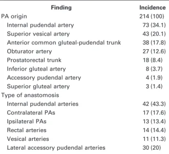
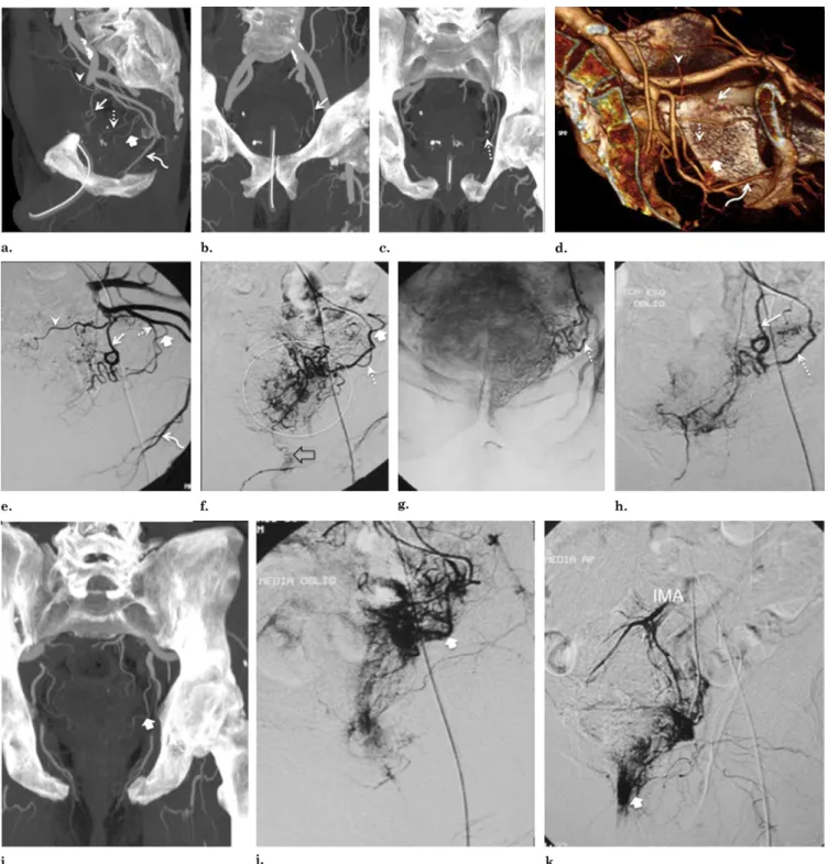
Figure 2. Rare prostatic artery (PA) origins. (a) Digital subtraction angiography (DSA) with same-side anterior oblique projection (35°) and caudal/cranial angulation (10°) of a left-side PA (straight arrow) arising from the inferior gluteal artery (curved arrow). The dotted circle marks prostate gland opacification. (b) DSA with same-side anterior oblique projection (35°) and caudal/cranial angulation (10°) of a right-side PA (straight arrow) arising from the superior gluteal artery (curved arrow). (c) Computed tomographic (CT) angiography with axial maximum-intensity projection (MIP) reformat shows PA (straight arrow) arising from a lateral accessory pudendal artery (curved arrow). (d) CT angiography with coronal MIP reformat shows PA (straight arrow) arising from a lateral accessory pudendal artery (curved arrow). (e) Selective left-side DSA with same-side anterior oblique projection (35°) and caudal/cranial angulation (10°) shows two independent PAs (straight arrows) and one prostatorectal trunk (thick arrow) that bifurcates into the middle rectal artery (dotted arrows) and one small posterior/lateral PA (straight arrow) originating from the left-side lateral accessory pudendal artery, finishing as the dorsal artery of the penis (curved arrows). The dotted circle marks prostate gland opacification.
Table 1. PA Origin and Arterial Anastomosis Type
Finding Incidence
PA origin 214 (100)
Internal pudendal artery 73 (34.1)
Superior vesical artery 43 (20.1)
Anterior common gluteal-pudendal trunk 38 (17.8)
Obturator artery 27 (12.6)
Prostatorectal trunk 18 (8.4)
Inferior gluteal artery 8 (3.7)
Accessory pudendal artery 4 (1.9)
Superior gluteal artery 3 (1.4)
Type of anastomosis
Internal pudendal arteries 42 (43.3)
Contralateral PAs 17 (17.6)
Ipsilateral PAs 13 (13.4)
Rectal arteries 14 (14.4)
Vesical arteries 11 (11.3)
Lateral accessory pudendal arteries 30 (20) PA⫽ prostatic artery.
patients. Only patients treated with bilateral PAE were
included. Patients were excluded when bilateral selective
PA anatomy characterization with CT angiography and
DSA was not possible.
Pelvic CT Angiography Protocol
Pelvic CT angiography examinations were performed
with a 16-slice spiral scanner (GE Medical Systems,
Milwaukee, Wisconsin) in all patients in the supine
position before embolization to exclude patients not
suit-able for treatment and to identify the branching pattern of
the internal iliac artery and the arteries supplying the
prostate. Power settings were 100 –120 kV and 200 –300
mA, matrix of 512
⫻ 512 pixels, collimation of 16 ⫻
1.25 mm, slice thickness 1.25 mm, and pitch of 1.3.
Iodinated contrast agent injection (150 mL, 350 mg
I/mL, rate of 5 mL/s) was performed with bolus
trigger-ing in the abdominal aorta above the renal arteries.
Figure 3. Images from a case with a solitary PA. (a) Pelvic CT angiography with sagittal MIP reformat shows a solitary PA (straight arrow) arising from the internal pudendal artery (curved arrow) above the sciatic notch on the right pelvic side. After its origin, the PA bifurcates into two PAs: anterior/lateral, vascularizing the central gland (open arrows); and posterior/lateral, vascularizing the peripheral gland (dotted arrow). The solid arrow marks the obturator artery. (b) Pelvic CT angiography with coronal MIP reformat shows a solitary PA (straight arrow) that bifurcates into two PAs: anterior/lateral (open arrow) and posterior/lateral (dotted arrow). (c) Pelvic CT angiography with 3D reformat shows a solitary PA (straight arrow) arising from the internal pudendal artery (curved arrow). After its origin, the PA bifurcates into two PAs: anterior/lateral (open arrow), and posterior/lateral (dotted arrow). The solid arrow marks the obturator artery. (d) Pelvic CT angiography with axial MIP reformat shows a solitary PA (straight arrow) that bifurcates into two PAs: anterior/lateral, vascularizing the central gland (open arrows), and posterior/lateral, vascularizing the peripheral gland (dotted arrows). Note the small anastomoses of the posterior/lateral PA to rectal branches (dotted arrows). (e) DSA of the right internal iliac artery performed with same-side anterior oblique projection (35°) and caudal/cranial angulation (10°) shows a solitary PA (straight arrow) arising from the internal pudendal artery (curved arrow). After its origin, the PA bifurcates into two PAs: anterior/lateral (open arrow) and posterior/lateral (dotted arrow). The solid arrow marks the obturator artery. (f) DSA after selective catheterization of the right-side PA with same-side anterior oblique projection (35°) and caudal/cranial angulation (10°). A solitary PA (straight arrow) bifurcates into two PAs: anterior/lateral, vascularizing the central gland (open arrows), and posterior/lateral, vascularizing the peripheral gland (dotted arrows). The solid arrow marks the small anastomoses to the inferior vesical artery, the dotted circle marks prostate gland opacification, and the curved arrow marks the rectal/perineal branches of the posterior/lateral PA. (g) Control DSA with same-side anterior oblique projection (35°) and caudal/cranial angulation (10°) after selective PAE of the right-side PA. The curved arrow marks the internal pudendal artery. (Available in color online atwww.jvir.org.)
Postprocessing
with
maximum-intensity
projections
(MIPs) and volume rendering with three-dimensional
(3D) reformats was performed by one of the
interven-tional radiologists who was present for all PAE
proce-dures, with 5 years of experience in interventional
radi-ology and CT angiography and 2 years of experience
with PAE. Volumetric data was analyzed with 1.25 mm
thick axial reformats with special attention given to the
sagittal MIP and volume rendering with 3D reformats.
DSA Protocol
DSA was performed under local anesthesia in all patients via
a unilateral (usually the right side) or bilateral femoral
ap-proach with a 5-F, 11-cm-long hydrophilic sheath (Cordis,
Bridgewater, New Jersey). A Cobra-shaped catheter (C2F5;
Cordis) or Roberts uterine artery catheter (Cook,
Bloom-ington, Indiana) was introduced in the femoral artery to
catheterize the contralateral hypogastric artery. The catheter
tip was left in the proximal part of the anterior division, and
DSA was performed at three frames per second (3 mL/s)
with 6 mL nonionic contrast medium (Ioversol 350 mg
I/mL; Covidien, Dublin, Ireland) with a same-side anterior
oblique projection (35°) and caudal/cranial angulation
(10°). The findings of DSA, 3D volume rendering, and
sagittal MIP reformats of pelvic CT angiography were
correlated regarding the number of independent PAs and
their origin, direction, and termination. All PAs identified
on CT angiography and seen on DSA were selectively
catheterized with a Roberts uterine artery catheter or 2.5-F
microcatheter (Cantata [Cook] or Progreat [Terumo,
To-kyo, Japan]) and 0.016-inch guide wire (Sagitta [Cook] or
Glidewire GT [Terumo]). Selective PA DSA was
per-formed manually with 3–5 mL of contrast agent volume in
neutral and same-side anterior oblique (35°) and caudal/
cranial angulation (10°) projections. The contralateral
in-ternal iliac artery (usually the right) was selectively
cathe-terized after the catheter was reformed into a Waltman loop.
Selective PAE was performed with 100- or 200-
m
non-spherical polyvinyl alcohol particles (Cook).
PA Classification
All PAs identified were classified according to their origin,
direction, number of pedicles, termination with intraprostatic
branches, corkscrew appearance of capsular branches, and
anastomoses with surrounding arteries. Mean PA diameter
was measured by CT angiography and selective prostatic
DSA. Important anatomic variations relevant for PAE, such as
lateral accessory pudendal arteries, were identified and
char-acterized. To evaluate any possible relationships among
pa-tient age, prostate volume transformed), PSA values
(log-transformed), PA diameter, number of vascular pedicles, and
presence of the corkscrew pattern, we performed univariate
analyses with analyses of variance, Student t test, and Fisher
exact test as appropriate, and multivariate regression analyses.
Stata software (release 12; Stata, College Station, Texas) was
used for all analyses. Statistical differences were assumed with
a P value lower than .05.
RESULTS
Technical Results
During the study period (May 2010 to June 2011), we
evaluated 91 patients with pelvic CT angiography. We
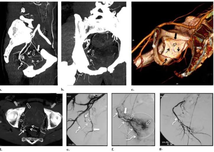
Figure 4. On the right pelvic side, there is a solitary PA (straight arrow) originating from the superior vesical artery (dotted arrows). The inferior vesical artery is shown by the solid arrow. (a) Pelvic CT angiography with sagittal MIP reformat. The straight arrow marks solitary PA, dotted arrows mark the superior vesical artery, the curved arrow marks anterior common gluteal-pudendal trunk, and the solid arrow marks the inferior vesical artery. (b) DSA of right internal iliac artery with the catheter tip in the proximal part of the anterior division was performed with same-side anterior oblique projection (35°) and caudal/cranial angulation (10°). The straight arrow marks solitary PA, dotted arrows mark the superior vesical artery, the curved arrow marks the anterior common gluteal/pudendal trunk, and the solid arrow marks the inferior vesical artery. (c) DSA after selective catheterization of the PA (arrow) with a microcatheter (the dotted circle marks prostate gland opacification).
excluded seven patients who underwent unilateral PAE
(8%) and two patients in whom selective PA
catheter-ization was impossible bilaterally (2%). Also, we
ex-cluded another seven patients (8%) based on the
evalu-ation of CT angiography alone as a result of tortuosity
and atherosclerotic changes in the iliac arteries and PAs.
Therefore, PA anatomy as identified on CT angiography
and DSA was compared in 75 patients (150 pelvic sides).
Bilateral selective PA catheterization was possible in all
pa-tients with the use of a microcatheter in 52 papa-tients (69%) and
a Roberts uterine artery catheter in the remaining 23 (31%).
Mean procedural DSA time was 86 minutes (range, 25–185
min) and mean fluoroscopic time was 27 minutes (range,
6 – 63 min). Embolization was performed with a mean volume
of 0.3 mL of polyvinyl alcohol, with 100-
m particles in 32
patients (43%) and 200-
m particles in 43 patients (57%).
Number of Independent PAs
and PA Origin
Based on the retrospective analysis of pelvic CT
angiogra-phy and selective DSA of the internal iliac arteries, it was
possible to identify the number of independent PAs and
their origin, trajectory, apparent capsular penetration, and
termination. There was one PA in 57% of pelvic sides (n
⫽
86) and two independent PAs in the other 43% (n
⫽ 64), for
a total of 214 PAs. Overall, we identified a mean of 2.9
⫾
0.9 independent PAs per patient (range, 2– 4 PAs) with a
mean diameter of 1.6 mm
⫾ 0.3.
The most frequent PA origin was the middle third of
the internal pudendal artery, above the sciatic notch
(34.1%; n
⫽ 73). Other common origins were a common
trunk with the superior vesical artery (20.1%; n
⫽ 43), the
anterior common gluteal/pudendal trunk (17.8%; n
⫽ 38),
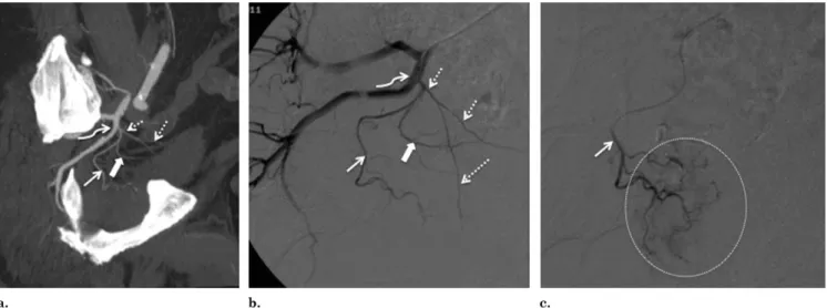
Figure 5. Solitary PA (straight arrows) originating from the obturator artery (solid arrows) on the left pelvic side. After its origin, the PA bifurcates into anterior/lateral and posterior/lateral PAs (straight arrows). (a) Pelvic CT angiography with sagittal MIP reformat. Straight arrows mark the PA, the solid arrow marks the obturator artery, and the curved arrow marks the internal pudendal artery. (b) Pelvic CT angiography with coronal MIP reformat. The straight arrow marks the PA and the solid arrow marks the obturator artery. (c) Pelvic CT angiography with three-dimensional (3D) reformat. Straight arrows mark the PA, the solid arrow marks the obturator artery, and the curved arrow marks the internal pudendal artery. (d) DSA of left internal iliac artery performed with same-side anterior oblique projection (35°) and caudal/cranial angulation (10°). Straight arrows mark the PA and the solid arrow marks the obturator artery. (e, f) DSA after selective catheterization of the left-side PA in neutral position before PAE. The straight arrow marks the PA and the solid arrow marks the obturator artery. (g) Control DSA in neutral position after PAE. The straight arrow marks the PA and the solid arrow marks the obturator artery. (Available in color online atwww.jvir.org.)
obturator artery (12.6%; n
⫽ 27), and a common trunk with
rectal branches (8.4%; n
⫽ 18). Rare origins were the
inferior gluteal artery (3.7%; n
⫽ 8), accessory pudendal
artery (1.9%; n
⫽ 4), and superior gluteal artery (1.4%; n ⫽
3;
Figs 1
and
2
,
Table 1
).
Solitary PAs
Solitary PAs arose from the internal pudendal artery,
superior vesical artery, common anterior
gluteal/puden-dal trunk, or obturator artery (
Figs 3
–
5
). Before reaching
the prostatic capsule, at a variable distance from their
origin, they bifurcated into two PAs, one anterior/lateral
and the other posterior/lateral. The anterior/lateral PA
followed an anterior and superior trajectory, penetrating
the prostatic capsule at approximately the 2-o’clock and
10-o’clock positions for the left and right sides,
respec-tively, vascularizing most of the central gland. The
pos-terior/lateral PA had a more posterior and inferior
tra-jectory, penetrating the prostatic capsule at
appro-ximately the 5-o’clock and 7-o’clock positions for the
left and right sides, respectively, vascularizing most of
the peripheral gland.
Two Independent PAs
When two independent PAs were identified, the anterior/
lateral PA had a more superior/proximal origin from the
common anterior gluteal/pudendal trunk close to, or with a
common origin with, the superior vesical artery,
vascular-izing most of the central gland. The posterior/lateral PA had
an inferior/distal origin from the internal pudendal artery
above the sciatic notch, vascularizing most of the peripheral
gland (
Figs 6
and
7
). In these individuals, there was a close
proximity between the vesical arteries and the anterior/
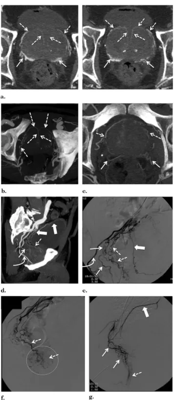
Figure 6. Images from a case with two independent PAs. (a) Pelvic CT angiography with axial MIP shows that capsular branches penetrate the capsule in four quadrants: anterior/lat-eral PAs (dotted arrows mark central gland vascularization) and posterior/lateral PAs (solid arrows mark peripheral gland vas-cularization) on each pelvic side. The apparent capsular pene-tration of the anterior/lateral PAs (dotted arrows) is made ap-proximately at the 2-o’clock and 10-o’clock positions for the left and right sides, respectively, and, for the posterior/lateral PAs, at the 5-o’clock and 7-o’clock positions for the left and right sides, respectively. Pelvic CT angiography with axial MIP shows (b) anterior/lateral PAs (dotted arrows) vascularizing most of the central gland and (c) anterior/lateral PAs (dotted arrows mark
central gland vascularization) and posterior/lateral PAs (solid ar-rows mark peripheral gland vascularization) on each pelvic side. Note the close proximity of the posterior/lateral PAs with the rectum. (d– g) Two independent PAs on the right pelvic side. (d) Pelvic CT angiography with sagittal MIP reformat shows the ante-rior/lateral PA (dotted arrow) with a more superior/proximal origin from a short common trunk with the superior vesical artery (solid arrows). The posterior/lateral PA (straight arrow) has an inferior/ distal origin from the internal pudendal artery (curved arrow) above the sciatic notch. (e) DSA of the right internal iliac artery performed with same-side anterior oblique projection (35°) and caudal/cranial angulation (10°). Dotted arrows mark the anterior/lateral PA, the solid arrow marks the superior vesical artery, straight arrows mark the posterior/lateral PA, and the curved arrow marks the internal puden-dal artery. (f) DSA with same-side anterior oblique projection (35°) and caudal/cranial angulation (10°) after selective catheterization of the anterior/lateral PA (dotted arrows). The dotted circle marks central gland opacification. (g) DSA with same-side anterior oblique projec-tion (35°) and caudal/cranial angulaprojec-tion (10°) after selective catheter-ization of the posterior/lateral PA. Note the overlapping vasculariza-tion between the peripheral prostatic gland (arrows) and rectal branches (dotted arrow marks anal blush) with retrograde opacifica-tion of the IMA (solid arrow).
Figure 7. Two independent PAs on the left pelvic side. (a) Pelvic CT angiography with sagittal MIP reformat. The anterior/lateral PA (straight arrow) has a more superior/proximal origin from a short common trunk with the superior vesical artery (arrowhead). The posterior/lateral PA (dotted arrow) has an inferior/distal origin from the internal pudendal artery (curved arrow) above the sciatic notch. Note the close proximity between the middle rectal artery (thick arrow) and the posterior/lateral PA (dotted arrow). (b) Pelvic CT angiography with coronal MIP reformat shows the anterior/lateral PA (arrow). (c) Pelvic CT angiography with coronal MIP reformat shows the posterior/lateral PA (arrow). (d) Pelvic CT angiography with 3D reformat. The straight arrow marks the anterior/lateral PA, the arrowhead marks the superior vesical artery, the dotted arrow marks the posterior/lateral PA, the curved arrow marks the internal pudendal artery, and the thick arrow marks the middle rectal artery. (e) DSA of internal iliac artery performed with same-side anterior oblique projection (35°) and caudal/cranial angulation (10°). The straight arrow marks the anterior/lateral PA, the arrowhead marks the superior vesical artery, the dotted arrow marks the posterior/lateral PA, the curved arrow marks the internal pudendal artery, and the thick arrow marks the middle rectal artery. (f) DSA with same-side anterior oblique projection (35°) and caudal/cranial angulation (10°) after selective catheterization of the posterior/lateral PA (dotted arrow). Note anastomoses with the rectal branches with retrograde opacification of the middle rectal artery (thick arrow), and anastomoses through small postcapsular branches to the internal pudendal artery (open arrow). Prostate gland opacification is seen inside the dotted circle. (g) DSA in neutral position after selective catheter-ization of the posterior/lateral PA (arrow) shows prostate gland opacification. (h) DSA with same-side anterior oblique projection (35°)
lateral PA and between the rectal arteries and the posterior/
lateral PA. It was easy to differentiate the anterior/lateral
PA from the vesical arteries: vesical arteries had a straighter
trajectory with a superior and more diffuse vascularization
to the bladder wall, whereas the PAs had a downward
undulating trajectory with a more globular and
circum-scribed vascularization below the bladder base.
Differenti-ating the rectal arteries from the posterior/lateral PA was
more difficult. The rectal arteries showed a more oblong
and vertical opacification to the rectal wall with a
promi-nent anal blush at the perineum, with retrograde filling of
the inferior mesenteric artery (IMA). Posterior/lateral PAs
had a more round and circumscribed appearance (
Figs 8
and
9
). These two circulations were frequently in close
proximity, which made it impossible to differentiate and
selectively catheterize them; on the contrary, the vesical
circulation was easier to separate from the prostatic
circu-lation by advancing the microcatheter distally to the vesical
artery origin.
Arterial Anastomoses
Based on CT angiography and selective prostatic DSA, it
was possible to evaluate the intraprostatic branches and
anastomoses with surrounding arteries. No significant
anas-tomoses were identified in 43% of pelvic sides(n
⫽ 64), and
anastomoses to adjacent arteries were documented in 57%
(n
⫽ 86). The most frequent types of anastomoses found
were to the internal pudendal arteries (43.3%), contralateral
(17.6%) and ipsilateral (13.4%) prostatic branches, rectal
arteries (14.4%), and vesical arteries (11.3%;
Table 1
). We
found two different types of anastomoses relevant to the
embolization technique: one through small pericapsular and
postcapsular branches, the other through large precapsular
branches that coursed alongside the anterior and lateral
aspect of the prostate from the base to the apex, terminating
in the perineum with anastomoses to the internal pudendal
artery (also called lateral accessory pudendal arteries).
Along their trajectory, these collateral vessels gave rise to
capsular prostatic branches (
Fig 10
). There were 30 pelvic
sides (20%) with lateral accessory pudendal arteries
repre-senting anastomoses between the PAs and the internal
pu-dendal arteries. We also found five pelvic sides (3.3%) with
lateral accessory pudendal arteries solely responsible for
the arterial blood supply to the corpora cavernosa in close
proximity with the PAs.
PA DSA Findings
The DSA findings of the PAs were very variable. A
cork-screw pattern of the capsular branches was found in 37% of
individuals (n
⫽ 28). In the remaining patients, a globular
circumscribed opacification was found below the bladder
base, without remarkable features (
Fig 10
).
Correlation between Anatomic
and Clinical Data
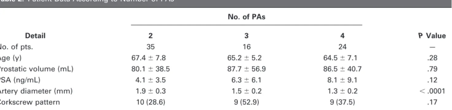
The number of PAs was correlated with the mean diameter
of the PAs (P
⬍ .0001); patients with only two PAs (one on
each pelvic side) had a larger mean PA diameter (1.9 mm)
than those with three (1.5 mm) or four (1.3 mm) PAs. The
number of PAs did not correlate with age, prostate volume,
PSA, or presence of the corkscrew pattern (
Table 2
). The
same results were obtained with ordered logistic regression.
The corkscrew pattern was associated with larger prostate
volumes (mean, 111.9 mL; P
ⱕ .0001) and higher PSA
values (8.1 ng/mL; P
⫽ .004) compared with the absence of
the corkscrew pattern (mean prostate volume, 67.0 mL;
mean PSA, 4.3 ng/mL;
Table 3
). In multivariate analysis
with logistic regression, only prostate volume was
indepen-dently associated with the corkscrew pattern (P
⫽ .01). We
found no correlations between PA diameter and patient age,
prostate volume, or PSA values.
DISCUSSION
We used CT angiography before DSA to exclude patients
with advanced atherosclerosis/tortuosity of the iliac arteries
or PAs (8% in the present study) and to allow us to draw a
vascular map of the internal iliac arteries and PAs. PAs on
DSA lack pathognomonic findings, and many small-sized
arteries may easily be mistaken for PAs based only on
DSA, leading to long procedural times and high radiation
dose or nontarget embolization. The use of CT angiography
before DSA reassures correct location before embolization,
and we believe the use of the anterior oblique projection
(35°) with caudal/cranial angulation (10°) in the initial DSA
of the internal iliac artery is fundamental, with excellent
correlation with the CT angiography sagittal MIP reformat,
allowing easy recognition of the PAs and avoiding multiple
DSA runs with different projections. The corkscrew pattern
of the PAs may also help to identify the PAs on DSA: this
was previously described in cadavers (
7
) and was present in
37% of individuals in the present study (n
⫽ 28). As
expected, this pattern was found in patients with larger
prostates and higher PSA values, as it is probably easier to
identify in these patients.
With preinterventional image guidance, PAE may be
performed with a mean procedural time of approximately
and caudal/cranial angulation (10°) after selective embolization of the posterior/lateral PA (dotted arrow). Note the retrograde filling of the anterior/lateral PA (straight arrow) through intraprostatic interpedicular anastomoses that enabled retrograde embolization of the anterior/ lateral PA. (i) Pelvic CT angiography with coronal MIP reformat shows the left middle rectal artery (arrow). (j) DSA of the left middle rectal artery (arrow) with same-side anterior oblique projection (35°) and caudal/cranial angulation (10°). (k) DSA of the left middle rectal artery in neutral position with rectal wall opacification, retrograde filling of the inferior mesenteric artery (IMA), and a prominent anal blush (arrow). (Available in color online atwww.jvir.org.)
90 minutes and a mean fluoroscopic time of
approxi-mately 30 minutes. However, even with
preinterven-tional image guidance, as many as 3% of cases result in
technical failure (ie, no embolization on either pelvic
side), and as many as 8% of patients may be amenable to
only unilateral embolization. From our experience, it is
very difficult to predict with complete accuracy if
bilat-eral embolization will be technically feasible based on
the CT angiography findings. Although this is not proven
in the present study, we believe PA diameter is not the
most important parameter for selective catheterization;
the angle of origin and the presence of atherosclerotic
plaques near the ostium of the PAs are more important
factors that may hamper selective catheterization,
espe-cially when the PAs arise from vesical or prostatorectal
arteries. Tortuosity of the iliac arteries and PAs may also
lead to technical difficulties.
Surprisingly, prostate volume did not correlate with
PA size in the present study. Patients with only one PA
on each pelvic side had larger PAs than those with more
than one PA on each pelvic side, but no other
associa-tions were found.
Two independent PAs were found in 43% of pelvic
sides (n
⫽ 64), which is a higher incidence than previously
reported rates (
7,8,10,16
). From our experience, the
in-creasing awareness of the possibility of more than one PA
on each pelvic side raised the suspicion for small branches
that may have been initially overlooked. In the present
study, we showed that the prostate has a dual blood supply,
as described by Bouissou and Talazac (
9
), with two PAs
that arose from a solitary PA or that arose independently.
We opted to use the term anterior/lateral PAs instead of
“cranial” PAs (
9
) because they penetrated the prostatic
capsule in the anterior/lateral quadrants, whereas we used
the term posterior/lateral PAs instead of “caudal” PAs (
9
)
because they penetrated the prostatic capsule in the
poste-rior/lateral quadrants. As described by Bouissou and
Ta-lazac (
9
), when these two PAs arose independently, the
anterior/lateral PA had a more proximal origin in close
proximity to the vesical arteries, whereas the posterior/
lateral PA had a more distal origin close to rectal branches.
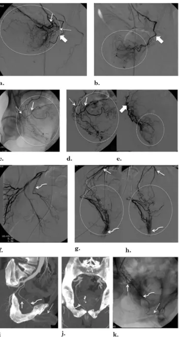
Figure 8. Illustrations of a potential pitfall of PAE, namely, neighboring arteries that may be confused with PAs: (a– e) ves-ical arteries and PAs, (f– h) rectal arteries, and (i– k) accessory pudendal arteries. (a) Left superior vesical artery (arrow) DSA with same-side anterior oblique projection (35°) and caudal/ cranial angulation (10°) shows diffuse bladder opacification (in-side circle). The dotted arrow marks the inferior vesical artery and the solid arrow marks PA. (b) After selective PA (solid arrow) DSA with same-side anterior oblique projection (35°) and caudal/cranial angulation (10°), prostate opacification is seen (inside circle). (c, d) Diffuse bladder opacification (inside circle) after selective DSA with same-side anterior oblique pro-jection (35°) and caudal/cranial angulation (10°) of the superior (arrow) and inferior (dotted arrow) right vesical arteries arising from a common trunk. (e) Selective right-side PA (solid arrow) DSA with same-side anterior oblique projection (35°) and cau-dal/cranial angulation (10°) shows prostate opacification (inside circle). (f) DSA of the right middle rectal artery (curved arrow) with same-side anterior oblique projection (35°) and caudal/ cranial angulation (10°). (g) Selective DSA with same-side an-terior oblique projection (35°) and caudal/cranial angulation (10°) of the right middle rectal artery shows anal blush (curved arrow) and retrograde filling of the IMA (arrow). Diffuse rectal
opacification is seen inside the circle. (h) Selective DSA in neu-tral position of the right middle rectal artery shows anal blush (curved arrow) and retrograde filling of the IMA (arrow). Diffuse rectal opacification is seen inside the circle. (i) Pelvic CT angiog-raphy with sagittal MIP reformat of the right pelvic side. The curved arrow marks the right accessory pudendal artery that courses near the prostate and ends as the dorsal artery of the penis (dotted arrow). The straight arrow marks the PA arising from the accessory pudendal artery. (j) Pelvic CT angiography with coronal MIP reformat. The curved arrow marks the right accessory pudendal artery. (k) DSA of right accessory pudendal artery (curved arrow) performed with same-side anterior oblique projection (35°) and caudal/cranial angulation (10°). The dotted arrow marks the dorsal artery of the penis; the straight arrow marks the PA.
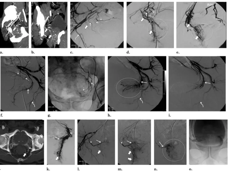
Figure 9. Vesical, rectal, and prostatic arteries. (a) Pelvic CT angiography with sagittal MIP reformat of the right pelvic side. The anterior/lateral PA (straight arrow) arises from a short common trunk with the vesical arteries (dotted arrows). There is a common prostatorectal trunk (thick arrow) supplying the posterior/lateral PA and running downward into the perineum adjacent to the prostatic capsule (middle rectal artery). The curved arrow marks the internal pudendal artery. (b) Pelvic CT angiography with sagittal MIP reformat of the right pelvic side. The straight arrow marks the anterior/lateral PA, dotted arrows mark vesical arteries, and thick arrows mark the common prostatorectal trunk. (c) DSA of the right internal iliac artery performed with same-side anterior oblique projection (35°) and caudal/cranial angulation (10°). The straight arrow marks the anterior/lateral PA, dotted arrows mark the vesical arteries, the thick arrow marks the common prostatorectal trunk, and the curved arrow marks the internal pudendal artery. (d) DSA after selective catheterization of the common prostatorectal trunk (thick arrows) with same-side anterior oblique projection (35°) and caudal/cranial angulation (10°). There is diffuse prostatic and rectal opacification with anastomoses and retrograde filling of the anterior/lateral prostatic arterial pedicle (straight arrow) and vesical arteries (dotted arrow) and of the IMA, rendering embolization unsafe. Prostate gland opacification is seen inside the dotted circle. (e) DSA after selective catheterization of the common prostatorectal trunk (thick arrows) in neutral position. Prostate gland opacification is seen inside the dotted circle, and there is retrograde filling of the IMA. (f–i) Vesical and prostatic arteries in another patient (left pelvic side). (f) DSA of the internal iliac artery performed with same-side anterior oblique projection (35°) and caudal/cranial angulation (10°). The PA (straight arrow) arises from the vesical arteries (dotted arrows). The curved arrow marks the internal pudendal artery. (g) Selective DSA of the vesical arteries (dotted arrows) in neutral position shows diffuse bladder wall opacification. (h) Selective DSA with same-side anterior oblique projection (35°) and caudal/cranial angulation (10°) of the PA (straight arrow) before PAE. The curved arrow marks the internal pudendal artery. Prostate gland opacification is seen inside the dotted circle. (i) Control angiogram with same-side anterior oblique projection (35°) and caudal/cranial angulation (10°) after PAE. The straight arrow marks the PA and the curved arrow marks the internal pudendal artery. (j) Axial CT angiography scan shows a common prostatorectal trunk on the left side bifurcating into the posterior/lateral PA (straight arrow) and the middle rectal artery (thick arrow). (k) Selective DSA with same-side anterior oblique projection (35°) and caudal/cranial angulation (10°) of the common prostatorectal trunk on the left side (arrowhead) shows diffuse rectal/anal opacification (thick arrow) and prostatic opacification (straight arrow), which was difficult to isolate, rendering embolization unsafe. (l) DSA of the right pelvic side with same-side anterior oblique projection (35°) and caudal/cranial angulation (10°) shows a common prostatorectal trunk (arrowhead) that bifurcates into the middle rectal artery (thick arrow) and the PA (straight arrow). (m) DSA with same-side anterior oblique projection (35°) and caudal/cranial angulation (10°) of the common prostatorectal trunk (arrowhead) that bifurcates into the middle rectal artery (thick arrows) and the PA (straight arrow). (n) DSA with same-side anterior oblique projection (35°) and caudal/cranial angulation (10°) of the PA (arrow) shows prostate gland opacification (inside dotted circle). In these situations, it is possible to isolate the prostatic circulation from the rectal circulation by advancing the microcatheter distally. (o) DSA in neutral position shows prostate gland opacification after embolization.
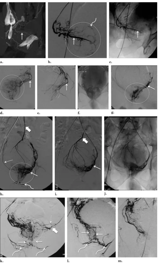
Figure 10. DSA findings of PAs and arterial anastomoses. (a) Sagittal MIP CT angiography shows a right-side PA (straight arrow). (b, c) Selective DSA with same-side anterior oblique projection (35°) and caudal/cranial angulation (10°) of the right-side PA (straight arrow). The dotted circle marks prostate gland opacification, and a corkscrew pattern is shown (curved arrow). (d) Selective DSA with same-side anterior oblique projection (35°) and caudal/cranial angulation (10°) of a left-side PA (straight arrow) before PAE. The dotted circle marks prostate gland opacification. (e) Selective DSA with same-side anterior oblique projection (35°) and caudal/cranial angulation (10°) of a left-side PA (straight arrow) after PAE. (f) DSA in neutral position shows prostate gland opacification after bilateral PAE. (g) Selective DSA (neutral position) of a left-side PA (straight arrow) shows a more diffuse and heterogeneous pattern of prostatic glandular opacification (dotted circle). (h) DSA of the left-side PA (straight arrow) in neutral position before PAE shows diffuse
The vesical and rectal arteries may represent pitfalls to
PAE, and may be confused with PAs. Here we showed
DSA findings that differentiate PAs from vesical or rectal
arteries.
The anterior/lateral PA was shown to be the main
supply to the central gland, and therefore should probably
be the preferred artery to embolize when two independent
PAs are present. Embolization of one of the two PAs might
be enough because of the intraprostatic interpedicular
anas-tomoses that may be found, as previously described by
Bouissou and Talazac (
9
). When only one PA is found, we
usually leave the catheter tip before its bifurcation to allow
embolization of both PAs. Further studies in patients with
two independent PAs are needed to compare embolization
of both PAs versus only one PA.
In the present study, findings of PA origin were
somewhat different from previously published results in
cadaveric specimens (
7,8,10,17
). These differences may
be explained by distinct anatomic terminology and
tech-niques used in cadaveric versus in vivo studies. It was
stated (
7
) that, in most individuals, 41.5%–74.3% of the
prostate gland is supplied by a common prostate/vesical
arterial trunk (also named the inferior vesical artery) that
gives rise to the inferior vesical artery and the PA.
However, Clegg (
7
) reported that the so-called inferior
vesical artery might terminate supplying the prostate
without any branches to the bladder. We also found that
many of the arteries supplying the prostate had no
rela-tion to the vesical arteries, and therefore decided to
replace the term “inferior vesical artery” with the term
“PA,” which may or may not have inferior vesical
branches.
Significant anastomoses may be identified on DSA
in as many as 60% of individuals, usually to the perineal,
middle rectal, or anal branches of the internal pudendal
artery, as previously described (
7
). From our experience,
in the presence of small-sized anastomoses through
cap-sular branches, a slow and controlled infusion may avoid
distal embolization with nontarget ischemia to the
blad-der, rectum, anus, or corpus cavernosum. Large (2–3
mm) precapsular branches may represent lateral
acces-sory pudendal arteries. In cases in which this artery ends
as the dorsal artery of the penis—and is therefore solely
responsible for arterial blood supply to the corpora
cav-ernosa, as found in 3.3% of pelvic sides in the present
series—the best option for embolization is to selectively
embolize all prostatic branches, without compromising
prostatic glandular opacification (inside circle) with anastomoses to rectal branches, with retrograde filling of the IMA (thick arrow) and anastomoses to the contralateral PA (dotted arrow) and internal pudendal artery (curved arrow) through small postcapsular branches. (i, j) DSA of the left-side PA (straight arrow) in neutral position after PAE. The thick arrow marks the IMA. (k) Selective DSA of the right-side PA in neutral position shows small pericapsular and postcapsular anastomoses (straight arrows) to the internal pudendal artery (curved arrows) and anastomoses to contralateral PA (thick arrow) and inferior vesical artery (dotted arrow). Prostatic glandular opacification is seen inside the circle. (l) DSA of the right-side PA in neutral position before PAE shows multiple arterial anastomoses through larger precapsular branches that course alongside the anterior and lateral aspect of the prostate from the base to the apex (straight arrows), terminating in the perineum—also called lateral accessory pudendal arteries—with anastomoses to the internal pudendal artery (curved arrows). Prostatic glandular opacification is seen inside the circle. (m) DSA of the right-side PA in neutral position after PAE.
Table 2. Patient Data According to Number of PAs
Detail No. of PAs P Value 2 3 4 No. of pts. 35 16 24 — Age (y) 67.4⫾ 7.8 65.2⫾ 5.2 64.5⫾ 7.1 .28 Prostatic volume (mL) 80.1⫾ 38.5 87.7⫾ 56.9 86.5⫾ 40.7 .79 PSA (ng/mL) 4.1⫾ 3.5 6.3⫾ 6.1 8.1⫾ 9.1 .12 Artery diameter (mm) 1.9⫾ 0.3 1.5⫾ 0.2 1.3⫾ 0.2 ⬍ .0001 Corkscrew pattern 10 (28.6) 9 (52.9) 9 (37.5) .17
PA⫽ prostatic artery, PSA ⫽ prostate-specific antigen, SD ⫽ standard deviation.
Values presented as mean⫾ SD where appropriate. Values in parentheses are percentages.
Table 3. Patient Data According to Presence or Absence of the Corkscrew Pattern
Detail Corkscrew Pattern P Value Present Absent No. of pts. 28 47 — Age (y) 66.5⫾ 7.3 65.7⫾ 7.1 .63 Prostatic volume (mL) 111.9⫾ 52.8 67.0 ⫾ 24.3 ⬍ .0001 PSA (ng/mL) 8.1⫾ 8.5 4.3⫾ 4.5 .004 Artery diameter (mm) 1.6⫾ 0.3 1.6⫾ 0.4 .58 PSA⫽ prostate-specific antigen, SD ⫽ standard deviation. Values presented as mean⫾ SD where appropriate.
this artery. If this artery terminates as an anastomotic
arch, as found in 20% of pelvic sides in the present
series, with the termination of the internal pudendal
artery, another option is to use 2–3-mm microcoils or
Gelfoam torpedoes to exclude this anastomosis and then
embolize the entire artery with particles. Accessory
pu-dendal arteries may represent pitfalls to PAE and may be
confused with PAs; the main DSA findings in the present
study may help to differentiate them.
Anastomoses with contralateral PAs may be present
in as many as 20% of individuals, which might explain
why some patients have good clinical results even when
only unilateral embolization is achieved (
6
). Further
studies comparing clinical outcomes between patients
with unilateral and bilateral PAE are needed to assess
this issue.
The limitations of the present study include the
absence of cone-beam CT during selective DSA of the
PAs, which may be helpful to certify correct catheter
position before embolization, especially in the initial
learning curve or in doubtful cases; however, we do not
believe routine use is necessary if CT angiography has
been performed and the operators have experience in
PAE. Other limitations relate to the classification of the
PA origin that may vary depending on the anatomic
classifications used. Also, small anastomoses may go
undetected with conventional angiography, and we did
not use wedged catheter DSA runs. Future studies
ad-dressing anastomosis evaluation with the use of
intrap-rocedural cone-beam CT or with cadavers may also help
with this subject. It would be very important to identify
the diameter of the small anastomoses to guide the
choice of embolic material size to avoid extraprostatic
embolization. In the present study no evaluation was
performed of how the knowledge gained from CT
an-giography affected the embolization technique.
PAE has been shown to be a promising new
mini-mally invasive therapeutic option for BPH; however, to
perform the procedure in a safe manner without
nontar-get embolization to periprostatic tissues, precise
knowl-edge of the PA anatomy and imaging findings is required.
In this work, we describe the vascular anatomy of the
prostate by using CT angiography and DSA, focusing on
the most relevant aspects for PAE.
REFERENCES
1. Mauro MA. Can hyperplastic prostate follow uterine fibroids and be managed with transcatheter arterial embolization? Radiology 2008; 246:657–658. 2. Sun F, Sánchez FM, Crisóstomo V, et al. Benign prostatic hyperplasia:
transcatheter arterial embolization as potential treatment—preliminary study in pigs. Radiology 2008; 246:783–789.
3. Jeon GS, Won JH, Lee BM, et al. The effect of transarterial prostate embolization in hormone-induced benign prostatic hyperplasia in dogs: a pilot study. J Vasc Interv Radiol 2009; 20:384 –390.
4. DeMeritt JS, Elmasri FF, Esposito MP, Rosenberg GS. Relief of benign prostatic hyperplasia-related bladder outlet obstruction after transarterial polyvi-nyl alcohol prostate embolization. J Vasc Interv Radiol 2000; 11:767–770. 5. Carnevale FC, Antunes AA, da Motta Leal Filho JM, et al. Prostatic artery
embolization as a primary treatment for benign prostatic hyperplasia: preliminary results in two patients. Cardiovasc Intervent Radiol 2010; 33:355–361. 6. Pisco JM, Pinheiro LC, Bilhim T, Duarte M, Mendes JR, Oliveira AG.
Prostatic arterial embolization to treat benign prostatic hyperplasia. J Vasc Interv Radiol 2011; 22:11–19.
7. Clegg EJ. The vascular arrangements within the human prostate gland. Br J Urol 1956; 28:428 – 435.
8. Clegg EJ. The arterial supply of the human prostate and seminal ves-icles. J Anat 1955; 89:209 –216.
9. Bouissou H, Talazac A. Arterial vascularization of the normal and the pathological prostate. Ann Anat Pathol 1959; 4:63–79.
10. Ambrósio JD, De Almeida JS, De Souza A. Origin of prostatic arteries in man. Rev Paul Med 1980; 96:52–55.
11. Slojewski M, Czerwinski F, Sikorski A. Microangiographic imaging of the prostate. BJU Int 2002; 89:776 –778.
12. Duclos JM, Chanzy M, Alexandre JH. Prostatic vascularisation. Arch Anat Pathol 1972; 20:355–358.
13. Ambrósio JD, De Almeida JS, De Souza A. Apparent penetration of the arteries into the prostatic capsule in man. Rev Paul Med 1980; 95:98 – 100.
14. Yamaki K, Saga T, Doi Y, et al. A statistical study of the branching of the human internal iliac artery. Kurume Med J 1998; 45:333–340. 15. Bilhim T, Casal D, Furtado A, Pais D, O’Neill JE, Pisco JM. Branching
patterns of the male internal iliac artery: imaging findings. Surg Radiol Anat 2011; 33:151–159.
16. Bilhim T, Pisco JM, Furtado A, et al. Prostatic arterial supply: demon-stration by multirow detector angio CT and catheter angiography. Eur Radiol 2011; 21:1119 –1126.
17. Chen XQ. Anatomic study of the arteries and nervous plexus of the prostate and their clinical significance. Zhonghua Wai Ke Za Zhi 1989; 27:373–375.