DOI: 10.1111/j.1468-3083.2008.02941.x
JEADV
Blackwell Publishing Ltd
ORIGINAL ARTICLE
Hansen’s disease in Portugal: multibacillary patients treated
between 1988 and 2003
S Medeiros,*† MG Catorze,‡ MR Vieira†
†Dermatology Department, Curry Cabral Hospital, ‡Dermatology Department, Centro de Dermatologia Médico-Cirúrgica, Lisbon, Portugal
*Correspondence: S Medeiros. E-mail: [email protected]
Abstract
Background There is an estimate low incidence of patients with Hansen’s disease in Portugal. Following the 1982 World
Health Organization (WHO) recommendations, extended multidrug therapy (MDT) was started for multibacillary (MB) patients. Patients were then treated with rifampicine (RFP), clofazimine (CLF) and dapsone (DDS) for a minimum of 2 years or until smear negativity. The aim of this study was to evaluate MDT efficacy in our patient population.
Methods Retrospective and descriptive study of 102 MB patients who underwent MDT from 1988 to 2003.
Results The number of new MB patients has gradually increased since 1960, the first year of our consultation, due
mostly to a rise in imported cases. Overall, 34% of the subjects were immigrants, mainly from former Portuguese Colonies. Forty-six patients had previously received monotherapy with DDS (mean duration of this treatment, 22 years). Relapse after MDT occurred in 9 cases (8.8%), but importantly, all relapsed cases were smear negative at least on one occasion after the end of treatment, suggesting these were true relapses rather than treatment failures.
Conclusions Despite the 2-year WHO-MDT regimen, patients with MB disease clearly face the possibility of relapse.
We propose that any reduction in the duration of therapy such as the recently proposed 6-month standard MDT is likely to increase the relapse rate even further. Important issues for future consideration are the needs to identify those at risk of relapse and in need of alternative antimicrobial treatment with a prolonged clinical follow-up.
Received: 1 April 2008; Accepted 19 May 2008
Keywords
Hansen’s disease, multibacillary patients, multidrug therapy, relapse
Conflicts of interest
None declared
Introduction
Hansen’s disease is caused by Mycobacterium leprae which causes selective damage to the skin and peripheral nerves. There is a wide variety of clinical findings in this disease. In 1966, Ridley and Jopling created a classification based on polar forms.1 The illness spectrum ranges from tuberculoid disease to lepromatous disease, characterized by few skin lesions, and a marked cell-mediated immune response to the mycobacteria in the former, and multiple lesions, the greatest number of bacilli and low-immunity in the latter. Borderline leprosy, as it name implies, has features that are intermediate between the two ends of the spectrum and refers to 3 forms: borderline lepromatous (BL), borderline-borderline (BB) and borderline-tuberculosis (BT). Sequelae may be multiple and variable mainly in late diagnosed and treated patients, affecting skin, nerves, eyes and musculoskeletal system. The introduction of dapsone was a major breakthrough in the treatment of Hansen’s disease. Nevertheless, documentation of primary and secondary
resistance to this drug and to other pharmacological agents led to the current recommendation that only regularly scheduled com-bined treatment could prevent resistance and subsequent treatment failure. Abandoning dapsone monotherapy in 1982, the World Health Organization (WHO) recommended instead, a minimum of a 2-year regimen (within stipulated 36 months) of rifampicin (RFP), dapsone (DDS) and clofazimine (CLF) for cases of multi-bacillary (MB) Hansen’s disease, continued, wherever possible until smear negativity.2,3 In 1998, with mounting evidence that less than 24 months of therapy was effective, recommended duration was reduced to 1 year.4,5 Significant controversy was recently generated following a proposal for a reduction in the length of treatment to 6 months with a standardized MDT regimen for all Hansen’s disease patients.6
Despite the WHO declaration that Hansen’s disease would be eliminated as a public health problem (defined as a registered prevalence rate of less than 1 case per 10 000 population) by 2000,7
30 Medeiros et al.
new cases continue to be diagnosed even outside endemic areas,8,9 and the illness remains far from being eradicated.10 Relapse rate is the single most important measure of MDT efficacy,11 and it ranges from below 1% to over 20% in various recent studies, depending upon duration of follow-up, percentage of patients with a high bacterial burden, type of treatment received, medical expertise, treatment compliance and the relapse criteria included in each study.5,8–10,12–14 Unfortunately, there is no clear consensus in these studies as regards the features which most effectively identify a relapse. There is currently no WHO recommendation on follow-up,3 and patients cease to be considered as active cases in many clinical centres after MB treatment is completed. In a country with low incidence rates, patients may experience a delay in referral to a diagnostic centre and also in accurate diagnosis of relapse. The majority of Hansen’s disease patients in Portugal are treated at our clinic in the Curry Cabral Hospital in Lisbon allowing for adequate prompt therapy after diagnoses and for an accurate diagnosis of relapse. Our consultation is a reference consultation for this disease and we receive patients from all Portuguese regions. Additionally, some of these patients came from a previous leprosarium located in Coimbra region, Leprosaria Rovisco Pais.
In this study, our purpose was to evaluate MB patients under-going extended MDT with a particular emphasis on the incidence of true relapses after adequate therapy.
Materials and methods
We performed a retrospective, descriptive and analytical study at Hansen’s disease Consultation in Curry Cabral Hospital. Weekly appointments by two Dermatology specialists treated Hansen’s disease patients and their contacts, mainly from central and littoral regions of Portugal (Fig. 1).
Detailed clinical evaluation included complete dermatological and neurological examination and all patients were referred to Ophthalmologic and Ear, Nose and Throat departments during the first examination. Bacteriological evaluation consisted of nasal scrapings and skin smears samples every 6 months. Slit-skin smears were obtained with a scalpel from pinched skin lesions, the earlobe or other cold skin areas. All smears were sent to the hospital’s micro-biology department for analysis. Each patient underwent a biopsy of the skin during the first visit and subsequently, when required. In some cases, a nerve biopsy was required for diagnosis. Nerve conduction studies were carried out in the majority of cases. The Ridley-Jopling classification was used to classify all patients.
The implementation of WHO multidrug therapy (MDT) began in 1988 with a monthly supervised treatment programme of at least 2 years or until negative skin smear was obtained: drug therapy consisted of RFP 600 mg with CLF 300 mg once monthly, and CLF 50 mg associated to DDS 100 mg daily. Our hospital control programme consisted in a twice year follow-up for 12 years after completion of MDT intended to detect delaying reactions, relapses and prevent disease sequelae. Following this period,
routine evaluation was discontinued but open access to the hospital clinic was provided whenever required.
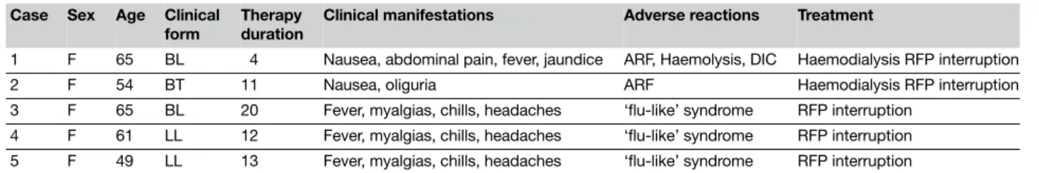
In order to evaluate MDT efficacy, retrospective analysis of clinical records of all subjects (both ‘new’ and ‘old’ patients) was undertaken after the introduction of MDT and until 2005 (in order to include a 2-years follow-up of those patients treated up to 2003). Of the 292 patients detected during this period, the majority had MB forms (230 patients). Of these MB forms, 118 were not selected to initiate MDT because they were ‘old’ patients that had already undergone a period of treatment with DDS in excess of 20 years. MDT was started on a total of 112 patients. Among these 112 patients, 102 MB patients concluded extended-MDT, with five patients either lost to follow-up or non-compliant with regular therapy. Adverse reactions to rifampicine were responsible for the complete interruption of this drug in the other five cases: three patients had a ‘flu-like’ syndrome and two patients developed renal failure (Table 1; data previously reported).15
The following were studied in the 102 MB patients treated with extended MDT: demographic data (age, gender, country and city of origin, current residence), time between onset of initial
Hansen’s disease in Portugal 31
symptoms and start of therapy, clinical presentation, previous monotherapy with dapsone and its duration, bacterial index (BI) before and after extended-MDT, treatment duration until smear negativity, clinical reactions and their management, relapses, bacterial index and histopathologic diagnosis at the relapse, characteristics of lesions on relapses, presence of sequelae (ocular, otorrinolaringologic, neurological and bone) and duration of follow-up.
Relapse was defined as patients remaining symptom-free after MDT until smear negativity but developing:
• Skin and/or nerve lesions clinically and/or histopathologically consistent with active Hansen’s disease using the Ridley-Jopling classification.1
• BI of 2+ in one or more smear sites.
Statistical analysis
Continuous variables were tested for normal distribution with normality tests (Shapiro-Wilk if < 50 cases, Kolmogorov-Smirnov if ≥ 50 cases). Most continuous variables had a non-parametric distribution. Bivariate analysis between independent and dependent variables was made using non-parametric tests: Spearman’s corre-lation analysis for continuous variables, two-tailed chi-squared test with Yates correction and Fisher’s exact test, whenever applicable, for categorical variables and Mann-Whitney’s test to compare categorical and numerical variables.
Quantitative and qualitative data of baseline characteristics of population were expressed as median [1st–3rd quartiles] or mean ± standard deviation (SD) and percentage (95% confidence inter-vals, 95% CI), respectively.
Bivariate analysis was made using Kaplan-Meier and logrank test: relapse survival rates were compared between groups (sex, bacillary skin index ≤/> 2, previous DDS therapy and steroid therapy of reactions), as well as mean survival time and 95% CI. P-values less then 0.05 were considered statistically significant.
Data from each case were recorded on standardized report forms, entered into a database and analysed using SPSS software 13.0 for Windows® (SPSS Inc., Chicago, IL).
Results
The 102 MB patients were registered at our clinic between 1968 and 2003 and the year of diagnosis ranged between 1934 and 2003.
The number of Hansen’s disease patients increased in recent years (Fig. 2).
Table 2 provides the major baseline characteristics of our cohort, comparing patients with relapse vs. those without relapse.
Table 1 Adverse reactions to monthly Rifampicin schedule in the treatment of leprosy (n = 5 cases).15
Case Sex Age Clinical
form
Therapy duration
Clinical manifestations Adverse reactions Treatment
1 F 65 BL 4 Nausea, abdominal pain, fever, jaundice ARF, Haemolysis, DIC Haemodialysis RFP interruption
2 F 54 BT 11 Nausea, oliguria ARF Haemodialysis RFP interruption
3 F 65 BL 20 Fever, myalgias, chills, headaches ‘flu-like’ syndrome RFP interruption
4 F 61 LL 12 Fever, myalgias, chills, headaches ‘flu-like’ syndrome RFP interruption
5 F 49 LL 13 Fever, myalgias, chills, headaches ‘flu-like’ syndrome RFP interruption
ARF, Acute renal failure; DIC, disseminated intravascular coagulopathy.
Figure 2 Hansen’s disease year of diagnosis (n = 102).
Table 2 Patients baseline characteristics
Relapse ( n ==== 9) No relapse ( n ==== 93) P-value Median [Q1–Q3] Age at diagnosis 27 [20–49] 30 [24–53] P > 0.05 Age at start MDT 55 [37–64] 55 [36–62] P > 0.05 n (%) Male gender 3 (33.3) 51(54.8) P > 0.05 Clinical form LL 5 (55.6) 63 (67.7) P > 0.05 BL 4 (44.4) 30 (32.2) P > 0.05 Steroid therapy 6 (66.7) 25 (26.8) P > 0.05 Previous DDS therapy 3 (33.3) 38 (40.9) P > 0.05 BI ≥ 4 2 (22.2) 24 (25.8) P > 0.05 Non Portuguese 3 (33.3) 33 (35.5) P > 0.05
32 Medeiros et al.
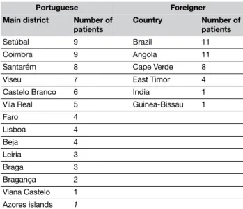
Of the 102 patients followed up, 52.9% were male with a mean age of diagnosis of 30.5 years. Sixty-six (65.3%) were Portuguese and 36 foreigners (34.7%): 11 patients from Brazil (mainly from the states of Goiás and Minas Gerais), 11 from Angola, 8 from Cape Verde, 4 from East Timor, one from India and another one from Guinea-Bissau (Table 3).
Thirty patients already had a family history of the disease. Two-hundred and ninety-nine (299) contacts of the 102 patients were observed, but only one with an indeterminate form of Hansen’s disease was diagnosed.
Time elapsed between the onset of Hansen’s disease symptoms and diagnosis had a median of 12 months ranging from 5 to 36 months. In 74.2% of patients, it was less than 24 months, and the maximum interval registered was 22 years.
Using the Ridley-Jopling classification, 68 patients were classi-fied as having a lepromatous form of the disease and 34 patients as borderline lepromatous. The majority of patients (74.5%) had reduced BI (≤ 3+) in mucous and skin smear prior to MDT.
Forty-one subjects (40.9%) had been previously treated with DDS monotherapy for varied periods, and the mean duration of therapy was 22 years [13.3–28.5] with the longest period of previous DDS monotherapy being 36 years. Earlier monotherapy had a high association with a lower bacillary index (P< 0.0001) before MDT. In 45% of cases, MDT duration was for 2 years, and in 34·3% of cases, it exceeded 2 years but was less than 5 years. In 20.6% of cases, MDT was more than 5 years. Median therapy duration was 29 months [IQ 24–60]. The longest period of treatment was 10 years in three cases. Duration of MDT until smear negativity was below 24 months in 74.1% of cases, and the longest period of time until negative testing was 96 months (Fig. 3).
MDT duration until smear negativity was not influenced by previous DDS therapy (P= 0.369) nor by duration of current one
(P= 0.837, rho = 0.034). But the association between initial bacte-riological index and the duration of MBT up to BI negativity (P= 0.0001, rho = 0.716) was statistically significant. Fig. 4 shows that patients with higher BI before MDT were associated with longer periods of therapy until smear negativity with the inverse applying to lower BI cases.
In what concerns follow-up, three patients were followed to death (1, 10 and 17 years after MDT completion) and 31 patients completed the 12-year biannual follow-up. The mean follow-up time for all patients was 5.5 years,2–8 with 74 patients followed after 2000 and 40 patients were still in follow-up in the year 2005.
Table 3 Demographic distribution of patients
Portuguese Foreigner
Main district Number of
patients
Country Number of
patients
Setúbal 9 Brazil 11
Coimbra 9 Angola 11
Santarém 8 Cape Verde 8
Viseu 7 East Timor 4
Castelo Branco 6 India 1
Vila Real 5 Guinea-Bissau 1
Faro 4 Lisboa 4 Beja 4 Leiria 3 Braga 3 Bragança 2 Viana Castelo 1
Azores islands 1 Figure 3 Time to smear negativity with MDT (months).
Figure 4 Relation between initial BI and the duration of MDT until
smear negativity (thick bars represent mean, upper and lower lines of the boxes represent IQR: percentile 75 and 25).
Hansen’s disease in Portugal 33
Reactions
Reactions occurred in 41 subjects:
• Twenty-one patients had reversal reactions (15 cases were classified as an upgrading reaction and 6 cases as a down-grading reaction).
• Twenty patients had erythema nodosum leprosum.
In 29 patients (70.7%), reaction occurred after initiation of MDT and in 12 patients (29.3%) before MDT. In the post-therapeutic reaction group, this episode occurred in 96%16 cases within 24 months after initiation of MDT.
Previous monotherapy was significantly associated with fewer reactions (25% with DDS, 49.1% without DDS, P= 0.02, two-tailed Fisher exact test).
Thirty-one patients were treated exclusively with corticosteroids (n= 17) or in association with non-steroidal anti-inflammatory drugs (n= 5), thalidomide (n= 7) or pentoxifiline (n= 2). One patient was treated only with non-steroidal anti-inflammatory drugs and thalidomide was the only drug used with nine cases.
Sequelae
The majority of patients presented sequelae: 96% with neurological (nerve enlargement, ‘papal hand’ and ‘claw hand’ flexion, sensory-motor neuropathy, foot drop); 29.7% with otorrinolaringologic (‘saddle nose’, rhinitis, anosmia) and 36.6% with osseous sequelae (bone resorption, neurotrophic ulcers). Ocular sequelae (lagoph-talmos, keratitis, episclerites or blindness) occurred in 12.9% of patients.
There was no statistical correlation between previous medi-cation with DDS and the appearance of sequelae (Chi-squared test, P= 0.81).
Relapses
Relapses occurred in nine cases (8.8%) without significant com-orbidity (Table 2). Six patients were Portuguese, two Brazilian and one Angolan. All patients with relapse had zero BI on MDT completion, indicating that these are relapsed cases rather than treatment failures. Five cases were classified as having an LL form and four cases were BL. The female/male ratio was 3 : 1. Age of relapsed patients ranged from 20 to 72 years at the beginning of treatment without predominance of any single age group. Relapsed cases were treated with mean MDT duration of 3.3 years [IQ 2 – 4]. The duration between end of treatment and relapse varied from 24 months to 11.5 years, with the mean of this period for all patients being 5 years [IQ 3–8.9].
Skin and mucous smear at onset and on relapse were available in all cases. BI detected in these patients prior to MDT was not high: in five cases the highest BI recorded in skin or mucous smear was between 2+ and 3+, with other patients presenting values between 4+ and 6+, with BI at relapse similar to levels before treatment.
Relapsed patients most commonly presented maculo-papular type lesions. Plaques and papules were seen in four cases. These
clinical manifestations could be closely compared at the onset of MDT.
No association was observed between relapse and previous DDS therapy (P =0.29).
Relapse cases were not associated with peripheral neuropathy or disability and we also found that risk of relapse was similar both for those who were administered steroids during treatment and those who were not (P =0.577).
To compare groups regarding relapse rates bivariate analysis was made using Kaplan-Meier and log-rank test (Table 4): relapse survival rates were compared between groups (sex, bacillary skin index ≤/> 2, previous DDS therapy and steroid therapy for reactions). From the comparative factors studied, only bacillar index ≤ 2 (P= 0.014) and previous DDS therapy (P= 0.049) were statistically associated with a more prolonged time until relapse (Figs 5 and 6).
Discussion and conclusions
A comparison between this study and an earlier one conducted at the Curry Cabral Hospital between 1980 and 189917 shows that imported new cases of the disease have increased in the past few years. While in the earlier study, imported cases represent 23% of the total number, in the present study, these contribute with 34%. This comparison with the previous study also revealed a shift in imported patient origin from African Speaking African Countries to Brazil, coinciding with the major waves of immigration into the country over the past years. The majority of Portuguese patients continue to originate from central coastal regions. We conclude that Portugal has a low incidence of Hansen’s disease with a
Figure 5 Relapse-free survival curves in patients with bacillary skin
index ≤ 2 and > 2. The difference in relapse times between the two groups was statistically significant (P = 0.014).
34 Medeiros et al.
significant contribution from immigration. As a result, national awareness of the disease is poor and early diagnosis and identifi-cation of relapse continue to present significant challenges.8,9
In comparing the subgroup of DDS patients with new cases beginning MDT, it is interesting to note that monotherapy and its duration had no influence on the period necessary to reach smear negativity after MDT initiation. As previously documented in a study at Karigiri Hospital in India,13 it also had no impact on the appearance of sequelae or relapses. Monotherapy was, on the other hand, significantly associated with fewer reactions and a lower initial BI (before start of MDT).
Also of statistical significance was the relationship between the bacteriological index and the duration of MBT up to BI negativity.
Another notable finding was that despite initiation of MDT in our patients, we continued to observe severe reaction episodes and sequelae and that 25·9% of our patients were smear positive even after 24 months of MDT.
Growing evidence suggest that a high initial BI is associated with elevated subsequent relapse rates.12,14,18 Although our relapse rate was high (8.8%), our patients did not present a high initial bacterial load, being the relapse rate similar for LL or BL, in accordance to published reports for both subgroups.14,18 Gelber et al. suggested that males could have as much as twice the risk of relapse compared to females.12 Conversely, we found that in the group of relapses only 3 patients were males. The risk of relapse, in both studies, was the same for those given steroids during treatment and those that were not. Additionally, in the relapse survival rates comparison we found that bacilar index ≤ 2 and previous DDS therapy were statistically associated with a more prolonged time until relapse.12 The elapsed time after extended MDT for relapses varied from 24 months to 11.5 years, with a mean of 5.8 ± 3.6 years. According to other studies, a follow-up of seven to 10 years is more appropriate for drawing final conclusions on relapse rates.13 As our follow-up time was relatively short, we consider that continuation of this study may reveal an increased relapse rate.
Our present findings and earlier studies show that attaining smear negativity is not a guarantee of absence of future progressive relapse.13,14,18 Relapse cases may be either due to persistent micro-organisms or to re-infection;19 therefore, in the absence of any method to prove re-infection and in a low-incidence country, it is reasonable to assume that the nine reported cases are persisters.
While we acknowledge that early WHO’s treatment strategies have given better access to the diagnosis of Hansen’s disease, with more than 10 million patients treated and free of charge MDT drugs now,10,20 we believe that the recent WHO’s policy of considering this disease as a health problem which has been almost entirely eliminated, has encouraged countries to further reduce vital invest-ment and research into this pathology10,20,21 and waned enthusiasm for vaccines research.20
Table 4 Influence of sex, skin bacillar index, previous DDS therapy, and corticotherapy for reactions in respect to survival time to relapse
(Kaplan-Meier survival estimates)
Comparative factor Comparative
levels
Mean for survival time to relapse Cumulative proportion surviving at the time (Kaplan-Meier estimates)
P-value
(log-rank test)
Estimate 95% CI
Lowerbound Upper bound 1 year 2 years 5 years 10 years
Sex Male 170.552 145.964 195.139 0.978 0.923 0.686 0.686
Female 160.891 136.034 185.748 0.980 0.952 0.714 0.714 0.347
Skin bacillar index ≤ 2 181.283 165.386 197.180 0.979 0.979 0.979 0.857
≥ 3 115.697 97.095 134.300 0.976 0.864 0.864 0.691 0.014
Previous dapsone therapy Yes 180.035 163.083 196.987 0.971 0.861 0.861 0.861
No 118.849 103.117 134.581 0.960 0.930 0.000 0.000 0.049
Corticotherapy Yes 131.570 92.041 171.099 0.933 0.837 0.777 0.777
No 143.750 122.957 164.543 0.750 0.750 0.750 0.750 0.292
Figure 6 Relapse-free survival curves in patients with or without
previous DDS therapy. The difference in relapse times between the two groups was statistically significant (P = 0.049).
Hansen’s disease in Portugal 35
MDT duration was, in our study, over 2 years in 55% of cases. In our opinion, the 24 months treatment duration proposed by the WHO may be insufficient, and it is probably not appropriate to reduce this period further to the proposed six-month standard MDT.10 There have also been various recently reported cases of multidrug-resistant strains of M. leprae.16,22–25 Despite the success of WHO-MDT, patients with MB disease are clearly at risk of relapse. In our opinion, there’s still a need to identify those at risk, to improve infrastructures for long-term follow-up and also to develop effective and more appealing therapies for MB Hansen’s disease.3,5,21
Acknowledgements
The authors gratefully acknowledge Dr Israel Macedo and Dr Pedro Aguiar for the precious contribution with the statistical analysis of this work. We also present a sincere thank to Dr Prates for his teachings in Hansen’s Disease.
Dr Israel Macedo (MD): Pediatry Department, Maternidade Alfredo da Costa, Lisbon.
Dr Pedro Aguiar: Escola Nacional de Saúde Pública.
Dr Prates (MD): Dermatology Department, Hospital Curry Cabral, Lisbon.
References
1 Ridley DS, Jopling WH. Classification of leprosy according to immunity.
Int J Lepr 1966; 34: 255–273.
2 Chemotherapy of Hansen’s disease for control programmes. Report of a WHO study group. World Health Organ Tech Rep Ser 1982; XX: 675. 3 WHO WORLD HEALTH ORGANIZATION. Chemotherapy of leprosy
for control programs, report of a study group s.n.t. [Technical Report Ser 675], 1982.
4 WHO Expert Committee on Hansen’s disease. World Health Organ Tech
Rep Ser 1998; 874: 1–43.
5 Villahermosa LG, Fajardo TT, Abalos RM et al. Parallel assessment of 24 monthly doses of rifampin, ofloxacin and minocycline versus two years of world health organization multi-drug therapy for multi-bacillary Hansen’s disease. Am J Trop Med Hyg 2004; 70: 197–200.
6 Ji B, Saunderson P. Uniform MDT (U-MDT) regimen for all Hansen’s disease patients: another example of wishful thinking. Lepr Rev 2003;
74: 2–6.
7 Hansen’s disease resolution WHA 44.9. 44th World Health Assembly, 1991.
8 Murray CK, Joyce MP, Longfield RN. Short report: treatment failure in Hansen’s disease. Am J Trop Med Hyg 2003; 68: 233–234.
9 Ustianowski AP, Lockwood DNJ. Hansen’s disease: current diagnosis and treatment approaches. Curr Opin Infect Dis 2003; 16: 421–427.
10 Anonymous. Global Hansen’s disease situation, 2005. Wkly Epidemiol Rec 2005; 34: 289–295.
11 WHO Hansen’s disease unit, division of control of tropical diseases. Risk of relapse in Hansen’s disease. WHO document (WHO/CDT/LEP/94.1) WHO, Geneva, 1994.
12 Gelber RH, Balagon VF, Cellona RV. The relapse rate in MB Hansen’s disease patients treated with 2-years
of WHO-MDT is not low. Int J Lepr other Mycobact Dis. 2004; 72: 493–500. 13 Norman G, Joseph G, Richard J. Relapses in multibacillary patients treated with multi-drug therapy until smear negativity: findings after twenty years.
Int J Lepr other Mycobact Dis. 2004; 72: 1–7.
14 Cellona RV, Balagon MV, Dela Cruz EC et al. Long-term efficacy of 2-year WHO multiple drug therapy (MDT)
in multibacillary (MB) Hansen’s disease patients. Int J Lepr other Mycobact
Dis. 2003; 71: 308–319.
15 Catorze MG, Vieira R, Nunes P. Adverse reactions to intermittent rifampin schedule in the treatment of Leprosy. Epidemiological aspects of the casuistics in the Department of Dermatology of a Lisbon Hospital, 1988–1998. Trab Soc Port Derm Ven 2000; 58: 215–218.
16 Shetty VP, Uplekar MW, Antia NH. Primary resistance to single and multiple drugs in Hansen’s disease – a mouse footpad study. Lepr Rev 1996;
67: 280–286.
17 Marques Pinto G, Rodrigues AM, Vieira R et al. Consulta de Doença Hansen, do Hospital de Curry Cabral (1980–1988). Trab Soc Port Derm Ven 1989; 47: 101–109.
18 Shetty VP, Wakade AV, Ghate SD, Pai VV, Ganapati RR, Antia NH. Clinical, histopathological and bacteriological study of 52 referral MB cases relapsing after MDT. Lepr Rev 2005; 76: 241–252.
19 Waters MFR. Relapse following various types of multidrug therapy in multibacillary Hansen’s disease. Lepr Rev 1995; 66: 1–9.
20 Moschella SL. An update on the diagnosis and treatment of Hansen’s disease. J Am Acad Dermatol 2004; 51: 417–425.
21 Lockwood DNJ, Suneetha S. Hansen’s disease: too a complex disease for a single elimination paradigm. Bull World Health Organ 2005;
83: 230–234.
22 Cambau E, Perani E, Guillemin I, Jamet P. Multidrug-resistance to dapsone, rifampicin, and ofloxacin in Mycobacterium leprae. Lancet 1997; 349: 103–104.
23 Maeda S, Matsuoka M, Nakata N et al. Multidrug resistant Mycobacterium
leprae from patients with Hansen’s disease. Antimicrob Agents Chemoter
2001; 45: 3635–3639.
24 Matsuoka M, Kashiwabara Y, Liangfen Z et al. A second case of multidrug-resistant Mycobacterium leprae isolated from a Japanese patient with relapsed lepromatous Hansen’s disease. Int J Lepr Other
Mycobact Dis 2003; 71: 240–243.
25 Matsuoka M, Kashiwabara Y. Mycobacterium leprae isolate resistant to dapson, rifampicin, ofloxacin and sparfloxacin. Int J Lepr other Mycobact