FROM MUCOR IA VANICUS: KINETIC
MODELLING
OF pH AND TEMPERATURE
DEPENDENCIES
.VICTOR M. BALCÃO, TERESA A. OLIVEIRA and F. XAVIER MALCATA*
Escola Superior de Biotecnologia, Universidade Católica Portuguesa, Porto, Portugal
(Received 27 November 1996; Revised 8 July 1997)
The present communication reports experimental and modelling work pertaining to the independent roles of pH and temperature on deactivation of a crude lipase from Mucor javanicus. Experimental data oflipolytic activities were generated by a classic pH-stat assay on a triolein emulsion following incubation at several pH values for a fixed time, or at several temperatures for various times; postulated models were then fitted by nonlinear fitting to such data. The pH-dependence data were best fit by assumption of three forms of enzyme with increasing states of protonation, with pKa values of 6.2 and 11.3, respectively, where only the intermediate form is stable within the time frame considered. The thermal-dependence data were best fit by assumption of parallel steps of deactivation and rearrangement, with activation energies of 228.8 and 221.7 kJ mol~l, respectively.
Keywords: Enzyme; pH; Lipolytic activity; Mathematical modelling; Batch reactor; Activation
energy; Stability .
INTRODUCTION
Lipases (or acylglycerol acylester hydrolases, EC 3.1.1.3) are enzymes that catalyze deavage of ester bonds of acylglycerols with concomitant
* Corresponding author. Address for correspondence: Escola Superior de Biotecnologia, Rua Dr. António Bernardino de Almeida, P-4200 Porto, Portugal.
46 V.M. BALCÃO et ai.
consumption of water molecules (hydrolysis), as well as its reverse reaction under microaqueous conditions (ester synthesis); if such two basic processes are combined in a sequential fashion, a general set of reactions commonly termed interesterifications is obtained (Balcão and Malcata, 1996a; Balcão
etal.,1996b).
Application of these enzymes in biotechnological processes is limited frequently by: their rates of deactivation. Enzyme deactivation is usually associated with denaturation of the native protein molecule as a result of modifications of the secondary, tertiary, and/or quaternary structure. Sev-eral environmentatfactors promote such changes in the enzyme structure that reduce its catalytic activity, viz. pH, temperature, and physico-chemical nature of the reaction medium.
ln most models of deactivation, an active lipase undergoes a reversible or irreversible structural change (i.e. temperature-induced conformational transition) or chemical charige (e.g. deamidation or hydrolysis of side groups of amino acid residues, or destruction of disulfide bonds) to produce a catalytically inactive form (Ahern and Klibanov, 1985); such processes are frequently characterized by first-order kinetics (Bailey and Ollis, 1986), and the activation energies associated therewith often range from ca. 75 to
145kJ mol-l(Malcata et aI., 1992b). Although such simple first-order
deacti-vation models have been widely reported in the literature, they are not adequate to describe the behaviour of enzymes in a number of instances (Bailey and Ollis, 1986; Henley and Sadana, 1986); hence, more complex models of enzyme deactivation have been proposed (Malcata et aI., 1992b). These models can in general be viewed as multi-step mechanisms which comprise irreversible or reversible rearrangements between various active forms of enzyme coupled with first-order irreversible or reversible deactiva-tion of labile forms (Henley and Sadana, 1986); since, as enzymes, lipases contain basic, l1eutral, and acidic amino acid residues, such active formsmay be accounted forby various prototropic variants in mutual equilibria at any given pH. Such ionizable groups often constitute part of the active site and are frequently involved in general acid-base catalysis (Bailey and Ollis, 1986);hence, the catalytic activity ofthe lipase is expected to change in a bell-shaped form with pH of incubation (Segel, 1993).
Conformational stability of lipases is an important technological factor, and so attempts to predict the variation in catalytic performance brought about by changes in pH, temperature, and processing time are in order, especially encompassing those enzymes that will most likely be utilized in practice; this research work attempted to meet this goal via mathematically simulating the stability of a commerciallipase.
MATERIALS Enzyrne
The lipase used was a commercial crude powder kindly supplied by Amano
Pharmaceutical (Nagoya, Japan) and obtained from the mold Mucor
javanicus (MlOTM). The lipase was used without further purification.
Chernicals
Citric acid monohydrate, o-phosphoric acid (85%, v/v),Coomassie brilliant blue G-250, ethanol for spectroscopy (UVASOL@), albumin fraction V (from bovine blood serum), sodium dodecyl sulphate, ,8-mercaptoethanol, o-phthaldialdehyde, tris(hydroxymethyl)-aminomethane, potassium hydrox-ide, sodium hydroxhydrox-ide, sodium tetraborate, dihydrated calcium chlorhydrox-ide, potassium chloride, sodium chloride, and methanol were obtained from Merck (Darmstadt, Germany). Boric acid, trihydrated dipotassium hydro-genophosphate, Sigma Lipase Substrate@, and Wide Molecular
Weight-Range SigmaMarkerTM protein standards were purchased from Sigma
(St. Louis, MO, USA). Hydrochloric acid (37%) was obtained from
PRONALAB (Lisbon, Portugal). Native PAGE minige1s and PhastGel
SDS buffer strips were purchased from Pharmacia LKB Biotechnology (Uppsala, Sweden). Prior to use, tap water was subject to successive steps of reverse osmosis, adsorption, deionization, microfiltration, and photooxida-tion in a Milli-Q Plus 185 water purificaphotooxida-tion system (Molsheim, France) to a final conductivity of 18.2MOcm-l.
t
Processing EquiprnentA 200 mL jacketed glass beaker (Schott, Duran, Germany) was used to carry out the pH-stat assays. The setup comprised a constant temperature bath equipped with a mechanical agitator and a digital temperature controller (model VC) from Julabo Labortechnik (Seelbach, Germany) with external recirculation through the jacketed glass beaker, and a constant temperature bath equipped with a recirculation cooler (model UL TRA TEMP FP40- VC, Julabo Labortechnik) for incubation of the lipase solutions.
Analytical Equiprnent
pH measurements were carried out with a Crison micro pH 2002 unit (Barcelona, Spain). Spectrophotometric measurements were carried out in a UV-VIS spectfopÍ1otometer model UV-1203 (Shimadzu, Kyoto, Japan).
48 V.M. BALCÃO et ai.
Calorimetric studies were carried out in a differential scanning calo rimeter (Shimadzu), which comprised a detecto r (DSC-50) and a thermal analyzer (TA-50l). Electrophoretic analyses were carried out in a PhastSystem unit (Pharmacia LKB Biotechnology, Uppsala, Sweden).
EXPERIMENTAL PROCEDURES
Preparation of Stock Solutions
Aqueous Buffers
The tris(hydroxymethyl)aminomethane-HCI (Tris) buffer (pH 8.0) was pre-pared according to Dawson et ai. (1969) with modifications: 50mL of a 5mmoljL solution ofTris were poured into a glass beaker, and 29.2mL ofa 5 mmoljL solution of HCI were then added, as well as NaCI and CaCl2 up to a final concentration of 40mmoljL and 20mmoljL, respectively. The resulting solution was diluted to 100mL with deionized water, and the resulting buffer was adjusted to pH 8.0 using NaOH. All other buffers were prepared according to the procedures by Dawson et ai. (1969), viz. McIlvaine buffer (at pH 4, 5, 6, and 7), Clark & Lubs buffer (at pH 9 and 10), and hydroxide-chloride buffer (at pH 12).
~
Coomassie Solution
The stock solution of Coomassie was prepared according to the procedure by Robyt and White (1990) with modifications: 100mg of Coomassie brilliant blue G-250 were dissolved in 50mL of99.9% ethanol, added to 1O0mL of 85% o-phosphoric acid, and the resulting sOlution was then diluted to 1 L
with deionized water.
t
o-Phthaldialdehyde Stock Solution
The OPA solution was prepared on a daily basis by mixing 50 mL of 100mM of sodium tetraborate aqueous solution with lOmL of 10% (wtjwt) sodium dodecylsulphate aqueous solution, 80 mg of o-phthaldialdehyde previously dissolved in 2 mL of methanol, and 0.2 mL of ,6-mercaptoethanol, and diluting to a final volume of 100mL with déionized water.
Experimental Methods
Protein Assays
The amount of protein contained in the crude lipase preparation was determined by the Coomassie method (Balcão et ai., 1996c). This method
(also known as Bradford method) basically consisted in adding 0.5 mL aliquots of solution of crude lipase in McIlvaine buffer to 4.5 mL of Coomassie stock solution, followed by measurement of absorbance at 595nm (using disposable cuvettes) after 5 mino Plain McIlvaine buffer was used as blank. The protein content was expressed as equivalent bovine serum albumin via utilization of a calibration curve in the range 0-500 /lg/mL.
Electrophoresis Assays
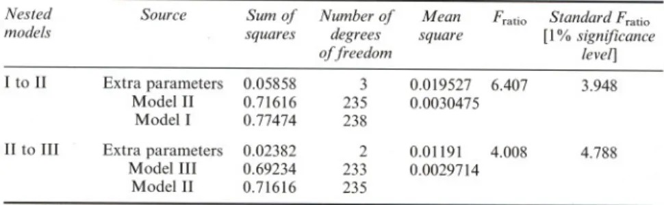
Sodium dodecylsulphate polyacrylamide gel electrophoresis (SDS-P AGE) was performed on the crude commercial lipase preparation, as well as on molecular weight standards, using the PhastSystem unit and PAGE minigels (50 mm height x 43 mm width x 0.45 mm thickness) of 12.5% polyacryla-mide. The buffer system in the gels was 0.112M in acetate and 0.112M in Tris (pH 6.5). The gels were run for 30 min at a constant electrical current of 10mA using PhastGel SDS buffer strips consisting of 2% Agarose, 0.2M tricine, 0.2 M Tris, and 0.55% SDS (pH 8.1). SigmaMarkerTM protein stan-dards were run in lanes parallel to that of crude lipase, and consisted of myosin (205 kDa), ,6-galactosidase (116 kDa), phosphorylase-b (97 kDa), fructose-6-phosphate kinase (84 kDa), albumin (66 kDa), glutamic genase (55 kDa), ovalbumin (45 kDa), glyceraldehyde-3-phosphate dehydro-genase (36 kDa), carbonic anhydrase (29 kDa), trypsinogen (24 kDa), trypsin inhibitor (20 kDa), a-lactalbumin (14.2 kDa), and aprotinin (6.5 kDa). After electrical resolution of the sample proteins, the gels were transferred to the development section of the PhastSystem unit and stained with Coomassie Blue.
J
Lipolytic Activity AssaysThe lipolytic activity of the crude lipase powder was determined via a modified version ofthe classic pH-stat method. For each assay, Sigma Lipase SubstrateTM (50% v/v olive oil emulsion stabilized with an emulsifier and further preserved with 0.1 % sodium azide) was diluted to 1: 5 with Tris-HCl buffer. A 40 mL volume of this mixture was transferred to a 200 mL jacketed glass beaker kept at 40°C in which a pH glass electrode was immersed. The emulsion's temperature was allowed to equilibrate for ca. 10min, and then the pH ofthe emulsion was brought to ca. 8.0 by adding a few drops ofKOH. A volume of 1.0mL of the sample (ca. 20 mgcrudelipasepowder/mLTris-HClbuffer) was pipetted into the thermally equilibrated substrate, and the pH was , rapidly brought to ca. 8.0 via addition of several droplets of KOH. At this point, the stopwatch was started, the titrating solution of KOH (0.05 moI/L) was added for 10min from a burette so as to maintain the pH of the reacting
50 V.M. BALCÃO et ai.
mixture at 8.0, and the titre was calculated from the concentration and total volume of KOH used. For the blank, a similar procedure was applied but I mL of Tris-HCI buffer with no enzyme was added instead. The lipolytic activity was expressed in LU (lipolytic units), where I LU is defined as the number of micromoles of total free fatty acids released per min and per mg of lipase powder acting for lOmin (time period during which the process reaction curve was reduced to its linear part) on the aforementioned olive oil emulsion at pH 8.0 and 40°C.
pH Stability Assays
To a buffered solution at a desired pH, ca. 1O0mg of crude lipase were dissolved in 5 mL of the corresponding buffer, and the resulting lipase solution was incubated at 20°C (temperature at which virtually no thermal deactivation of the commerciallipase preparation could be observed) for ca.
10mino After thorough mixing, I mL of lipase solution was withdrawn and assayed for lipolytic activity using the pH-stat method. Eight pH values were tested, viz. 4, 5, 6, 7, 8, 9, 10, and 12, and for each pH value the experiments were carried out in triplicate.
~
Thermal Stability Assays
For each experiment at a given temperature, a known amount of crude lipase powder (ca. 400 mg) was poured into a 50 mL glass flask, and dissolved in 20mL of Tris-HCI buffer (pH 8.0). After complete solubilization, I mL samples were taken and assayed for lipolytic activity; these values were taken as the initial activity. The flask containing the remaining solution was immediately placed in a water bath preset at the desired temperature, and samples were taken every 20 min (throughout a total time period of 5 h); the remaining lipolytic activity was determined using the pH-stat method. Five temperatures were tested, viz. 20, 30, 35, 40, and 50°C, and for each temperature the experiments were carried out in triplicate.
t
Calorimetric Assays
For each differential scanning calorimetry assay, two different patterns were tested. In the first pattern, ca. 10mg of crude lipase were weighed directly in a high-pressure aluminum cell which was then duly sealed. A blank was also prepared simply by sealing air inside a cell. The sample was then cooled to ca. -lOoC using liquid nitrogen, maintained at this temperature for ca. 1min, and then heated to 120°Cat a rate of 2°Cjmin. In the second pattern, 200 mg of crude lipase were dissolved in 1mL of McIlvaine buffer (pH 7.0), and 30llL ofthis solution were withdrawn with a microsyringe and poured into a
high-pressure aluminum cell which was then sealed. A blank was also prepared simply by sealing 30l1L of plain McIlvaine buffer (pH 7.0) inside another high-pressure cell. The sample was then cooled to ca. -lO°C using liquid nitrogen, maintained at this temperature for ca. 1min, and then heated to 1O0°Cat a rate of 2°C/min.
Proteolytic Activity Assays
t
The search for (tentative) proteolytic activity in the crude lipase preparation was done following the o-phthaldialdehyde method (Church et ai., 1983), modified later by Sousa and Malcata (1996), using bovine serum albumin as substrate. The assay comprised the addition of 300 I1Lof solution of crude lipase, prepared by dissolving 10mg of crude preparation in 5mL of sodium phosphate buffer (50 mmol/L, pH 8.0), to 2.7 mL of a 0.2% (wt/wt) solution of bovine serum albumin in sodium phosphate buffer (50 mmol/L, pH 8.0), and incubation of the resulting mixture at 37°C in a shaking bath for 60 mino Samples (200I1L) were then withdrawn at various time intervals (O,3, 5, 10, 15,20, 25, 30, 35, 40, 45, 50, 55 and 60min), added to 2mL of the stock solution containing o-phthaldialdehyde (to transform the primary amines of proteins, peptides, and free amino acids into fluorescent adducts), {3-mercaptoethanol (to reduce disulphide bridges), and sodium dodecyl sulphate (to terminate proteolysis and ensure full exposure of a-amino groups), and their absorbance was measured, after 2 min of reaction, at 340 nm in 10mm quartz cuvettes. Three blanks were used in the calculation of the corrected absorbance (A*): a blank for the substrate, consisting of 900 I1Lbovine serum albumin solution and 1O0l1Lsodium phosphate buffer (at the apropriate pH); a blank for the lipase, consisting of 100I1Lof solution of crude lipase and 900l1L of sodium phosphate buffer (at the appropriate pH); and a blank for the reagent, consisting of2000 I1Lof o-phthaldialdehyde and 1000l1L of sodium phosphate buffer (at the appropriate pH).
EXPERIMENTAL RESULTS
The protein content ofthe crude lipase powder was ca. 9.7:!: 1.7% (w/w) (as equivalent bovine serum albumin). The results pertaining to stability of crude lipase with respect to pH are depicted in Fig. 1. The results pertaining to stability of crude lipase with respect to time and temperature are plotted in Fig. 2. The thermograms associated with denaturation of crude lipase are presented in Fig. 3. The electrophoretograms of the crude lipase (see Fig. 4) have shown that its molecular weight is ca. 26.1 :!:0.7 kDa. The evolution of the corrected absorbance (A*) accounted for by the fluorescent adducts ofthe
4
FIGURE 1 Variation ofmaximum rate with pH ofincubation; experimental values (O) and theoretical curve (-).
1.2
Q,j ...E
1.0
=
e 0.8
....
~8 0.6
"O~
0.4
....
-
~S 0.2
-o
Z 0.0
O
O(
60
120
180
240
Incubation time (min)
300
FIGURE 2 Variation of normalized maximum rates with incubation time at the various temperatures tested; experimental values (20°C (O), 30°C (.), 35°C «», 40°C (.), and 50°C (O)) and theoretical curves (-).
52 V.M. BALCÃOet ai. ,.--, 7.0 ---<I> 6.01 O ê Q,j ...
-
E5.0
- '"e e
4.0
::s.
=
e....
3 o
.... =
'-'
.
-;:;2.0
....
-
o
1.0
e
0.0
O
2
4
6
8
10
12
14
Incubation pH
-0.095 Q.I ;... "CQ.I
= i::
;...=
U,.o Q.I --... -=~.~
-0.105 ;;;
5.
,.o..:: OJJ-0.110 "C""SQ.lu-.. -e:;E~
-0.115~
I::S
,.o-- '-' ~ Q.I "-'-~
~c..
Q.I.-
=::--0.050~
=:: I:: ~ -0.052~ ~
-~
-0.054.a.~
~ o [JJ ..,_o
.
056
~ r: ~~c..
-0.058~~
-0.060S~
(JO~ "O -0.062~ 'E.
Q. ~ -0.064 ~'-' S' 90 100 -0.100 I'I/IIA..,
Irf111
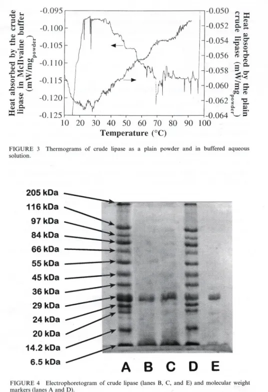
-0.125 10 20 30 40 50 60 70 80 Temperature (°C)FIGURE 3 Thermograms of crude lipase as a plain powder and in buffered aqueous solution.
t
205 kDa 116 kDa97kDa
84 kDa 66 kDa 55 kDa 45 kDa 36 kDa 29 kDa 24 kDa 20 kDa 14.2 kDa 6.5 kDaA
B
c
D
E
FIGURE 4 Electrophoretogram of crude lipase (lanes B, C, and E) and molecular weight markers (lanes A and D).
54 V.M. BALCÃO et aI.
primary amines of proteins, peptides, and free amino acids throughout time is displayed in Fig. 7; folIowing fit ofthe data to a linear model, the estimate encountered for the slope was 0.00005::!::0.00094 (correlation coefficient: 0.033).
MODELLING
Kinetic Model for Stability of Lipase with respect to pH
~ The active sites of enzymes are frequently composed of ionizable groups which must be in the proper ionic form to maintain the conformation of the active site able to bind substrate and thus catalyze the reaction (Segel, 1993). In this research effort, assessment of the pH -dependent stability of the crude lipase powder was performed in two consecutive steps, viz. incubation of a lipase buffered solution at a given pH folIowed by assaying of the residual activity of said soluble lipase. In the development of a suitable kinetic mechanism, the lipase was assumed to undergo quasi-equilibrium steps of rearrangement resulting from proton transfer and complexation with substrate, and irreversible steps of unimolecular deactivation. These elemen-tary steps are depicted in Fig. 5.
Ouring incubation at a given pH in the absence of substrate (see Fig. 5(a)), lipase was considered to undergo protonation/deprotonation. If one assumes that (i) three possible consecutive protonation states are possible for the lipase, and (ii) alI three protonated forms of lipase are able to undergo irreversible deactivation, then the total concentration of lipase forms can be calculated from:
d
- dt {[Eo]+ [EJ]+ [E2]}= ko[Eo]+ kdEJ] + k2[E2],
t
(1) where t denotes time. The definition of protolysis constants, viz.
K] = [EJ][H+]
[E2] , (2)
K2 = [Eo][H+]
[EJ] , (3)
and the overalI mass balance to active sites, viz.
Ed~ Eo +H+ Ed tkl K2 Kj k2 o( )o E 1 + H+ o( )o Ez ~ Ed
a
b
.
k2 Ed~E2 +SK,tl
aKs Ykp o( )o Ez. S )o Ez + PilaK',
H+ kl + Ed~EI +SK,tl
H+ Ks ) ~ + kp ~EI +P EI.Si1 Ki'
H+ H+ ko + ~Ks +Ed~ Eo + S o( )o Eo.S Okp ~ Eo + P
FIGURE 5 Molecular mechanism postulated for lipase incubated in the absence (a) and presence (b) of substrate.
.
allow Eq. (1) to be rewritten, after integration, as[E]tot(tinc)= {[Eo]+ [Ed + [E2]}(t = tine)
= [
E] .{
_
[ (koK2/[H+]inJ + kl + (k2[H+Lnc!Kd ] . t-}tot,O exp (K2![H+]ine)+ 1 + ([H+]inc!KI) me,
(5) where subscript 'inc' denotes at the end of incubation and subscript 'O' at the beginning thereof.
During incubation at the reference pH in the presence of substrate (see Fig. 5(b)), all three aforementioned forms of 1ipasewere assumed to be able to bind substrate and yield product according to a general Dixon- Webb
methodology. The corresponding rate expression has been derived elsewhere (Segel, 1993) for intact enzyme; if the enzyme has been already subject to
56 V.M. BALCÃO et ai.
TABLE I Definition of lumped parameters in the pH-stability model
Parameter Definition
w {J b[H+JlaKe.) + 1+ (8Ke2/.B[H+])
max,O'(Ke2/.B[W])+ I + ([WJlaKed {Jmax,o kp' [E]tot,o
deactivation as depicted in Eq. (5), then the resulting expression is
{ [
(koK2/lO-PH) + k\ + (k21O-pH / Kd
] }
19max
=
W . exp - (K2/ lO-pH) + 1 + (lO-pH/ K\) . tine,4
(6) where W is a lumped parameter defined in Table I (where a, {3,
"
and (j aredimensionless constants depicted in Fig. 5(b), and kp is the catalytic constant associated with reaction catalyzed by the intermediate form oflipase); ko, k\, and k2 are elementary kinetic constants associated with deactivation of ali protonated states of (free) lipase, K\ and K2 are dissociation constants associated with consecutive states of protonation of lipase, pH refers to the
pH prevailing during incubation, and tineis the incubation time at that pH
(viz. 10min). In the derivation of Eq. (6) advantage was taken from the fact that the pH-stat assays were carried out using such a high concentration of substrate and for such a little time that the rate expression could reduce to pseudo-zero-order (i.e., Ks
«
[S]).Kinetic Model for Stability of Lipase with respect to Temperature
In its most general form, the substrate-independent thermal deactivation of
enzymes can be described by a multi-step mechanism which involves only unimolecular steps (Henley and Sadana, 1986; Malcata, 1991; Van der Padt
et ai., 1992; Malcata et ai., 1993) to yield the following general rate
.
expresslOn:
ã{t n},
=
a~ l e-{3n,11+
a~2e-{3n.21+ . . . +a~n -le-{3n.n-11+
{I - an,l -a n,2 - an,n- 1}e-{3n,nl, (7)where ã is the activity of the lipase normalized by its initial value, t is time elapsed, and the an,;'s and the {3n,;'sare adjustable parameters functionally dependent on the intrinsic activities of each enzyme form and the first-order kinetic constants of rearrangement between, and deactivation of, said enzyme forms.
FIGURE 6 Schematic representation of unimolecular models considered for lipase deactivation.
In attempts to model data taken at various times but at a constant (optimal) pH, three levels of nesting of the aforementioned mechanism for enzymatic thermal deactivation were considered (and are depicted in Fig. 6): Mechanism I, which assumes that the native enzyme form (E1) may be deactivated to an inactive form (Ed) with rate constant kl; Mechanism lI, which assumes that the native enzyme form may be rearranged to another active (but stable) species(EI)with rate constant kal, but may be deactivated in parallel to an inactive form (Ed) with rate constant kl; and Mechanism 11I, which assumes that the native enzyme form may be rearranged to another active (but stable) species(EI)with rate constant kaj, and both these forms may be deactivated to inactive forms (Ed, and Edj, respectively) with rate constants kl and kdj, respectively. The lumped rate expressions associated with these three postulated mechanisms are mathematically depicted below.
M echanism I ã{t}
=
e-!3I,lt , (11,1= kl. (8) M echanism II ã{t} = ü2,le-!32,lt+ {I- ü2,I}, (12,1= kal+
kl. (9) M echanism III ã{t} = ü3,le-!33.lt + {I - ü3,I}e-!33,2t, (13,1= kal+
k1, (13,2= kdl. (10)El
El
kal
El
El
kal
El
kl
tkl
tkl
tkdl
Ed
Ed
Ed
Edl
58 V.M. BALCÃO et ai.
Postulating Arrhenius temperature dependencies for the elementary first-order kinetic constants depicted in Eqs. (8)-(10), and centering the reciprocal temperatures with their median value (Tm) in order to improve convergence and decrease interparameter correlation in the numerical procedure required by nonlinear regression fitting, one will obtain the following form of the rate expressions:
Model I
ã = exp{ - [w. exp( -~{ ~-
)m})]
. t}. (11) 4Model II
ã
=
a2,l . exp{ - [w. exp{ -~(~ - )m) }+ X' exp{ -,(~- )m) }] . t} + {1 - a2,J}. ( 12)
ModelIlI
ã
=
a3,l . exp{ - [w. exp{ -~(~ - )m) } + X' exp{ -,(~ - )m) }] . t}+ {1 - a3,J}. exp{ - [8. exp{ - W(~- )m) }] . t}, (13)
where the definitions ofthe lumped parameters (a, w, ~, x, " 8, and w) in the three nested models can be found in Table lI. It is apparent from comparison
TABLE 11 Definition of lumped parameters in temperature stability models; ka,o, kd,o, and kd,1 are pre-exponential factors: Ea,d, Ea,l' and
Ead,1are activation energies; and R is the ideal gas constant 4
Model Parameter Definilion
lU kd,o exp{ - Ea,d/ RT m} Ea,d/ R 'P 11 a lU 'P X I a21 ka,o exp{- Ea,aI! RT m}
Ea,aI!R kd,o exp{ - Ea,d/ R Tm}
Ea,d/ R >
III a
lU
a31
ka,o exp{ - Ea,a I!R Tm}
Ea,aI!R kd,oexp{ -Ea,d/RTm} Ea,d/ R kdl,oexp{- Ea,dI!RT m} Ea,dI!R 'P X I [; \[1
between Figs. 5 and 6 that Model I corresponds to the middle portion of the mechanism depicted in Fig. 5(b) (as expected since only the optimum pH was considered), which implies that most enzyme is in the form possessing the intermediate state of protonation. Conversely, although Models 11 and 111 are also consistent with Fig. 5(b), they encompass long-term conformational rearrangement of lipase (to form Ê\) which was not postulated for the pH-dependent stability mechanism because it could not be experimental1y detected in the short time frame considered.
STATISTICAL ANALYSES
Model parameters were estimated by nonlinear regression using a General REGression package, GREG (Stewart et aI., 1992), at leveI 10. At this leveI, the program performs nonlinear, uniresponse regression analyses ofthe data using finite differences as approximants of the derivatives of the objective function with respect to each parameter, and using as objective function minimization of the sum of squares of the residuaIs between model and experimental data. Given starting estimates, this advanced regression soft-ware expands the objective function as a local quadratic, finds a solution for the feasible minimum of this quadratic expansion in terms of parameter values, and implements a weak line search for a smaller value of the objective function. The results of the regression analysis (as provided by the post-convergence report generated by GREG) for the mo deI postulated for the pH-dependent deactivation are tabulated in Table 111,whereas the results of the regression analysis for each one of the three nested models postulated for temperature-dependent deactivation are tabulated in Table IV.
In order to decide whether a simpler nested model (i.e., Models I or 11) rather than the full modeI (i.e., Model 111)fits the data set adequately, one
TABLE lU Parameter estimates and associated marginal infer-ence intervals for the parameters of the mo de! fitted to the data pertainirig to the pH-dependent deactivation of the crude com-merciallipase
Parameter Estimate ::I:2a
\[I ko kl k2 KI K2 5.60::1: 3.26 x 10⁻¹ 6.88 X 10⁻¹::1: 3.35 2.72 x 10⁻¹⁵::1: 00 7.48 x 10⁻²::1: 1.31 x 10⁻² 6.72 X 10⁻⁷::1: 5.19 X 10⁻⁷ 4.73 X 10⁻¹²::1: 2.43 X 10⁻¹¹
60 V.M. BALCÃO et ai.
TABLE IV Parameter estimates and associated marginal infer-ence intervals for the parameters of the three nested models fitted to the data pertaining to temperature-dependent deactivation of the crude commerciallipase
Model Parameter Estimate:!: 2a
'P 1.95 X 1O-3:!: 1.16 X 10⁻⁴ 2.67 X 1O4:!: 1.22 X 10³ 9.63 X lO-I:!: 1.65 X 10⁻² 5.70 X lO-li:!: 00 2.67 X 1O4:!: 00 2.05 X 1O-3:!: 1.30 X 10⁻⁴ 2.75 X 1O4:!: 1.34 X 10³ ~ w 11 a w 'P X I III a w 9.90 X 1O-2:!: 4.27 X 10⁻² 2.26 X lO-li:!: 00 6.55 X 1O4:!: 00 1.86 X 1O-6:!: 00 5.55 X 10⁴:!: 4.67 X 10³ 2.24 X 1O-3:!: 2.00 X 10⁻⁴ 2.89 X 10⁴:!: 1.76 X 10³ 'P X I {j \jI
has proceeded as in the Jinear case and used a Jikelihood ratio test (Draper and Smith, 1981). Because of the spherical normal assumption, this test leads to an assessment of the extra sum of squares due to the extra parameters involved in going from a partial to the full model (Bates and Watts, 1988);the results of the extra sum of squares analyses involved in going from Model I (2-parameter mo de!) to Model 11 (5-parameter model), and in going from Model 11 (5-parameter mo deI) to Model 111 (7-parameter mo deI) are summarized in Table V.
Since the data generated in this research effort include replications, it was possible to perform a test of lack of fit for the best (nested) mo deI found, i.e. that associated with Eq. (12). Such analysis (which is based on the fact that the replication subspace is always orthogonal to the subspace containing the averages of the replicated data and the model postulated) (Bates and Watts, 1988) proceeded via comparison ofthe ratio ofthe lack offit mean square to
~
TABLE V Extra sum of squares analyses for simpler and full model
Nested Source Sum of Number of Mean Fratio Standard Fratio
models squares degrees square [I % significance
of freedom levei]
I to 11 Extra parameters 0.05858 3 0.019527 6.407 3.948 Model 11 0.71616 235 0.0030475
Model I 0.77474 238
11to III Extra parameters 0.02382 2 0.01191 4.008 4.788 Model III 0.69234 233 0.0029714
TABLE VI Lack of fit analysis for the statistically selected model proposed for thermal deactivation (Model lI)
the replication mean square with the appropriate value of the F distribution. This analysis is depicted in Table VI.
DISCUSSION
Although several methods are available, the Coomassie method was selected for determination ofthe protein content ofthe crude lipase assayed because it is easy, reproducible, and much less susceptible to interfering substances than, e.g. the Lowry or the Folin-Lowry methods (Robyt and White, 1990). On the other hand, although glycerol trioleate is the most universal substrate for lipase (Brockerhoff and Jensen, 1974), a less expensive substitute (viz. olive oil) was used because the former is unduly expensive, even though only a minimum of 70% oleic acid residues can be guaranteed. Furthermore, for monitoring the rate of lipolysis via continuous titration of free fatty acids released, selection ofpH 8 to carry out the reaction was a consequence ofthe pKa of aliphatic acids (ca. 5), which results in alI of the fatty acids being dissolved in the aqueous phase in dissociated form, as required by quantitative assaying.
Calorimetric assays for the crude lipase (see Fig. 3) exhibit several peaks of heat absorption which are probably associated with unfolding of various proteins present therein. The electrophoretogram obtained for the crude lipase (see Fig. 4) shows only a major band for the crude lipase; as separation was effected on the basis of molecular weight, this implies that the proteins in the crude lipase powder would probably be a family of proteins with similar molecular weights.
As discussed by Lee et ai. (1989), lipases are apparent1y not sensitive to shear stress when acting in free form in a batch stirred tank reactor, and so it is not expected that our deactivation data reflected mechanical, rather than thermal or other forms of, deactivation. Furthermore, if one assumes that the increase of the inner area of the vortex caused by magnetic stirring is negligible and no extensive foam forms, then denaturation of lipase caused by adsorption onto the high surface tension air/water interface throughout our experiments can be assumed to be negligible. On the other hand, it can be
Source Nurnber of degrees Surn of Mean Fratio Standard Fratio[I % of freedorn squares square significance levei] Lack of fit 75 0.260074 0.00346765 1.216 1.562 Replications 160 0.456086 0.00285054
62 V.M. BALCÃO et ai.
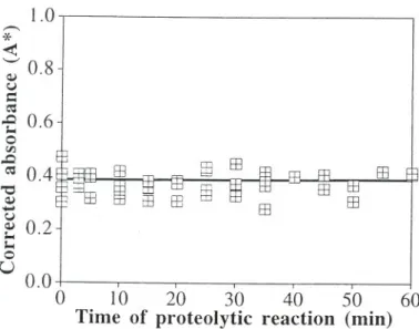
seen from Fig. 2 that the activity of the crude lipase decays faster and faster as temperature increases as would be expected from plain application of the Arrhenius law to the deactivation constants. The decay in lipase activity can safely be attributed solely to thermally driven changes in the three-dimensional conformation of the enzyme molecule because, in addition to the foregoing arguments, virtually no proteolytic activity could be found in the crude preparation assayed (see Fig. 7, where the null hypothesis that the concentration of products of proteolytic breakdown does not change with time was accepted at the I % leveI of significance).
The most common method of determining the temperature dependence of kinetic parameters is to independent1y fit data taken at each temperature, and then fit the resultant kinetic parameters to the Arrhenius relationship using a logarithmic/reciprocal plot. However, a better method is to fit the global mode! to the entire set of data points (as done in this research effort) because fitted parameters do not behave statistically as actual experimental data. On the other hand, centering the reciprocal absolute temperatures allowed mutual correlation between parameters be kept to a minimum, with off-diagonal elements of the normalized covariance matrix lying between -0.65 and +0.73 (results not shown).
Although convergence to best parameter estimates occurred in all fits, the confidence intervals for some parameters overlapped the null hypothesis
4 1.0 ,-.., .JEo ~ '-' 0.8Q) u c::: .~ ..o 0.6I-c o
'"
..o
~ 0.4 "'O Q)-
u
~
0.2 I-c o U 40.0
O
10
20
30
40
50
60
Time of proteolytic reaction (min)
FIGURE 7 Corrected absorbance produced by fluorescent adducts of primary amines in proteins, peptides, and free amino acids as a function of time of incubation of crude lipase with bovine serum albumin.
probably because of the limited range of the data set generated. Nonlinear fitting of the general mo deI for thermal deactivation to experimental data at various temperatures proceeded through consideration of increasing levels of nesting. Inspection of Table V indicates that Model 11should be selected to describe the experimental data because any further refinement of the fit resulting from a lower sum of squares of residuaIs is not statistically justified in view of the increased number of parameters. Therefore, postulation of an alternative active and stable form of lipase in the rate expression for deactivation is relevant, but assumption that this alternate form aIso deactivates irreversibly does not significant1y improve the fit of the data at all temperatures. These conclusions are in agreement with results reported elsewhere for a lipase produced by Aspergillus niger (Malcata et ai., 1992a). Since Model 11was selected to describe the data, further confirmation of the statistical adequacy of the fit was based upon a posteriori lack of fit analysis; inspection of Table VI does not raise any statistical queries as of the form of Model 11, on the 1% leveI of significance.
Inspection of Table IV indicates that the pre-exponential factor associated with kal is nonnegative (as required for physical significance) and that the activation energy associated therewith is 221.7kJmol-l; this value, which is much larger than that found for a similar rearrangement by Malcata et aI. (1992a) for a lipase from Aspergillus niger, clearly suggests that the process of rearrangement of the native active lipase to a more stable, active form involves rearrangement of several (at least 10) hydrogen bonds. Inspection of Table IV also indicates that the activation energy associated with the deactivation of the native lipase, kd, is 228.8 kJ moI-I; this value is of the order of magnitude of the activation energies associated with deactivation of enzymes (Malcata et ai., 1992a). Experimental studies with apure lipase from Mucor javanicus performed by Ogiso et ai. (1972) have resulted in activation energies for deactivation of this enzyme of ca. 26.6 kJ moI-I, which are rather low when compared with our case; however, our values for the activation energy associated with deactivation of the native lipase are of the same order of magnitude of those for a lipase from Chromobacterium
viscosum in an aqueous system at pH 8.0, viz. 161.5kJ mol-I (Prazeres et aI.,
1993), for pancreatic lipase, viz. 192.5kJ mol-I (Laidler and Bunting, 1973), and for Candida rugosa lipase, viz. 113.0-142.3 kJ mol-I (Shaw et ai., 1990). Departing from the values ofparameters w and cp,and X and I, for Model 11 in Table 11, the values for the elementary kinetic constants kal and kl, respectively, were calculated to range in the intervals 6.79 x 10⁻¹³-3.00 X 10⁻⁹ and 2.13 x 10⁻⁵-1.30 x lO-I min-I, respectively, for the temperature range 20-50°C. Therefore, deactivation of the native active form of lipase is apparent1y much faster than rearrangement of this form to
64 V.M. BALCÃO et ai.
another active species. The range of kl encompasses the values reported by Ogiso et ai. (1972) for the kinetic constant describing heat denaturation at 40, 50, and 60°C.
Experimental evidence (Neklyudov et ai., 1982) has indicated that serine and histidine residues form the active site of lipases, and that histidine must be in the proper ionic form to be able to abstract a proton from the hydroxyl group of serine (a preliminary step necessary to make a pair of electrons available for subsequent nuc1eophilic attack on the acyl moiety of the ester bond of the glyceride substrate); an amino acid residue containing a carboxylic acid as side group (i.e. aspartic acid or glutamic acid) in the c1ose vicinity of the active site has also been implicated in the formation of a three-dimensional pocket where the substrate must bind before its ester bond can be attacked by lipase. If the aforementioned His and AspjGlu residues are not in the proper ionic form, physical binding and subsequent chemical transformation of substrates will be prevented. It is thus expected that the
effect ofpH upon the stability ofthe lipase (Bailey and Ollis, 1986) displays a
behaviour at least similar to that depicted in Fig. 1, although the values for pK1 and pK2 (viz. 6.2 and 11.3, as obtained from Table lU) are somewhat higher than those usually associated with such residues (Segel, 1993). Inspection of the data generated by nonlinear fitting of the stability model (viz. Eq. (6)) to the experimental data (see Fig. 1) shows that the optimum pH in terms of stability occurs at ca. 8.2, which is in agreement with the optimum pH for a similar lipase réported by Ogiso et aI. (1972). (Since such fit possesses a correlation coefficient above 0.98, it is expected that it should not be considerably affécted by the absence of a datum at pH 11, which was due to an unavoidable experimental problem.)
Inspection of Table lU shows that the value obtained for kl is very small, virtually zero, which implies that the intermediate enzyme form in the model depicted in Fig. 5(a) is very stable; conversely, ko and k2 are much higher than kJ, which implies that the two puta tive enzyme forms Eo and E2 are quite prone to inactivation. The rate of deactivation of form E1, at pH 8, will only be accurately estimated at longer reaction times, as apparent from inspection of the values of X and r in Table IV, which yield the range 2.13 x 10⁻⁵ -1.30 x lO-lmin-1 for kl in the temperature range 20-50°C.
~
~
CONCLUSIONS
From this research it has been possible to simulate the kinetic stability of a lipase of industrial importance at various temperatures and pH values; such
simulation is important in attempts to design biochemical processes for which the aforementioned lipase is a potential candidate. It was conc1uded that (i) only thermal deactivation played a relevant role in terms of the performance of such crude enzyme, which could be described via parallel steps of deactivation and rearrangement (with activation energies of ca. 230 and 220 kJ moI-I, respectively), and that (ii) the observed stability behaviour was consistent with an assumption of three forms of active enzyme with increasing states of protonation (with pK values of ca. 6 and 11), where the intermediate form is particularly stable.
Acknowledgements
The lipase assayed was a gift from Amano Pharmaceutical (Nagoya, Japan). Partial funding for this research effort was provided through grants by FLAD (Portugal; Project Lipase-catalyzed interesterification of butterfat with
olive oil) and by Institut CANDIA (France; Project Modification de la matiére grasse par des lipases immobilisées sur un réacteur à membrane).
Funding for author V.M. Balcão was provided through a Ph.D. fellowship by JNICT (Portugal; programs CIENCIA BD/2091/92-IF and PRAXIS XXI BD/5317/95). The authors are grateful to Df. M.J. Sousa for the help concerning proteolytic assays.
References
Ahern, T.J. and Klibanov, A.M. (1985) The mechanism of irreversible enzyme inactivation at 1O0°C. Science, 228, 1280-1284.
Bailey, J.E. and Ollis, D.F. (1986) Biochemical Engineering Fundamentais. McGraw-Hill, New York, USA, pp. 135-144, 148-152.
Balcão, V.M. and Malcata, F.X. (1996a) Reactors with immobilized lipase. Mathematical modelling. In: Engineering Of7With Lipases, Ed., F.x. Malcata. Kluwer Academic Publish-ers, Dordrecht, The Netherlands, pp. 435-454.
Balcão, V.M., Paiva, A.L. and Malcata, F.x. (1996b) Bioreactors with immobilized lipases: state-of-the-art. Enzyme Microb. Technol., 18, 392-416.
Balcão, V.M., Vieira, M.C. and Malcata, F.x. (1996c) Adsorption of protein from several commerciallipase preparations onto a hollow-fiber membrane module. Biotechnol. Progr.,
12,164-172.
Bates, D.M. and Watts, D.G. (1988) Nonlinear Regression Analysis and its Applications. Wiley Interscience, New Y ork, USA.
Brockerhoff, H. and Jensen, R.G. (1974) Lipolytic Enzymes. Academic Press, New York, USA, p.20.
Church, F.C., Swaisgood, H.E., Porter, D.H. and Catignani, G.L. (1983) Spectrophotometric assay using o-phthaldialdehyde for determination of proteolysis in milk and isolated milk proteins. J. Dairy Sei., 66, 1219-1227. .
Dawson, R.M.C., Elliott, W.H., Elliott, D.C. and Jones, K.M. (1969) Data for Biochemical
Research. Oxford Science Publications, Oxford, UK, p. 427.
66 V.M. BALCÃO et ai.
Henley, J.P. and Sadana, A. (1986) Deactivation theory. Biotechnol. Bioeng., 28, 1277-1285.
Laidler, K.L. and Bunting, P.S. (1973) The Chemical Kinetics of Enzyme Action. Oxford University Press, London, UK.
Lee, Y.-K. and Choo, C.-L.(1989) The kinetics and mechanism of shear inactivation of lipase from Candida cylindracea. Biotechnol. Bioeng., 33, 183-190.
Malcata, F.x. (1991) Hydrolysis ofbutterfat by immobilized lipase using three-phase membrane reactors, Ph.D. thesis, University of Wisconsin - Madison, USA.
Malcata, F.X., HiU, c.G. and Amundson, C.H. (1992a) Hydrolysis of butteroil by immobilized lipase using a hoUow fiber reactor: IV. Effects of temperature. Biotechnol. Bioeng., 39,
1097-1111. .
Malcata, F.x., Reyes, H.R., Garcia, H.S., Hill, c.G. and Amundson, C.H. (l992b) Kinetics and mechanisms of reactions catalyzed by immobilized lipases. Enzyme Microb. Technol., 14,
426-446.
Malcata, F.x., Hill, c.G. a~d Amundson, C.H. (1993) Hydrolysis of butteroil by immobilized lipase using a hoUow fiber reactor: Part V. Effects of pH. Biocatalysis, 7, 177-219. Neklyudov, A.D., Shvedov, B.D. and Tsibanov, V.V. (1982) Properties of a preparation of
immobilized Rhizopus oryzae 14-14 lipase. Appl. Biochem. Microhiol., 17, 378-382. Ogiso, T., Sugiura, M. and Kato, Y. (1972) Studies on bile-sensitive lipase. X. Preparation and
properties of carrier-bound Mucor lipase. Chem. Pharm. Buli., 20, 2542-2550.
Prazeres, D.M.F., Garcia, F.A.P. and Cabral, J.M.S. (1993) Temperature, pH and media influence on lipase stability: In: Stability and Stabilization of Enzymes, Eds., W.J.J. van den Tweel, A. Harder and R.M. Buitelaar. EIsevier, Amsterdam, The Netherlands, pp. 445-450. Robyt, J.F. and White, B.J. (1990) Biochemical Techniques - Theory and Practice. Waveland
Press, Chicago, USA.
Segel, LH. (1993) Enzyme Kinetics - Eehavior and Analysis of Rapid Equilibrium and Steady-State Enzyme Systems. Wiley, New York, USA.
Shaw, J.-F., Chang, R.-C., Wang, F.F. and Wang, Y.J. (1990) Lipolytic activities of a lipase immobilized on six selected supporting materiais. Biotechnol. Bioeng., 35, 132-137.
SoÍlsa, M.J. and Malcata, F.x. (1996) Effects of processing conditions on the caseinolytic activity of crude extracts of Cynara cardunculus L. Food Sei. Tech. Int., 2, 255-263. Stewart, W.E., Caracotsios, M. and Sorensen, J.P. (1992) GREG Software Package
Documenta-tion. University of Wisconsin, Madison, USA.
Van der Padt, A., Sewalt, J.J.W., Ágoston, S.M.L and Van't Riet, K. (1992) Candida rugosa lipase stability during acylglycerol synthesis. Enzyme Microb. Technol., 14, 805-812.
4
.~