Ana Cristina Rafael Joyce Coutinho
Development and characterization of pH-sensitive
fucoidan-chitosan nanoparticles for oral delivery of
methotrexate to lung cancer cells
Dissertação do 2º Ciclo de Estudos Conducente ao Grau de Mestre em Química Farmacêutica
DISSERTAÇÃO DE MESTRADO REALIZADA SOB A ORIENTAÇÃO DE:
Professora Doutora Maria de La Salette de Freitas Fernandes Hipólito Reis Dias Rodrigues
COORIENTAÇÃO DE:
Professor Doutor Carlos Manuel Magalhães Afonso
DE ACORDO COM A LEGISLAÇÃO EM VIGOR, NÃO É PERMITIDA A
REPRODUÇÃO DE QUALQUER PARTE DESTA DISSERTAÇÃO.
ACCORDING TO THE LEGISLATION, THE REPRODUCTION OF ANY
PART OF THIS DISSERTATION IS NOT AUTHORIZED.
AUTHOR DECLARATION:
Under the terms of the Decree-Law no 216/92, of October 13th, is hereby declared that the author afforded a major contribution to the conceptual design and technical execution of the work and interpretation of the results included in this dissertation. Under the terms of the referred Decree-Law, is hereby declared that the following articles/communications were prepared in the scope of this dissertation.
The results presented in this dissertation are part of the following scientific communications:
Selected oral communications:
A. Joyce, S. Costa Lima, S.Reis. "Development and characterization of
pH-sensitive fucoidan-chitosan nanoparticles for oral delivery of methotrexate and bioactive xanthone derivatives". 11th Meeting of Young Researchers of University of Porto, Porto, Portugal, 7-9th February 2018.
This work was developed in Laboratório de Química Aplicada in partnership with Laboratório de Química Orgânica e Farmacêutica, Departamento de Ciências Químicas, Faculdade de Farmácia da Universidade do Porto. This research was supported through national funds provided by Foundation for Science and Technology (FCT) and European Regional Development Fund (ERDF) and COMPETE under the project PT2020 UID/MULTI/04378/2013. This work also received financial support from the European Union (FEDER funds through COMPETE POCI-01-0145-FEDER-016790) and National Funds provided by FCT through project PTDC/MAR-BIO/4694/2014.
AGRADECIMENTOS
Esta dissertação é representativa não só de todo um ano de esforço, dedicação e aprendizagem, mas também representa um ano de aventuras e agradáveis surpresas. Aqui, encontra-se o culminar de um projeto que me foi proposto e que eu abracei sem qualquer arrependimento, e que não seria possível de concretizar sem a ajuda de um número considerável de pessoas.
À Professora Salette Reis, gostaria de expressar o meu maior agradecimento, por me ter acolhido no seu grupo de investigação ao qual eu posso chamar de segunda família. Muito obrigada pela orientação, disponibilidade e entusiasmo e também por todas as oportunidades que me proporcionou. Para além de Professora e orientadora, é imensamente amiga, sempre com os melhores conselhos e boa disposição.
Ao Professor Carlos Afonso, também gostaria de agradecer o incentivo e apoio demonstrados ao longo deste mestrado, bem como a atenção e disponibilidade com que sempre acompanhou o meu percurso académico.
De maneira muito especial, gostaria de mostrar a minha admiração e gratidão à Dra. Sofia Costa Lima. Por toda a dedicação, pela tamanha sabedoria que tanto me quis transmitir, pelas horas de trabalho despendidas a meu favor e por todas as vezes que me fez rir quando me sentia cansada. Obrigada pela sua boa disposição, por me apontar sempre o caminho certo e pela confiança depositada no meu trabalho para a elaboração deste projeto.
Gostaria também de agradecer a todos os meus Professores que me acompanharam ao longo destes 2 anos, em especial à Professora Madalena Pinto por todo o conhecimento prestado.
Aos meus colegas desta jornada que ficaram amigos, Daniela, João e Francisca, obrigada por toda a preocupação e amizade.
Obrigada a todos os meus colegas do Laboratório de Química Aplicada, em especial aos meus companheiros do Laboratório de Biofísica Molecular e Biotecnologia (MB2). Andreia M., Andreia G., Rita, João, José, Joana, Priscila, Yong e Catarina obrigada por me aceitarem na vossa família. Obrigada pela amizade e por toda a ajuda prestada.
À Isabel, um enorme obrigada por tudo, não há palavras para descrever a tua energia. Obrigada por todos os momentos de salvaguarda quando eu me sentia completamente perdida. Vais ser uma investigadora enorme, tenho a certeza.
Aos meus amigos e companheiros de percurso académico, tenho uma sorte imensa em ter-vos do meu lado. Obrigada por todos os lab-meetings, mesmo quando eu sou a única da biotecnologia. À Maria e à Marta, pela confiança, incentivo, compreensão,
x amizade e companheirismo. Obrigada por todos os momentos, todas as pausas e pela constante partilha.
Às amigas de sempre e para sempre, Sara, Joana, Filipa, Inês C., Sandra, Inês R., Rita e Daniela, agradeço por todos os maus caminhos, jantares e gargalhadas.
À minha “outra” família, a da Zara, que pouco ou nada sabe sobre a vida de um investigador. Paula, Cristiana, Sara, Mafalda e Vânia, obrigada pela imensa amizade. Agradeço também à Mafalda e ao Luís por conseguirem sempre gerir o meu horário de forma a tornar possível aguentar esta aventura. Graças a vocês sou mais responsável.
À minha família, em especial à minha mãe, por todas as lições valiosas, por todo o amor e dedicação que tem comigo durante toda a minha vida. Obrigada por incutirem em mim todos os valores em que acreditam.
INDEX
1 INTRODUCTION ... 3
1.1 The Marine Biosphere ... 3
1.2 Marine polysaccharides ... 4
1.2.1 Organisms source ... 4
1.2.2 Biomaterial for drug delivery systems ... 5
1.2.3 pH sensitive drug delivery systems ... 7
1.3 Nanoparticles based on marine polysaccharides ... 9
1.3.1 Chitosan ... 9
1.3.2 Fucoidan ... 11
1.3.3 Fucoidan/Chitosan nanoparticles ... 13
1.4 Factors influencing the impact of nanocarriers ... 18
1.4.1 Size ... 18
1.4.2 Surface charge ... 19
1.4.3 Surface Polarity ... 19
1.4.4 Stimuli responsive systems ... 20
1.5 Lung cancer therapy ... 20
1.6 Oral administration route ... 22
2 AIM AND STRATEGY ... 27
3 MATERIALS AND METHODS... 31
3.1 Materials ... 31
3.2 Methods ... 31
3.2.1 Preparation of Fucoidan/Chitosan Nanoparticles ... 31
3.3 Characterization of Fucoidan/Chitosan Nanoparticles ... 32
3.3.1 Particle Size and Polydispersity Index ... 32
3.3.2 Zeta Potential ... 32
3.3.3 Entrapment Efficiency and Loading Capacity ... 33
xii
3.3.5 Transmission Electron Microscopy ... 34
3.3.6 Fourier Transform Infrared Spectroscopy ... 34
3.3.7 pH responsiveness ... 35
3.3.8 In Vitro drug release studies ... 35
3.3.9 In vitro assessment of mucosadhesive property ... 36
3.4 Cell Studies ... 36
3.4.1 In vitro viability assay ... 37
3.4.2 Cellular uptake assay ... 38
3.4.3 Apoptosis assay ... 39
3.5 Statistical Analysis ... 40
3.6 Chemical Structures ... 40
4 RESULTS AND DISCUSSION ... 43
4.1 Production of Fucoidan/ Chitosan nanoparticles ... 43
4.2 Characterization of Fucoidan/ Chitosan nanoparticles ... 44
4.2.1 Storage stability ... 45
4.2.2 Morphology analysis ... 47
4.2.3 Fourier Transform Infrared Spectroscopy analysis ... 47
4.2.4 pH responsiveness ... 49
4.2.5 In vitro drug release studies ... 51
4.2.6 In vitro assessment of mucoadhesion properties ... 52
4.3 Cell Studies ... 53
4.3.1 In vitro viability assay in fibroblasts ... 53
4.3.2 FC nanoparticles and MTX effect on lung cancer cells ... 54
4.3.3 Cellular uptake assay ... 57
4.3.4 Apoptosis assay ... 60
5 CONCLUSIONS AND FUTURE PERSPECTIVES ... 65
ABSTRACT
The vast history of traditional medicine is one of the most important examples of how natural products can be safe and efficient. Considering the marine environment, which is an extraordinary reservoir of bioactive natural compounds, its exploitation can provide various compounds with wide biotechnological and biomedical applicabilities. Marine polysaccharides such as fucoidan (from algae), and chitosan (from crustaceans) are biocompatible, and have important biological properties such as anti-inflammatory, anti-viral and anti-cancer activities, and potential application in the nanotechnology field. Methotrexate is an antimetabolite immunosuppresive and antifolate drug, with poor aqueous solubility, widely used in several cancer therapies (e.g. breast, lung, leukemia and osteosarcoma) and inflammatory diseases (e.g. rheumatoid arthritis and psoriasis). Regardless of methotrexate efficacy, it has been associated with several side effects, and drug delivery systems may contribute to overcome these limitations.
In the present work, fucoidan and chitosan were exploited for the development of pH-sensitive nanoparticles for the oral drug delivery of methotrexate. The polysaccharides were mixed in a ratio of 5:1, respectively, and several characterization parameters were assessed in both 5F1C and methotrexate-loaded 5F1C nanoparticles. These polymeric systems were also tested in fibroblasts for biocompatible assessment, and in a human lung carcinoma cell to assess methotrexate anti-cancer activity.
The size of the developed nanoparticles was around 300 nm, the zeta potential value was negative (ca. -30 mV), revealing low tendency to aggregate. Empty 5F1C nanoparticles destabilized at physiological pH, but methotrexate-loaded 5F1C nanoparticles exhibited a 100-nm increase in size, through the evaluated pH range, without disintegration, revealing resistance through the GIT. When stored at room temperature, both formulations were stable in a period of 6 weeks, but the methotrexate loaded nanoparticles loss their stability upon 4 weeks when storage at 4ºC. The 5F1C nanoparticles showed no cytotoxicity at 0.49 mg mL-1 for the fibroblasts and reduced lung cancer cells (A549) viability to 40% after 72 hours. The methotrexate loaded nanoparticles showed a time-dependent cytotoxicity for the fibroblasts. Regarding the A549 cell line, both plain methotrexate and drug loaded nanoparticles showed an anti-proliferative effect, mediated by an apoptotic process, though the nanoparticles were 7-fold more effective.
In sum, fucoidan/chitosan nanoparticles were successfully developed and characterized for the delivery of methotrexate, with promising antiproliferative activity towards lung cancer cells.
RESUMO
A história da Medicina Tradicional mostra a importância dos produtos naturais e da sua segurança e eficiência. A exploração do ambiente marinho, em particular, os polissacarídeos marinhos, como o fucoidan (obtido a partir de algas), e o quitosano (obtido de crustáceos) são moléculas biocompatíveis, com atividade biológica, nomeadamente: anti-inflamatória, antiviral e anticancerígena, e potenciais aplicações na nanotecnologia. O metotrexato é um composto imunossupressor e antifolato, com baixa solubilidade aquosa, utilizado no tratamento de várias cancros e doenças inflamatórias. Independentemente da eficácia do metotrexato, ele está associado a vários efeitos colaterais, e os sistemas de administração de medicamentos podem contribuir para superar essas limitações. Assim, neste trabalho, polissacarídeos marinhos foram utilizados no desenvolvimento de nanopartículas sensíveis ao pH para a administração oral de metotrexato. O fucoidan e o quitosano foram misturados numa proporção de 5:1, respetivamente, e caracterizados por vários parâmetros físico-químicos. Estes sistemas foram estudados em fibroblastos e células de cancro do pulmão para avaliar viabilidade e efeito antiproliferativo, respectivamente.
O tamanho das nanopartículas desenvolvidas foi de cerca de 300 nm, o valor do potencial zeta foi negativo (cerca de -30 mV), revelando pouca tendência para agregar. As nanopartículas vazias desestabilizaram-se a pH fisiológico, mas as nanopartículas contendo metotrexato exibiram um aumento de 100 nm, ao longo dos valores de pH estudados, sem sofrer desintegração, revelando estabilidade ao longo do TGI. Quando armazenadas à temperatura ambiente, ambas as formulações permaneceram estáveis por um período de 6 semanas, mas as nanopartículas contendo o metotrexato perdem sua estabilidade após 4 semanas quando armazenadas a 4ºC. As nanopartículas não apresentaram citotoxicidade na concentração de 0,49 mg mL-1, para os fibroblastos e reduziram a viabilidade das células do cancro do pulmão. Nos fibroblastos, as nanopartículas contendo o metotrexato mostraram uma citotoxicidade dependente do tempo. Nas células do cancro do pulmão tanto o metotrexato livre como incorporado nas nanopartículas apresentaram efeito antiproliferativo, mediado por um processo apoptótico. Em suma, as nanopartículas de fucoidan/quitosano foram desenvolvidas com sucesso e caracterizadas para a administração de metotrexato, apresentando promissora atividade antiproliferativa em células de cancro do pulmão.
Palavras-chave: apoptose, absorção celular, sistemas de entrega de fármacos,
INDEX OF FIGURES
FIGURE 1:POLYSACCHARIDES FROM MARINE ALGAE GROUPED ACCORDING TO THEIR ELECTROSTATIC NATURE.ADAPTED FROM
CARDOSO, ET AL [4]. ... 5
FIGURE 2:ILLUSTRATION OF PH VARIATION THROUGHOUT THE HUMAN BODY, ADAPTED FROM BAZBAN-SHOTORBANI ET AL (2017)[36]. ... 7
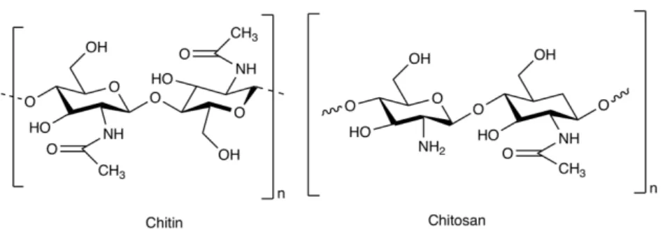
FIGURE 3.CHEMICAL STRUCTURE OF CHITIN AND CHITOSAN. ... 9
FIGURE 4.CHEMICAL STRUCTURE OF FUCOIDAN. ... 11
FIGURE 5.ILLUSTRATION OF FUCOIDAN/CHITOSAN NANOPARTICLES FORMATION BY ELETROSTATIC INTERACTIONS. ... 13
FIGURE 6:SCHEMATIC ILLUSTRATION OF THE EPR EFFECT. ... 18
FIGURE 7.CHEMICAL STRUCTURES OF METHOTREXATE (1) AND FOLIC ACID (2). ... 21
FIGURE 8.ILLUSTRATION OF NANOPARTICLE FORMULATIONS ADME MECHANISMS FOLLOWING ORAL ADMINISTRATION, ADAPTED FROM GRIFFIN ET AL (2016)... 23
FIGURE 9:FIGURE REPRESENTATIVE OF A HEALTHY AND AN APOPTOTIC CELL WITH MARKERS FOR APOPTOSIS DETECTION... 39
FIGURE 10:SCHEMATIC REPRESENTATION OF THE 5F1C-MTX NANOPARTICLES PREPARATION PROCESS. ... 43
FIGURE 11:STORAGE STABILITY OF 5F1C(A,C AND E) AND 5F1C-MTX(B,D AND F) FORMULATIONS AT ROOM TEMPERATURE (DARK GREY) AND AT 4ºC(LIGHT GREY).PARAMETERS AS SIZE (A,B),PDI(C,D) AND ZETA POTENTIAL (E,F) WERE DETERMINED FOR 6 WEEKS.EACH RESULT REPRESENTS MEAN ± STANDARD DEVIATION FOR N=6 MEASUREMENTS. ... 45
FIGURE 12:TEM IMAGES OBTAINED FOR THE DESIGN NANOPARTICLES.BOTH FRESHLY PREPARED NANOPARTICLES ARE IN PH3.0 (A–5F1C AND B–5F1C-MTX).SCALE BAR:200 NM. ... 47
FIGURE 13:FTIR SPECTRA OF CHITOSAN, FUCOIDAN AND 5F1C NANOPARTICLES.THE CHARACTERISTIC CHEMICAL GROUPS ARE PROPERLY IDENTIFIED. ... 48
FIGURE 14:FTIR SPECTRA OF MTX,CHITOSAN,FUCOIDAN AND 5F1C-MTX NANOPARTICLES.THE CHARACTERISTIC GROUPS ARE PROPERLY IDENTIFIED. ... 48
FIGURE 15:IN VITRO RELEASE OF 5F1C-MTX NANOPARTICLES UNDER DIFFERENT PH CONDITIONS:A– PH5.0 AND B– PH7.4 AT PHYSIOLOGICAL TEMPERATURE (37ºC).DATA POINTS CORRESPOND TO THE MEAN ± STANDARD DEVIATION FOR N=3 REPLICATES (N=2 MEASUREMENTS OF MTX RELEASE PER REPLICATE). ... 51
FIGURE 16:VIABILITY OF L299 FIBROBLASTS EXPOSED TO 5F1C AND 5F1C-MTX NANOPARTICLES.CELL VIABILITY WAS DETERMINED BY THE MTT ASSAY AFTER 24 HOURS (A) AND 72 HOURS (B) INCUBATION WITH EMPTY NANOPARTICLES (l) OR MTX-LOADED NANOPARTICLES (¨).EACH RESULT REPRESENTS THE MEAN ± STANDARD DEVIATION FOR N=4 REPLICATES OF 3 ASSAYS.HORIZONTAL DOTTED LINE INDICATES ACCEPTED LIMIT OF VIABILITY (80%). ... 53
FIGURE 17:VIABILITY OF L929 FIBROBLASTS EXPOSED TO FREE MTX.CELL VIABILITY WAS DETERMINED AFTER 24 HOURS (OPEN TRIANGLES) AND 72 HOURS (DARK TRIANGLES) BY MTT ASSAY.EACH RESULT REPRESENTS THE MEAN ± STANDARD DEVIATION FOR N=4 REPLICATES OF 3 ASSAYS.HORIZONTAL DOTTED LINE INDICATES ACCEPTED LIMIT OF VIABILITY (80%). ... 54 FIGURE 18:VIABILITY OF A549 EXPOSED TO 5F1C AND 5F1C-MTX NANOPARTICLES.CELL VIABILITY WAS DETERMINED BY MTT
xviii
RESULT REPRESENTS THE MEAN ± STANDARD DEVIATION FOR N=4 REPLICATES OF 3 ASSAYS.HORIZONTAL DOTTED LINE INDICATES ACCEPTED LIMIT OF VIABILITY (80%). ... 55
FIGURE 19:VIABILITY OF A549 UPON EXPOSURE TO PLAIN MTX.CELL PROLIFERATION WAS DETERMINED BY MTT ASSAY AFTER
72 HOURS.EACH RESULT REPRESENTS THE MEAN ± STANDARD DEVIATION FOR N=4 REPLICATES OF 3 ASSAYS.HORIZONTAL DOTTED LINE INDICATES ACCEPTED LIMIT OF VIABILITY (80%). ... 55
FIGURE 20:GROWTH OF A549 CELLS UPON INCUBATION TO 5F1C AND 5F1C-MTX NANOPARTICLES.CELL PROLIFERATION WAS DETERMINED BY SRB ASSAY AFTER 72 HOURS INCUBATION WITH EMPTY NANOPARTICLES (l) AND MTX-LOADED NANOPARTICLES (¨).EACH RESULT REPRESENTS THE MEAN ± STANDARD DEVIATION FOR N=4 REPLICATES OF 3 ASSAYS.
HORIZONTAL DOTTED LINE INDICATES ACCEPTED LIMIT OF VIABILITY (80%). ... 56
FIGURE 21:GROWTH OF A549 AFTER EXPOSURE TO MTX.CELL PROLIFERATION AFTER 72 HOURS WAS DETERMINED BY SRB ASSAY.EACH RESULT REPRESENTS THE MEAN ± STANDARD DEVIATION FOR N=4 REPLICATES OF 3 ASSAYS.HORIZONTAL DOTTED LINE INDICATES ACCEPTED LIMIT OF VIABILITY (80%). ... 56
FIGURE 22:CELLULAR UPTAKE OF 5F1C-FITC NANOPARTICLES IN A549 CELL LINE AFTER 2 HOURS INCUBATION AT 37ºC.FLOW CYTOMETRY VALUES REPRESENT THE MEAN ± STANDARD DEVIATION FOR N=4 REPLICATES FOR ONE ASSAY. ... 58
FIGURE 23:CELLULAR UPTAKE OF 5F1C-FITC NANOPARTICLES (0.25 MG ML-1) IN A549 CELL LINE FOR 6 HOURS INCUBATION AT
37ºC.FLOW CYTOMETRY VALUES REPRESENT THE MEAN ± STANDARD DEVIATION FOR N=3 REPLICATES FOR ONE ASSAY. . 59
FIGURE 24:FLOW CYTOMETRY HISTOGRAMS OF THE CELLULAR UPTAKE TIME KINETICS OF 5F1C-FITC NANOPARTICLES (0.25 MG ML-1) FOR 6 HOURS INCUBATION AT 37ºC. ... 59
FIGURE 25:APOPTOSIS MEDIATED BY MTX.THE % OF A549 APOPTOTIC CELLS AFTER 24 HOURS INCUBATION WITH FREE AND
5F1C-LOADED NANOPARTICLES AND EMPTY 5F1C NANOPARTICLES.DATA EXPRESSED AS MEAN ±SD(N=4). ... 60
FIGURE 26:FLOW CYTOMETRY ANALYSIS OF APOPTOSIS IN A549 CELL LINE TREATED WITH 5F1C AND 5F1C-MTX
NANOPARTICLES AND FREE MTX.CELLS WERE STAINED WITH ANNEXIN V-FITC AND 7-AAD AND ANALYZED BY CYTOMETRY.
INDEX OF TABLES
TABLE 1:PROPERTIES OF DIFFERENT CHITOSAN/ FUCOIDAN CARRIERS. ... 16
TABLE 2:MASS OF POLYMER AND METHOTREXATE ADDED TO EACH FORMULATION ... 43
TABLE 3:CHARACTERIZATION OF 5F1C AND 5F1C-MTX NANOPARTICLES. ... 44
TABLE 4:LIST OF CHARACTERISTIC PEAKS OF THE COMPOUNDS AND NANOPARTICLES DETERMINED BY FTIR[87]. ... 49
TABLE 5:THE CHARACTERISTICS OF 5F1C AND 5F1C-MTX NANOPARTICLES UNDER DISTINCT PH CONDITIONS. ... 50
TABLE 6:MUCOADHESION ASSAY OF 5F1C AND 5F1C-MTX, TO SIMULATE THE MUCUS LAYER. ... 52
TABLE 7:SATURATED CELLULAR UPTAKE VALUE (YMAX) AND UPTAKE RATE CONSTANT (K) FOR THE STUDIED NANOPARTICLES... 58
ABBREVIATIONS
7-AAD 7-aminoachinomycin D
Abs Absorvance
CS Chitosan
DDS Drug Delivery System
DHFR Dihydrofolate reductase
DLS Dynamic Light Scattering
DMEM Dulbeccos Modified Eagle Medium
EE Entrapment Efficiency
ELS Electrophoretic Light Scattering
EMA European Medicines Agency
EPR Enhanced permeability and retention
FDA Food and Drug Administration
F Fucoidan
FA Folic acid
FC Fucoidan/ Chitosan
FITC Fluorescein isothiocyanate
FITR Fourier-transform infrared spectroscopy
GIT Gastrointestinal Tract
HBSS Hanks Balanced Salt Solution
LC Loading Capacity
MTT 3-[4,5-dimethylthiazol-2-yl]-2,5-diphenyltetrazolium bromide
MTX Methotrexate
MW Molecular Weight
NPs Nanoparticles
PBS Phosphate Buffer Saline
PDI Polydispersity Index
SEM Scanning Electron Microscopy
SRB Sulforhodamine B
TEM Transmission Electron Microscopy
1 Introduction
1.1 The Marine Biosphere
Being a spacious aquatic ecosystem, the marine environment is considered to be one of the most important sources of natural bioactive compounds with extremely rich biodiversity capacity. Oceans cover about 71% of earth surface and are mainly composed by salt water and represent a vast source of natural substances. Sea reconnaissance as a renewable source of bio-compounds has a positive impact on the development of new systems with wide biotechnical and biomedical applicabilities [1]. When compared to bioactive compounds extracted from terrestrial life forms, marine biomaterials have shown a higher incidence of bioactivity and chemical innovation which make marine organisms an important source of structurally and biologically active secondary metabolites [2].
Over the past decades, marine natural drug discovery increased significantly leading to at least, eight related drugs being approved by the United States of America Food and
Drug Administration (FDA) and by the European Medicines Agency (EMA) [3]. Nowadays,
there has been a growing interest in many scientific areas related to the discovery of marine compounds due to their large biodiversity and simplicity on the extraction and purification processes.
Marine biomaterials are generally non-cytotoxic, biodegradable and biocompatible and such biological properties promote the discovery of a wide range of novel bioactive compounds with specific pharmacological properties being a fundamental keystone for the pharmaceutical industry [4, 5].
Nevertheless, due to the difficulties in reproducing the marine microenvironment at the laboratory, a high percentage of the marine biodiversity is still unexplored [6]. Furthermore, in order to recover microorganisms from high biodiversity environments, marine samples have to be subjected to several treatments with various cultivation techniques [7]. By applying these techniques, it is possible to gather a significant number of marine microbial strains with unique metabolic potential and also a high-quality natural product library containing novel structures [8].
Foremost, the origin of marine materials can be distributed in three main groups: polysaccharides, proteins, and lipids [9].
4
1.2 Marine polysaccharides
1.2.1 Organisms source
Marine algae are the main source of marine polysaccharides, but they can also be obtained from animal sources, such as the skeletons of crustaceans and cartilaginous fish tissue. Most polysaccharides are extracted from red, green or brown macroalgae. Seaweeds represent a different type of multicellular marine algae which are also a major source of polysaccharides.
Marine animals are also considered a source of marine polysaccharides providing two main categories of polymers: chitin-derived polymers and glycosaminoglycans (GAGs) [4]. These compounds can be described as a large and complex group of macromolecules with different biological properties between them [10, 11].
Polysaccharides are defined as polymeric carbohydrate structures formed by repeated monosaccharide units which are attached to each other by glycosidic bonds [12]. The polysaccharide classification is based on their primary or covalent structure which represents the sequence of the monomeric units along the chain. These repeated units are coupled by covalent chemical bonds which are not completely flexible, by limiting the monomers to a narrow range of orientations. Due to this characteristic, a polysaccharide chain is only able to adopt certain shapes, named “secondary structures”, depending on their primary sequence [13-17].
Marine polysaccharides are mainly used in food and cosmetic industries. Also, there is a great interest on using them as materials for the incorporation of bioactive agents, being widely applied in the synthesis of new pharmaceutical devices. As mentioned above, among the vast marine organism diversity, algae are the main source of marine polysaccharides, which can be divided accordingly to their electrostatic characteristics (Figure 1) [4]. Polysaccharides like alginate, carrageenan, and fucoidan are extracted from algae, while chitosan and hyaluronan (also known as hyaluronic acid) are obtained from animal sources. These extracts can exhibit different chemical structures and have important biological properties such as biocompatibility, biodegradability and anti-inflammatory activity as well as adhesive and antimicrobial activity. Properties such as shape, size and response to stimuli are dependent on pH and temperature.
1.2.2 Biomaterial for drug delivery systems
Biomaterials are known to offer various advantages for the encapsulation of genetic material and therapeutic agents. A drug delivery system (DDS) can be defined as a formulation or device that enables the insertion of a drug in the organism while improving its efficacy and ensuring its safety by controlling the rate, time and target of its release [18]. Nanomedicine and nanotechnology main purpose is to ensure the carrying and delivery of promising therapeutics in order to maximize their pharmacological activity and
polyanionic car b o xyl at ed su lf at ed Su lf at ed a n d c ar b o xy la te d neutral De cr ea si n g s u lf at e g ro u p s polycationic deacetylation
Figure 1: Polysaccharides from marine algae grouped according to their electrostatic nature. Adapted from Cardoso, et al [4].
6 overcome their limitations and possible drawbacks which may block the required effectiveness. In order to approach such therapeutic results, marine polysaccharides can be used to produce polymeric carriers by encapsulation of low drug/therapeutic agents dosages and release them at a scheduled time and location which could lead to a reduction of the pre-occurring side effects. These materials can also be used as signaling markers, through shell functionalization, capable of leading the delivery of a carrier to a specific location/active site as well as extend their function as DDS to diagnostic instruments [19, 20].
Accordingly, polysaccharides have intrinsic properties which are of great relevance on the field of drug delivery. They might be employed as/for:
i. Reproducible obtaining method from natural sources;
ii. Undergo chemical and enzymatic reactions in order to produce different materials;
iii. Biocompatibility, biodegradability and low immunogenic properties [16]; iv. Can be produced, conjugated and complexed with proteins, peptides and
other biomacromolecules [21];
v. Stimuli-responsive drug delivery systems [22, 23]; vi. Can be modified as gels;
vii. Can give rise to polymeric structures [24].
Marine polysaccharides DDS can be designed as gels or as nanoparticles. Gels are defined as consistent systems formed by a 3D polymeric network confining continuously water or a physiological media and can be formed by interchain linkages, crosslinks, which prevents the solubilization of the polymeric chains network through the fluid phase. In this type of system, drug delivery is controlled by physical processes, physicochemical processes and system parameters [25]. Polymeric nanoparticles have been limited in the past due to the rough conditions that are necessary to prepare them which could compromise drug stability and loading efficiency [26, 27]. Nowadays, there are several protocols with simple and precise methods of preparation that provide good solubility, stability, loading capacity and efficient crossing through cell membranes on a wide range of pH values.
1.2.3 pH sensitive drug delivery systems
Stimuli-responsive nanomaterials have been increasingly considered, over the last decades, as attractive vehicles for the transportation and release of drugs and genes owing to their capability to react to changes in the environmental parameters such as: temperature, pH, electric and magnetic fields, ultrasound, mechanical stress and biochemical stimuli [28-32]. Thereby, due to their nonlinear response to the variations occurring in the external environment they can be called smart or intelligent materials and have quality to be utilized as vehicles for drug delivery, catalysts, biosensors, and membranes [33-35].
There are several pH differences throughout the human body as illustrated in Figure 2 [37, 38]. For example, pH values throughout the digestive tract, range from 2 in the stomach up to 7 in the colon.
Figure 2: Illustration of pH variation throughout the human body, adapted from Bazban-Shotorbani et al (2017) [36].
Beyond that, at the tissue level, the pH profile of pathological tissues such as cancerous and inflamed tissues is different from that of the healthy tissues. Tumor tissues present pH values from 5.0 to 6.4 which is lower than the pH value surrounding normal tissues owing to metabolic glycolysis and lactic acid production. The same variation occurs at the cellular level where there are pH differences among lysosomes (pH from 4.5 to 5), endosomes (pH from 5.5 to 6) and cytoplasm (pH 7.4). Tumor cells have slightly lower extracellular pH values (6.5) because of their elevated metabolism in comparison with the
pH variation throughout the human body
Organ level Gastrointestinal tract stomach pH 1 - 2.5 colon pH 5.2 - 7.02 small intestine pH 5.26 - 7.35 Cellular level cytoplasm pH 7.4 lysossome pH 4 - 5 Tissue level Cancerous tissue tumor extracellular pH < 6.5 Normal tissue pH 7.4 Inflamed tissue pH < 7.4 pH sensitive drug delivery systems
▹ Oral protein delivery systems
▹ Oral and Colon specific drug delivery systems
▹ Injectable tumor-targeted drug delivery systems ▹ Anti-inflammatory drug
delivery systems
▹ Endosomal escape and cytoplasmic drug delivery systems
8 healthy cells. Due to the rapid proliferation of tumor cells, the local vasculature is often disorganized and insufficient to fulfil the nutritional and oxygen needs of their expanding population.
Thereafter, designing nanocarriers with the purpose of being responsive to specific pH value triggers them to be able to target a specific area in the human body and release their encapsulated drugs with maximum therapeutic impact and minimum side-effects [39, 40]. Biopolymers have been frequently used to design pH-responsive drug delivery systems. Due to the fact that polymers can be precisely adapted for their specific application and by associating that with their biocompability, biodegradability and biological functionality, synthetic and organic polymers ranging from the macroscale to the nanoscale, can be used as carriers [36].
The main factor for polymers to be pH-responsive is the presence of ionizable groups which are attached to the hydrophobic backbone of the polymer chain. These type of polymers belong to a class of polyelectrolytes with acid (e.g. carboxylic and sulfonic acids) or basic (e.g. amines, imidazole and pyridine) ionic functional groups which can be protonated or deprotonated as a response to the changes on the environmental pH, leading to the pH-induced release of the drug from the pH-sensitive carrier [33, 41, 42]. The pH value in which these changes occur is called transition pH or critical pH depending on the pKa value. As so, pKa of a polyelectrolyte is defined as the pH at which half of its ionizable groups are ionized [43, 44]. Therefore, it is possible to categorize any pH-responsive drug delivery systems based on the type of the polymeric carrier:
a) pH-sensitive hydrogels
b) pH-sensitive polymer-drug conjugates c) pH-sensitive polymeric micelles d) pH-sensitive dendrimers
1.3 Nanoparticles based on marine polysaccharides
According to the Encyclopedia of Pharmaceutical Technology and the Encyclopedia of Nanoscience and Nanotechnology, nanoparticles are well defined solid colloidal particles with sizes ranging approximatively from 1 to 1000 nm in which the active molecule or compound can be entrapped inside or adsorbed to their surface. The nanocarriers characteristics should be designed according to the intended application in order to attain an efficient action [45].
1.3.1 Chitosan
Chitosan (CS), shown in Figure 3, is a linear polysaccharide obtained by the deacetylation of chitin which is one of the most abundant natural polymers in the ecosystem. Chitin can be converted into chitosan by enzymatic or chemical processes. Presently, chitin is extracted from marine shell waste watercourses at industrial level, usually using chemical methods because of their low cost and suitability to mass production [9]. Depending on the source, chitin can occur in two allomorphs, the a and b forms. In the solid form, its chains are assembled by H-bonds network controlling chitin solubility, swelling and reactivity [46]. The a isoform is the most abundant, occurring in fungal and yeast cell walls, in lobster and crab tendons and, in shrimp shells. The b form is rare and is found in association with proteins in squid pens and in synthesized tubes by worms [47]. In spite of not being water soluble, chitin is usually converted into derivatives soluble in acidic conditions, such as CS soluble in acetic acid and carboxymethyl chitosan soluble in a range of acidic and alkaline solutions.
Figure 3. Chemical structure of chitin and chitosan.
The acetylation degree reflects the balance between the two types of residues and differentiates chitin from CS. When after the deacetylation process the outcome has molar percentage lower than 50% mol the product is named chitosan and becomes soluble in
10 acidic aqueous solutions [46]. In fact, this limit of molar percentage depends on the distribution of acetyl groups along the chains, being possible to obtain a complete characterization of the polymer which may differ from the starting material. CS physical properties depend on the degree of acetylation and on the acetyl group distribution along the chains.
Owing to the amine groups present in the structure, and pKa value close to 6, chitosan major drawback is its poor solubility at physiological pH values being positively charged in acidic environments and neutral in alkaline pH values [48]. Regarding oral delivery, the high solubility of CS at acidic pH values is a major drawback. In relation to its cationic nature and high charge density in a solution, CS forms stable ionic complexes with multivalent water-soluble polyanions under mild physiological conditions [49, 50]. CS possess many beneficial properties such as biocompatibility, biodegradability, safety and interesting biological activities which attract much attention mostly for biomedical applications such as synthesis of nanoparticles, beads and capsules for controlled drug delivery and also membranes, films and scaffolds for tissue engineering and regenerative medicine [51]. CS polymer exhibit good antimicrobial and antioxidant properties, a broad spectrum of activity, and lower toxicity towards several cell lines [52]. CS-based nanoparticles, especially the low molecular weight (MW) ones, can penetrate bacteria cell wall, combine with DNA and inhibit mRNA synthesis and DNA transcription [53, 54]. On the other hand, high-MW CS-based nanoparticles can interact with cell surface and consequently alter cell permeability [54]. Regarding its antioxidant activity, researchers have reported that CS acts as an antioxidant by scavenging oxygen radicals such as hydroxyl as well as highly stable 2,2-Diphenyl-1-(2,4,6-trinitrophenyl)hydrazyl (DPPH) radicals. Park et al., have also demonstrated that low-MW CS are more active than high-MW [55].
Mucoadhesive polymers have been usually applied on the fabrication of NPs for oral administration [56, 57]. Considering the entire advantages of using biodegradable and biocompatible mucoadhesive polymers the ones that stand out exhibit increased residence time in the intestine and thus prolonged contact with the intestinal surface membranes which leads to an improvement on the absorption [58].
Mucoadhesion is a characteristic of CS that enhances paracellular drug transport via transient opening of the tight junction between epithelial cells as well as making a great candidate for controlled drug release in oral delivery [59-61]. Mucous membrane or mucous layer consist of mucous secreting epithelial cells and is present at the inner side of many organs of the human body, such as in the gastrointestinal tract (GIT) and upper respiratory tract. Water builds more than 95% of the total mucous weight, making it very hydrated. The name of the layer is derived from mucin glycoprotein, which gives structure and
adhesiveness to the mucous. The thickness of mucosal surfaces is quite varying at different places.
Most of polymers which interact with mucins in the gut are hydrophilic and positively charged when in the gut environment. CS has hydroxyl and amine groups that can give rise to hydrogen bonding-mediated interactions with the components of the mucus and has been studied for mucosal drug delivery namely in: oral [61], buccal [62], and nasal[63] [64]. The positive charges on the chain are able to interact with sialic and sulfonic acids of the mucus layer by strong electrostatic interactions, therefore exhibit favorable cell adhesion due to the attraction to the negative charge of the cell membrane [65].
In addition to its mucoadhesive properties, CS have attracted much interest also for its ability to act as permeation enhancers and as efflux transporter P-glycoprotein inhibitor, already demonstrated in vitro and in vivo [66].
1.3.2 Fucoidan
Fucoidan, shown in Figure 4, is a heparinoid anionic sulfated polysaccharide with a substantial quantity of L-fucose and sulfate ester groups, which is mainly extracted from natural non-toxic brown seaweed, although there are other sources resulting in different polymeric compositions [67, 68]. Fucus vesiculosus is the most common algae species used to extract the simplest polymer of the entire group having only L-fucose and sulfate units [69, 70]. Structurally, fucoidan has a backbone of α-(1–3)-linked fucose units or, it is composed by repeating disaccharide units of α-(1–3)- and α-(1–4)-linked fucose residues with O-2 arm. Depending on the structure of the main chain, fucoidan can be sulfonated at O-4, O-2, or at both positions of the fucose units. Beyond that, some type of fucoidan can be both sulfated and acetylated [69].
Figure 4. Chemical structure of fucoidan.
In spite of having, until today, a broad range of reported MW (from 5 to several hundred kDa), Balboa et al., have suggested that low MW fucoidan fractions are more biocompatible than high MW. The same author has identified fucoidan to exhibit some newsworthy pharmacological effects such as: antithrombotic, antitumoral, antiviral, immunomodulatory, antioxidant and anti-inflammatory activity [71]. In fact, fucoidan has a
12 wide variety of biological activities being the anticoagulant action the most studied. Many researchers have reported that its anticoagulant activity could be directly related to the sulfate content and position, MW and sugar composition [72]. Thrombin plays an important role in thrombosis and so its inhibitor has become the main content of studies on antithrombotic drugs. However, some researchers have reported that the anticoagulant properties of fucoidan was determined by thrombin inhibition whose anticoagulant activity was similar to heparin [73]. In order to be able to bind the thrombin, fucoidan requires a long sugar-chain and a comfortable conformation [68]. Concerning fucoidan antitumor activity and comparing with synthetic drugs, the natural products have attracted increasing attention of patients for their biological activities and lower side effects and it has been reported that fucoidan has a cytotoxic effect via enhancing immunity on tumor cells but not on healthy cells [74].
Some authors have reported fucoidan as a pH-sensitive polymer, mainly due to the acidic functional groups in the structure and also by the total number of negatively charged groups on the chain that can urge a response to changes in external pH [75, 76]. Rocha de Souza et al., have reported that fucoidan from Fucus vesiculosus has an inhibitory effect on the formation of hydroxyl and superoxide radicals [77]. Fucoidan from Fucus vesiculosus is composed by 44.1% of fucose, 26.3% of sulfate and 31.1% of ash [78]. This type of polymer has a relatively simple chemical composition, but most of fucoidans have a complex composition.
Regarding the use as DDS, the main benefit of the systematic study of this bioactive compound is the discovery of new chemical modification methods aiming to harness specific biological activities inducing changes in their affinity to specific drugs. Thereby, it has been possible to increase fucoidan ability to encapsulate drugs increasing their release efficacy, either by chemical reactions or by interactions with other polymers [79]. Kurosaki
et al., reported the first application of fucoidan as matrix material for the synthesis of drug
delivery system regarding the delivery of protein-based drugs [80]. In this study, the positive zeta potential of the polyplexes enabled the complexation with the negatively charged fucoidan by electrostatic interactions mediated by the sulfate groups. It is possible to modulate zeta potential values in nanoparticles with sizes around 200 nm varying the used polymer ratios [81].
1.3.3 Fucoidan/Chitosan nanoparticles
Polymeric nanoparticles, such as fucoidan/ chitosan nanoparticles (Figure 5) are colloidal vehicles in which the drug is entrapped, encapsulated, or absorbed aiming to improve drug absorption and pharmacokinetics (Figure 5) [82]. These class of nanoparticles is mainly used for the controlled delivery of drugs in several cells, such as RAW 264.7 cells (mouse macrophage cell line), human primary macrophages and HepG2 (liver hepatocellular carcinoma) [83-85]. According to Le Droumaguet and co-workers nanocarriers developed for drug delivery should ideally be: (i) biocompatible and biodegradable for safe administration, (ii) able to avoid the immune response, and (iii) decorated by ligands to target cells or tissues [86].
In general, there are three fundamental steps to take into consideration during the production of nanomaterials for oral drug delivery:
I. The nanomaterial must be able to survive the harsh conditions of the GIT; II. Once administrated, the DDS must reach its target location;
III. The pharmacokinetics of the free drug must be maintained.
Figure 5. Illustration of Fucoidan/Chitosan nanoparticles formation by eletrostatic interactions.
The main advantage of using the combination of the marine polysaccharides fucoidan and CS is due to their intimate contact with the mucus and consequently longer resistance times at the absorption site [87]. Several reports have described their applicability as DDS are summarized on Table 1.
Accordingly to Yi-Cheng Huang, the ammonium groups of CS interact ionically with the sulfate groups of fucoidan during sonication to form the polyelectrolyte complex [88]. Regardless of the drug release profile and the association with the protein, these class of nanoparticles have the great disadvantage of requiring the use of aggressive conditions
14 that are not applied in a simple polyelectrolyte complexation [81]. Indeed, Yi-Cheng Huang research group have been intensely working on the development of Fucoidan/Chitosan nanoparticles. In 2011 firstly reported Fucoidan/Chitosan pH sensitive nanoparticles and studied the possibility of being used as oral DDS [70]. In this work, it was used the ionic-gelation method at room temperature with different weight ratios of polymers to synthetize the nanoparticles named: C1F1, C1F5 and C5F1. The obtained nanoparticles had a diameter range of 200 to 300 nm at a pH of 1.2 and the C/F nanoparticles became larger and unstable as the pH increased. In 2013 Huang et al., developed several weight ratios of biodegradable C/F nanoparticles loaded with the basic fibroblast growth factor (bFGF) [89]. Experimental results showed that the particles had approximately 200 nm, were able to control the release of bFGFs and had a protector effect of bFGF depended on the weight ratio C/F in nanoparticles. In vitro cell studies demonstrate that C/F nanoparticles were no cytotoxicity to PC12 cells. In 2014 the research group used the C1F1 ratio to evaluate its potential for the oral delivery of 99m Tc-methylene produced by the simple polyelectrolyte self-assembly method [88]. The nanoparticles size was approximately 380 nm and revealed a significant pH-sensitive behavior being stable at pH 2.5 and decomposed at pH 7.4. As for the cellular work, the outcome of the trans-epithelial electric resistance (TEER) in a Caco-2 monolayer shows that C1F1 are able to open the cell tight junction.
Also, in 2014 the research team developed C/F nanoparticles by varying the CS amount with several weight ratios (from C1F1 until C5F1) using simple polyelectrolyte self-assembly method and evaluated their potential for antioxidant oral delivery [90]. The particles presented a spherical shape and a size range from 230 to 250 nm. As for the stability, they maintained compactness and stability for 25 days in a phosphate buffer saline solution with pH ranging from 6.0 to 7.4. The antioxidant effect was confirmed by scavenging activity, reducing the concentration of intracellular ROS and superoxide anion (O2-). The nanodelivery systems were able to control the release of the antibiotic Gentamicin with an initial burst effect followed by a slow drug release. The cellular studies were performed in A549 lung cancer cell line and the cell viability assay revealed an absence of toxicity after the exposure to the formulations containing up to 3 mg nanoparticles per milliliter. Focusing on the delivery of Gentamicin, Huang and co-workers reported the development of different weight ratios C/F nanoparticles for its pulmonary delivery by presenting a biphasic release feature [91]. The particles had a size range from 370 to 420 nm, zeta potential values from +38 to +42 mV, and were able to encapsulate approximately 95% of Gentamicin. Since the nanoparticles were able to provide a controlled release of the antibiotic the system is able to deliver it in a localized region leading to an increase of local drug concentration.
Pinheiro et al., reported the development of C/F multilayer nanocapsules, prepared through layer-by-layer method for the controlled delivery and release of bioactive compounds [92]. The nanovehicle was composed by 10 chitosan/fucoidan layers and consequent formation of a multilayer film on polystyrene. The C/F particles morphology and size analysis revealed an average diameter of approximately 90 nm. The encapsulation efficacy of poly-L-lysine (PLL) was approximately of 45% depending on the initial PLL concentration used. This work allowed the clarification of the release mechanisms of these drug delivery systems which was found to be pH-dependent in the two pH values (pH 2 and pH 7) within the human GIT.
Ho and co-workers, have also developed C/F multilayers formed using the layer-by-layer method on poly(dimethylsiloxane (PDMS) substrates [93]. The formation of the layer-by-layers and zeta potential was verified by atomic force imaging confirming the formation with consistent charge switching as each polysaccharide layer is adsorbed to the PDMS substrate.
Kim et al., have developed C/F nanoparticles for the encapsulation of red ginseng to improve its antithrombotic activity [94]. The particles were prepared using various antithrombotic materials, including fucoidan. C/F nanoparticles sizes were approximately 440 nm and provided a sustained release of the 40% of red ginseng encapsulated in an acidic environment and improved ginsenoside solubility by approximately 122.8%. Therefore, the authors reported that is expected the increase of the stability of ginsenosides in the GIT. In vivo test results performed in rabbit and ex vivo rat platet revealed aggregation of red ginseng. Although red ginseng exhibited no effect on in vivo carrageenan induced mouse tail thrombosis, C/F nanoparticles demonstrated significant effects on the anticoagulation activity of fucoidan.
16
Table 1: Properties of different chitosan/ fucoidan carriers.
C/F ratio Synthesis Method pH Size (nm) Zeta Potential (mV) Encapsulated drug Encapsulation efficiency (%) Administration route Ref C1F1, C1F5, C5F1 ionic-gelation 1.2 200 to 300 -39.43 to +24.00 Curcumin 85.2 to 92.1 oral [70] C1F1 simple polyelectrolyte self-assembly
1.2 to 7.4 361.40 to 1009.5 -19.03 to +23.03 99m Tcmethylene diphosphonate n.i. oral [88]
C1F1, C2F1, C3F1, C4F1, C5F1 simple polyelectrolyte self-assembly
6.0 to 7.4 271 to 1252 -4.7 to +14.5 Gentamicin n.i. oral [90]
C3F1, C4F1, C5F1 ionotropic cross-linking 6.0 271.1 to 458.7 +0.11 to +41.8 Gentamicin > 90 pulmonary [91]
C10F1, C5F1, C1F1, C1F5, C1F10 ionotropic cross-linking n.i. 173 to 283.4 -52.5 to +33.9 Basic fibroblast
growth factor 80.9 to 90.5 n.i. [89]
C10F1 layer-by-layer assembly 2.0 to 7.0 ≈ 90 +26.0 poly-L-lysine 45.1 oral [92]
F/C multilayers layer-by-layer assembly on polydimethylsil oxane substrates.
n.i. n.i. +45 to +90 n.i. n.i. n.i. [93]
C/F ionic-gelation n.i. 440 to 1020 +2.7 to +4.9 Red ginseng extract 22.63 to 40.13 n.i. [94]
C5F1, C5F5, C1F5 ionotropic cross-linking n.i. 173.6 to 513.8 -49.8 to +33.9 Stromal
cell-derived factor 1 77.78 to 99.42 oral [95]
18
1.4 Factors influencing the impact of nanocarriers
Drug pharmaco-kinetics and phamaco-dynamics may be modulated and improved by the use of drug delivery systems. Size, surface potential, surface polarity, surface coating, targeting molecules are some of the factors that can influence the outcome of the nanocarrier.
1.4.1 Size
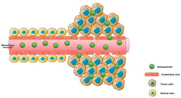
Nanoparticles (NPs) size can influence their biodistribution. In particular, following polymeric nanoparticles uptake into Caco-2 enterocytes and subsequent basolateral secretion into the interstitial area, bigger NPs tend to be taken up by the mesenteric lymphatic vessels leading to a changed systemic distribution, avoiding the first pass of hepatic metabolism [96]. The major underlying mechanism of the design of DDS is the enhanced permeation and retention (EPR) effect which, in 1986, was considered as a new concept in the drug delivery field [97]. Considering tumors, passive targeting via the EPR effect, illustrated in Figure 6, can be exploiting by engineered particles inboard specific size ranges [98, 99].
Figure 6: Schematic illustration of the EPR effect.
Nanoparticles size have an impact on their elimination and influence the degree of clearance through the reticuloendothelial system (RES) [100]. M Longmire et al. reported that for NPs that do not undergo renal clearance, the hepatobiliary system can be the
primary route of excretion because the primary function of the liver is to efficiently capture and eliminate particles ranging from 10 to 20 nm and accordingly nanoparticles included in this scale can often undergo rapid liver uptake [100].
1.4.2 Surface charge
The NPs surface is surrounded by a liquid layer called Stern layer where the ions bond strongly and, possesses a more diffuse outer zone where the ions are less tightly bound. In this diffuse layer the particle boundary with charged entity confers the charge on the nanoparticle. The charge on the NPs influences the stability and promotes aggregation in the intestine luminal fluids and, on the other hand, influence the absorption [101].
In addition, the charge density should also be noteworthy because it can be modified when in contact with the physiological medium. For example, depending on the nature of the charge, the electrostatic interactions with the mucus influences the result in prolonged residence time, or hinder the uptake due to the alternatively entanglement with the mucus [102]. Immediately after being absorbed into the systemic circulation, charged NPs specially the cationic ones, are known to interact with plasma proteins following by aggregation. Besides that, larger charged nanoparticles most likely undergo enhanced clearance via RES route preceding uptake into targeted tissues [103].
1.4.3 Surface Polarity
Uncoated NPs rapidly get proteins absorbed on the surface by opsonization, favoring phagocytosis and rapid clearance due to the high concentration of phagocytic cells in the liver [98]. In contrast, nanoparticles with lipophilic or amphiphilic surfaces can promote blending with the enterocyte membrane altering the membrane fluidity and enhancing permeability [101]. Some authors have also reported PEGylated NPs as a strategy for prolonged drug half-life absorption and decreased clearance [104].
PEGylating method has been frequently used with the purpose of enhancing nanoparticles in vivo stability. As so, PEGylated NPs are protected against aggregation in the intestine lumen decreasing the extent of enzymatic degradation [105-107]. Furthermore, even though PEGylation has been reported to promote mucus penetration the enhanced surface hydrophilicity can result in a decrease of the intestinal membrane permeability [108].
20
1.4.4 Stimuli responsive systems
Targeted drug delivery has been the major strategy for the development of successful drug carriers because of the many advantages associated with this approach. By matching a cell receptor with a proper ligand, it is possible to deliver a drug to its specific cell population. Also, if the disease microenvironment is known, like the acidic microenvironment of a tumor, it is possible to modify the NPs composition with pH sensitive molecules and exploit the EPR effect by synthetizing NPs with specific size ranges [109].
Fan et al. have developed a novel self-assembled polyelectrolyte nanoparticle complex by coating insulin loaded PGA-g-DA micelles with trimethyl chitosan (TMC) for oral delivery of proteins [110]. The formulation revealed to improve drug stability and had slower drug release. Following oral administration to diabetic rats, the NPs produced a hypoglycemic response and reduced serum glucose level showing high absorption of the nanoformulations.
1.5 Lung cancer therapy
Every year about 8.8 million people worldwide die of cancer, with respiratory tract carcinomas causing the highest number of deaths, according to the latest estimates by the World Health Organization.
Lung cancer is divided in two main types: small-cell lung carcinoma (SCLC) and non-small-cell lung carcinoma (NSCLC). From these, NSCLC regard over 80% of lung cancers in the world and is the less sensitive to surgery, and other types of therapy [111]. Several clinical studies have been developed for the treatment of lung cancer with many kinds of anticancer drugs can be used for this therapy, such as: adriamycin, paclitaxel, docetaxel, methotrexate, gemcitabine, etc. From all of these types of drug, methotrexate (MTX), shown in Figure 7, which is a cytotoxic drug, antimetabolite of folic acid was the selected drug for the development of the preset work.
MTX is known to interfere with the formation of DNA, RNA and proteins by blocking the action of dihydrofolate reductase leading to the inhibition of the metabolism of folic acid [112]. It has been widely used since the 1950s for the treatment of malignancies like childhood acute lymphocytic leukemia, lung and breast cancer and, for the therapy of auto-immune diseases such as rheumatoid arthritis, psoriasis and lupus [113, 114]. MTX is classified as class III in the Biopharmaceutical Classification System (high solubility and low permeability), which constitute a limiting property when the therapy requires the systemic absorption. Another limitation of MTX is its low oral bioavailability due to the action of the P-glycoprotein (P-gp) which acts as an efflux pump [115].
Methotrexate (Figure 7.1) has similar structure and physicochemical properties to those of folic acid (Figure 7.2). It has two carboxyl groups with pKa values of 4.8 and 5.5, both almost completely dissociated in physiological conditions. It is highly hydrophilic and so it is unable to cross biological membranes by simple diffusion implying the need of a carrier-mediated transport system for intestinal absorption, tissue distribution and renal excretion [116].
Figure 7. Chemical structures of methotrexate (1) and folic acid (2).
Methotrexate can be clinically administrated by different routes, such as: oral, intravenous, subcutaneous and intramuscular. Despite all this, MTX is one of a few chemotherapy agents that has been clinically administrated via oral route. MTX clinically efficacy is often compromised, by toxic dose-related side effects due to an excess of drug that have been usually administered leading to short half-life bloodstream and low bioavailability [117, 118].
The pharmaceutical nanotechnology enables the building of new nanostructures with different and improved properties, allowing the protection of drugs like MTX against premature degradation by the GIT. Until today, several MTX delivery systems have been reported for a suitable oral administration including MTX-loaded mesoporous silica nanoparticles [119, 120], solid lipid nanoparticles (SLNs) [121], self microemulsifying delivery system [122], proteinoid microspheres [123], gelatin microspheres [124], beta-casein nanoparticles [125], chitosan microspheres [126], and crosslinked guar gum microspheres [127]. Despite that, all these developed oral sustained release systems exhibit some problems namely: (i) non-ideal release profile, either very slow or too rapid; and (ii) toxic crosslinking agent (glutaraldehyde) used in the formulation released in the GIT. Since the solubility of MTX in water is very low (£ 1 mg mL -1), and considering the pKa value, is expected that the release (desorption) of loaded MTX from porous polymeric adsorbents is affected by the pH of the release medium. MTX is an ionizable compound and its solubility in an aqueous environment is affected by ionic strength. Once salts (mainly NaCl) that exist in the gastrointestinal fluid vary their concentration depending on the fasted
22 and fed states is expected that the release rate decreases in their presence. Thereby, the higher the salt concentration, the lower the release rate [128].
As described above, MTX is known to be an analogue of folic acid (FA) due to a high structural similarity and thereby, MTX is able to enter cells by reducing FA carrier, by membrane-association with FA receptor or proton coupled FA transport. MTX is responsible for the deactivation of the metabolism of disease cells by inhibiting the enzyme dihydrofolate reductase (DHFR) and so, it has a broad-spectrum clinical activity. The DHFR enzyme is involved in the DNA synthesis and leads to cell division. In cancer cells, which are in constant division, the need for DHFR is more and so the folate receptors are over expressed inducing an increase of targeting to the cells by MTX leading to the inhibition of DHFR [129].
1.6 Oral administration route
There are two major pathways for conventional polymeric DDS transport their cargo to the active site [130, 131]:
I. Active targeting in which the targeting agents, such as cell-specific ligands, are attached to the exterior of the carrier;
II. Passive targeting in which is valued the special characteristics of the physiological environment as well as the experimental procedures.
Following transport across the mucus layer the next significant barrier to absorption of NPs is the intestinal membrane. The extent of transcytosis will be influenced by cell adhesion, cellular uptake and intracellular trafficking and exocytosis [132].
Oral absorption of DDS can result in three potential scenarios: (i) nanocarriers can be absorbed intact along with the cargo into the systemic circulation; (ii) nanocarriers can be fused or directly interact with the intestinal membrane aiding the absorption of the cargo but without absorption of the nanocarrier; and (iii) the nanocarrier can prematurely release the cargo in the gut lumen prior to interacting with the intestinal membrane thus leading to degradation of the p/p [132].
As illustrated in Figure 8, it is important to recognize all the components and barriers occurring in the NPs pathway. The oral cavity is very rich in proteins, mucosal compounds, and bacterial flora, followed by the stomach, which is highly acidic, and finally the intestine, where the nanomaterials will be subjected to the activity of digestive enzymes, the presence of a mucus layer, and tight junctions. Once they have crossed the endothelial barrier, the nanomaterials reach the blood, where they can be taken up unspecifically by phagocytic cells or by specific target cells [133]. In addition, target sites for a drug can be distributed throughout the cytoplasm, nucleus, mitochondria, and lysosomes [134]. Fully understanding
of the mechanism of absorption, distribution, metabolism, and elimination of the nanoparticles is crucial in order to recognize its kinetics and dynamics throughout the human body. The effects of the mucus from the gastrointestinal barrier on intestinal absorption of NPs is related to its highly viscous nature which can reduce NPs diffusion, plus its negative charge which may hinder positively charged particles transportation [135].
Figure 8. Illustration of nanoparticle formulations ADME mechanisms following oral administration, adapted from Griffin et al (2016).
However, for an efficient oral delivery, nanomaterials must pass through pH value variation through the GIT. In the stomach where the pH is approximately 1.2–2.0 after a meal and 2.5–3.7 while fasting; in the duodenum where the pH value increases to 6.0–6.6, and then further increases to 6.6 - 7.0 in the proximal ileum. Therefore, as a potential oral delivery carrier, it is important to evaluate its characteristics at distinct pH values by simulating the pH environments in the GIT and at the intercellular spaces between the enterocytes.
Oral delivery is considered the most common and convenient route for drug administration with main advantages including patient acceptability and compliance, large surface area, rich blood supply, prolonged retention of drugs which allow for the effective drug absorption and lack of cross-infection [136]. Currently, oral pH-sensitive systems have gained attention not only for successful oral protein delivery, but also for treatment of local diseases associated with GIT, especially those associated with colon such as Crohns disease, ulcerative colitis, inflammatory diseases, infections, carcinomas, and colon cancers. However, the instability of a drug as it goes through the harsh GIT is one of the major limitations to its effectiveness.
Encapsulated anticancer drugs in polymeric pH sensitive nanoparticles can safely pass through the acidic environment of the stomach and reach their desired site of action, without major damage. This DDS can minimize side effects of anticancer drugs, prolong the therapeutic window, and increase the efficiency of oral chemotherapy [90].
2 Aim and Strategy
In designing a successful oral delivery system, the ruggedness of the GIT is a key limitation. Since the human organism acquires challenging physiological conditions, like a wide range of pH values throughout of the body, it triggers an interest in DDS development, as pH-sensitive nanoparticles might deliver the active compound in the target region of the human body.
Methotrexate is an antimetabolite immunosuppressive drug widely used in cancer therapy, and inflammatory diseases. To overcome the associated side effects, methotrexate could be loaded in drug delivery systems.
Considering chitosan and fucoidan inherent biological activities and their activity as efficient drug delivery systems, these marine polysaccharides were investigated in this work to develop an oral delivery system for methotrexate.
Thus, based on the fact that low molecular weight chitosan (LMWC) is advantageous in terms of biodegradability over high molecular weight chitosan (HMWC) and fucoidan were explored as pH-sensitive nanoparticles for drug delivery. Two specific objectives were defined for this research work:
1) to design and characterize pH sensitive fucoidan/chitosan nanoparticles;
2) to assess the applicability of methotrexate-loaded pH sensitive fucoidan/ chitosan nanoparticles for lung cancer treatment.
3 Materials and Methods
3.1 Materials
Fucoidan from Fucus Vesiculosus ≥95%, Low Molecular Weight Chitosan (molecular weight 50,000-190,000 Da, degree of acetylation: 75 – 85%), dimethyl sulfoxide and sodium chloride were supplied from Sigma-Aldrich (St Louis, MO, USA). Methotrexate was a gift from Excella (Feucht, Germany). Acetic glacial acid was obtained from VWR International LLC (Radnor, PA, USA). Double-deionized water was provided by an ultra-pure water system (Arium Pro, Sartorius AG, Göttingen, Germany). Mucins type II were purchesed from Sigma-Aldrich (St Louis, MO, USA).
The reagents were weighted in a digital analytical balance Kern ACJ/ACS 80-4 (Kern & Sohn; Balingen, Germany). pH measurements were achieved using a Crison pH meter GLP 22 with a Crison 52-02 tip (Crison; Barcelona, Spain).
For cell culture studies, L929 fibroblast cell line and A549 human lung carcinoma cell line was purchased from Cell Lines Service (CLS, Eppelheim, Germany). Dulbeccos Modified Eagles Medium (DMEM), fetal bovine serum (FBS), penicillin-streptomycin (10000 U/mL) mixture, trypsin-EDTA 0.25% (v/v) and Hanks Balanced Salt Solution (HBSS) were acquired from Gibco® (Invitrogen Corporation, UK).
3.2 Methods
3.2.1 Preparation of Fucoidan/Chitosan Nanoparticles
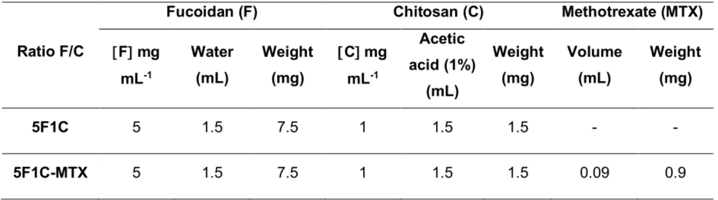
Fucoidan/ Chitosan (FC) nanoparticles were obtained by polyelectrolyte self-assembly method followed by ultrasonication at room temperature according to a previously described method [88].Chitosan was dissolved in 1% (v/v) acetic acid solution to obtain a chitosan-acetic acid solution of 1 mg mL-1, using an ultrasound bath to enhance the solubility. Fucoidan was dissolved in double-deionized water to obtain solutions of 5 mg mL -1. With these solutions was prepared a 5:1 ratio of the fucoidan/chitosan nanoparticles.
The preparation process initiates by mixing the two polysaccharides in a round-bottom glass tube under pulsed ultrasonication (pulse-on 3 seconds and pulse-off 7 seconds, to a total of 30 seconds with 100% range of motion) using a probe sonicator (VCX130, Sonics and Material Vibra-CellTM with a CV-18 probe; 115 Newtown CT, USA) to
promote the self-assembly of these compounds and produce the nanoparticles. After the self-assembly, the formulations were filtered using an 800 nm MinisartÒ®Syringe Filter

![Figure 2: Illustration of pH variation throughout the human body, adapted from Bazban-Shotorbani et al (2017) [36]](https://thumb-eu.123doks.com/thumbv2/123dok_br/15704122.1067777/29.892.193.763.528.896/figure-illustration-variation-human-body-adapted-bazban-shotorbani.webp)