Nature Cell Biology (Letter)
Drosophila CLASP is required for microtubule subunit
incorporation into fluxing kinetochore fibers
Helder Maiato
1, Alexey Khodjakov
1-3, and Conly L. Rieder
1-31 Lab of Cell Regulation, Wadsworth Center, N.Y.S. Dept. of Health, Empire State Plaza,
Albany, New York 12201-0509
2 Dept. of Biomedical Sciences, State Univ. of New York, Albany, N.Y. 12222 3 Marine Biological Laboratory, Woods Hole, MA 02543
Correspondence: Dr. C.L. Rieder, Lab of Cell Regulation, Division of Molecular Medicine, Wadsworth Center, P.O. Box 509, Albany, New York 12201-0509
Ph = 518-474-6774 Fax = 518-486-4901
The motion of a chromosome during mitosis is mediated by a bundle of
microtubules (MTs), termed a kinetochore fiber (K-fiber), which connects the kinetochore of the chromosome to a spindle pole. Once formed, mature K-fibers maintain a steady state length because the continuous addition of MT subunits onto MT plus ends at the
kinetochore is balanced bytheir removal at their minus ends within the pole. This
condition is known as “MT poleward flux”1. Chromosome motion and changes in position are then driven by changes in K-fiber length, which in turn are controlled by changes in the rates at which MT subunits are added at the kinetochore and/or removed from the pole2. A key to understanding the role of flux in mitosis is to identify the molecular players that drive it. Here we use Drosophila S2 cells expressing GFP-α tubulin, RNAi,
microsurgery and photobleaching to show that the kinetochore protein MAST/Orbit, the single CLASP orthologue in Drosophila, is an essential component for MT subunit
incorporation into fluxing K-fibers.
CLASPs belong to an expanding class of proteins that track on the plus ends of growing MTs (called +TIPs). They are thought to be involved in regulating MT dynamics during both interphase and mitosis3. In striking contrast with other +TIPs, CLASPs are unique because they bind to kinetochores throughout mitosis in the absence or presence of attached MTs4,5. We have previously shown that inhibiting CLASP function induces spindles to collapse, and causes abnormal chromosome congression, suggesting that this protein is involved in regulating MT dynamics, particularly within K-fibers5,6.
To examine this issue in detail we depleted MAST/Orbit in Drosophila S2 cells stably expressing GFP/α-tubulin (S2T cells7), by RNAi. This system has several advantages for such
studies. First, MAST/Orbit is the only Drosophila orthologue of CLASPs, which eliminates possible redundant functions between CLASP1 and 2. For nomenclature simplicity, we will use
the term ‘CLASP’ throughout the text for MAST/Orbit. Second, K-fibers in live S2 cells are robust and can be easily discerned (Fig. 1a). Also, spindle formation in these cells has been characterized in detail8. Importantly, in S2T cells the formation of K-fibers is often initiated by kinetochores. As a result, it is not unusual to find K-fibers that formed from the kinetochore without a relationship to an existing centrosome/aster (see Supplementary Information, movie S1a and ref. 8). Together these features provide a unique opportunity for studying how depleting CLASP impacts the formation and maintenance of K-fibers during mitosis in animals.
After 72 hrs of RNAi treatment S2T cells are depleted of CLASP (> 95%; see Fig. 1b and ref. 6). As in controls, in the absence of CLASP centrosomes were often positioned on opposite sides of the nucleus prior to nuclear envelope breakdown (NEB)(Figs. 1a,c). After NEB, K-fibers rapidly formed on chromosomes positioned between the separated centrosomes (cf Figs. 1a,c). However, unlike in controls, once formed in CLASP-depleted cells the spindle and its associated K-fibers gradually and progressively shortened (Fig. 1c and Supplementary Information, movie S1c). This confirms earlier conclusions that interfering with CLASP induces bipolar spindles to collapse, providing direct evidence for the shortening of K-fibers during this process. Interestingly, the initial rate of spindle collapse in CLASP-depleted cells was about 1.2 µm/min, or ~ 0.6 µm/min for each half spindle (N=5; Fig. 1e and Table I). However, this rate was not linear with time, implying the presence of a resistant force to spindle shortening. This resistance could arise from varying degrees of CLASP depletion, which could lead to heterogeneity in the severity of the phenotype. However, there is good evidence (e.g., see ref. 9) that both MT motor
molecules and the intrinsic dynamic properties of MTs are required to maintain spindle bipolarity. Thus, a variable resistance to collapse after CLASP RNAi more likely arises from varying structure of forming spindles combined with different activities of motor proteins like KLP61F/Eg5, members of the Kinesin-5 family10, which work to maintain spindle bipolarity.
This scenario is supported by the observation that spindle collapse in yeast STU1 mutants (the S.
cerevisiae CLASP) can be rescued by doubling the dosage of Cin8p, the counterpart of
KLP61F/Eg5 in this organism11.
Our initial observations confirmed that CLASP activity is not required for the genesis of K-fibers. In fact, it was not unusual in CLASP-depleted cells to see K-fibers form as described above for controls, via a mechanism intrinsic to kinetochores8 (Fig. 1c). However, as spindles collapsed in CLASP-depleted cells, mature K-fibers not connected to a spindle pole shrank as well (Fig.1d). This not only reveals that CLASP becomes essential for maintaining K-fibers after they reach a particular maturation stage, but also that K-fiber shortening in cells lacking CLASP occurs via a mechanism intrinsic to the kinetochore or K-fiber and not the spindle poles. In this regard we recently postulated that CLASPs regulate the dynamics of K-fibers by mediating MT subunit incorporation at the kinetochore5. If this is true, mature K-fibers in CLASP-depleted cells should not exhibit MT subunit flux (reviewed in12).
To directly test this prediction we photobleached short stretches of individual K-fibers in control and CLASP-depleted S2T cells. In control metaphase cells photobleached marks moved poleward at a rate of 0.70±0.15 µm/min (N=5; Fig. 2a,e and Table I). However, in CLASP-depleted cells the bleached mark remained stationary as the spindle gradually collapsed (Fig. 2b,e). CLASPs are components of the kinetochore corona, and are the last proteins known to become incorporated into this structure5,13. As a result, depleting CLASPs does not affect the targeting of other kinetochore proteins including CENP-A, CENP-B, CENP-C, CENP-E, Polo, Zw10, CLIP-170 and dynein5,6. Thus, it is likely that the absence of MT subunit flux in CLASP-depleted cells is due to the direct involvement of CLASP.
To confirm that the effects observed in CLASP-depleted cells are not due to a general perturbation of the kinetochore-MT interface we repeated our photobleaching experiments in
cells depleted of either cytoplasmic dynein, another corona protein14, or EB1, which is also a tracking protein delivered to the kinetochore on elongating MTs15. As previously reported, when dynein heavy chain is knocked down S2T cells still form bipolar spindles with properly aligned chromosomes that are delayed in metaphase7,8. However, as in plants, the spindle poles in dynein-depleted S2T cells were frequently splayed (Fig. 2c; see also ref. 8). As previously reported by others16, we also found that depleting S2T cells of EB1 led to a severe reduction of astral MTs and bipolar spindles that were shorter than those of controls (cf. Figs. 2a,d). Importantly, however, photobleached marks on K-fibers in both dynein- or EB1-depleted cells exhibited persistent poleward motion with rates similar to controls cf. Fig. 2a -e and Table I). Together, these results provide compelling evidence that the lack of MT subunit flux in CLASP-depleted cells is due specifically to the depletion of CLASP. This, in turn, means that CLASP is required for MT subunit addition into mature K-fibers, and in its absence spindles collapse because K-fibers progressively shorten by MT subunit loss at their minus ends. Here it is noteworthy that a similar phenotype occurs when vertebrate metaphase cells are treated with low doses of Taxol, which similarly inhibits subunit incorporation into kinetochore MT plus ends, but not their removal at the pole17. Nevertheless, since CLASP family members are also integral
centrosome components4,5, it is formally possible that CLASP depletion also affects MT minus end dynamics.
To examine this possibility, and to also directly determine the role of CLASP at kinetochores, we severed the connection between kinetochores and spindle poles in S2T cells by cutting mature K-fibers with a laser. As shown previously by us (ref. 8), and others for other systems18,19,20, when K-fibers in control cells were severed the fragment remaining connected to the pole (P-fragment) rapidly disassembled toward the pole at 21.80 ± 0.35 µm/min. In contrast,
the fragment remaining attached to the kinetochore (K-fragment) re-grew to its original length at 0.78 ± 0.17 µm/min (Fig. 3a, Table 1; Supplemental Information, movie S3a; and ref. 8).
As in control cells, when mature K-fibers were severed in CLASdepleted cells the P-fragments rapidly disassembled toward the spindle pole (Figs. 3b, Table 1, Supplemental Information, movie S3b). Thus, CLASP depletion does not modify the dynamics of free K-fiber MT plus ends created by laser microsurgery. However, in striking contrast to controls, in all 30 CLASP depleted cells examined the K-fragments did not change length (i.e., re-grow) during the course of the experiment, even as the spindle gradually collapsed (Fig. 3b).
We have previously shown that severed K-fiber re-growth in control S2T cells occurs because the new MT minus ends remain stable, while MT subunits continue to be incorporated into kinetochore MT plus ends8. Clearly, in control or CLASP-depleted cells the machinery for removing subunits from MT minus ends on K-fragments is not present. As a result, depleting CLASP has no discernibleimpact on MT minus-end dynamics. Accordingly, when we repeated these severing experiments in dynein- or EB1-depleted cells, the results were indistinguishable from the behavior of K-fragments in control cells (cf. Figs. 3a-e, Table I, Supplementary Information, movies S3c, d). Together these results reveal that K-fragments in CLASP-depleted cells do not elongate, or bleach marks do not move poleward, because CLASP is required for kinetochore-mediated MT subunit incorporation into mature (fluxing) K-fibers (see Fig. 4). This conclusion is consistent with the observation that CLASP binds to tubulin subunits in vitro (S. Gouveia, P. Sampaio, R. Reis, and C. E. Sunkel, unpublished).
In Drosophila and likely other systems21, K-fibers that form from the kinetochore do so via a two-step process. First, short MT stubs associated with the kinetochore corona begin to elongate by subunit addition at their kinetochore-attached plus ends. This continues until the fiber reaches its mature length, at which time this steady-state length is maintained until
anaphase onset. Both of these processes require subunit addition into MT plus ends at the kinetochore. However, our data reveal that these two processes occur by different mechanisms. Since K-fibers in CLASP-depleted S2T cells still form by MT elongation from the kinetochore, this process is independent of CLASP. In contrast, the mechanism that maintains the length of the K-fiber after it forms, which is based on MT subunit flux, clearly involves CLASP. This means that the properties of forming and mature K-fibers differ. In this regard it is possible that CLASP becomes important for kinetochore MT dynamics only after the flux machinery becomes engaged. For example, the switch between independent K-fiber formation, and CLASP-dependent K-fiber maintenance, may occur as the molecules responsible for removing subunits accumulate at MT minus ends and become operative.
KLP10A, a member of the Kinesin-13 family, has been implicated in K-fiber shortening during mitosis in Drosophila embryos by mediating MT subunit removal at the poles2. Its human orthologue, Kif2a, is also required for spindle bipolarity9. A molecular view is therefore emerging for how K-fibers elongate and shorten to allow for chromosome motion and position changes in mature spindles: CLASP is involved in regulating MT subunit incorporation into K-fibers at the kinetochore, while proteins like KLP10A/Kif2a regulate MT depolymerization at the poles (Fig. 4). Any imbalance in the activity of these proteins affects K-fiber length. This leads to changes in the position of the chromosomes relative to the poles and spindle equator, while also establishing spindle length. In crane flies and grasshopper spermatocytes, the shortening of K-Fibers occurs exclusively by enhancing the rate of subunit removal near the spindle pole without dampening the rate of incorporation at the kinetochore22,23. However, in vertebrates and
Drosophila, K-fiber shortening occurs primarily by dampening MT subunit incorporation at the
kinetochore without changing the rate of subunit removal at the pole24,25,26.Our data suggest that in these latter systems, switches between K-fiber shortening and elongation, required for
poleward and away from the pole kinetochore motion27,28, are regulated by changes in the functional state of CLASPs. Finally, we found that mature K-fibers not connected to a spindle pole shorten in CLASP-depleted cells. This implies that the stable MT minus ends of a growing K-fiber in S2T cells switch, at some point in their maturation, into a depolymerization state independent of their association with a centrosome. This likely occurs as the minus ends recruit molecular factors involved in MT subunit removal.
Materials and Methods
Growing and flattening S2T cells for time-lapse light microscopy, laser microsurgery and photobleaching
Drosophila S2T cells were a kind gift of Dr. R. Vale (Univ. Calif., San Francisco, CA).
The cells were flattened for live cell microscopy analyses, and laser microsurgery/photobleaching studies were conducted as previously detailed8. Time-lapse images were collected at 24-26 °C using either an Olympus IX70 or a Nikon Eclipse TE2000E DIC inverted light microscopes equipped, respectively, with CM350 or CoolSnap HQ cameras (Photometrics, Tucson, AZ). Time-lapse data sets were subsequently deblurred using the Softworx 2.5 (Applied Precision, USA) deconvolution algorithm. Image sequences were compiled with ImageJ 1.30 (NIH, USA) and processed for publication using Photoshop 6.0 (Adobe Systems).
RNAi
RNAi depletion of Drosophila CLASP, dynein and EB1 were conducted as described previously29 using published targeting sequences6,8,16. Cells were analyzed by light microscopy or by immunoblots 3 days after adding dsRNA to cultures, except for EB1 RNAi, where efficient
depletion could only be accomplished after a second treatment with fresh dsRNA for another 3 days (see Supplementary Information, Fig. S1).
Acknowledgements: We thank Drs. G. Goshima, S. Rogers and R. Vale (Univ. Calif., San
Francisco) for the S2T cells and EB1 antibodies, and Dr. C. Sunkel (Univ. of Porto, Portugal) for communicating results prior to publication. This work was supported by NIH grants GMS 40198 (to CLR), GMS 59363 (to AK), and postdoctoral research fellowship from Fundação para a Ciência e a Tecnologia of Portugal (SFRH/BPD/1159/2002 to HM).
Figure legends
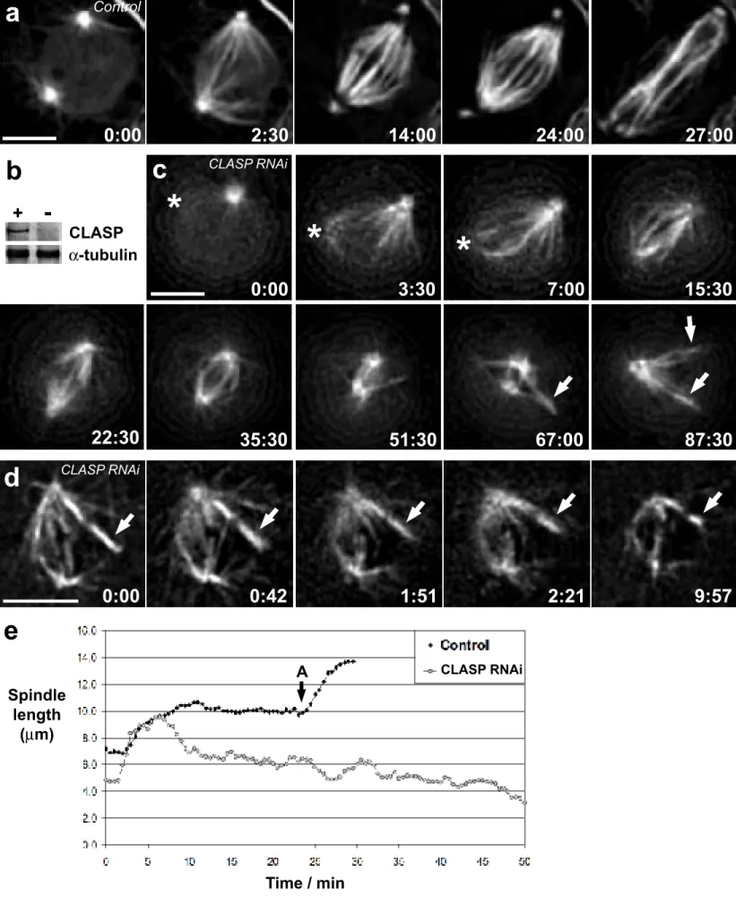
Figure 1. CLASP is required for maintaining spindle and kinetochore fiber length in Drosophila
S2T cells. (a) Mitotic progression in a control cell stably expressing GFP/α-tubulin. (b)
Western blot showing the depletion of Drosophila CLASP by RNAi (-) compared to control S2T cells (+). Endogenous α-tubulin levels are shown as a loading control of protein extracts derived from 106 cells per lane. (c) Spindle collapse in a Drosophila S2T cell depleted of CLASP by RNAi. Note that several kinetochore fibers were generated by kinetochores (arrows) as the spindle collapsed. Asterisks note a centrosome not visible in some focal planes during the early recording stages. (d) Mature K-fibers, not attached to a centrosome/spindle pole (arrow), also shorten as the spindle collapses in cells depleted of CLASP. Time in min:sec. Scale bar = 5 µm. (e) Spindle length over time for the control (a) and CLASP RNAi (c) depleted cells. The onset of anaphase in the control cell is indicated in the chart with an ‘A’. Movies of (a) and (c) can be found in the supplementary information.
Figure 2. CLASP is required for microtubule poleward flux. (a) Control cell showing the
poleward motion (microtubule subunit flux) of a photobleached region (arrow) on a K-fiber. (b) In cells depleted of CLASP, photobleached marks on K-fibers (arrow) do not move poleward as the spindle gradually collapses. (c, d) Photobleached regions on K-fibers in dynein- or EB1-depleted cells, respectively, flux poleward (arrow) at normal rates. Time in min:sec. Scale bar = 5 µm. (e) Kinetic traces of poleward flux, as assayed from the motion of photobleached regions on K-fibers, from 2 different cells per condition.
Figure 3. CLASP is required for subunit incorporation into K-fiber microtubules at their
kinetochore-attached plus-ends. (a) Control cell showing the re-growth (arrow) of a K-fiber after severing by laser microsurgery. (b) Similar to (a) but in a cell depleted of CLASP by RNAi. The severed K-fiber (arrow) does not re-grow even as the spindle gradually collapses. (c, d) K-fibers severed in dynein- or EB1-depleted cells, respectively, regrow (arrow) at the same rates seen in control cells. Time in min:sec. Scale bar = 5 µm. (e) K-fragment elongation curves after laser microsurgery for the cells shown in the previous panels. Movies of (a-d) can be found in the supplementary information.
Figure 4. Schematic depicting the role of CLASP at kinetochores. (a) CLASP (yellow) mediates
tubulin subunit (green) incorporation into kinetochore MT plus-ends at kinetochores. When a mature K-fiber is photobleached (top orange arrow b-d) the subunits move or “flux” poleward through the fiber and are removed near the poles. When a K-fiber is photobleached (bottom orange arrow in b) and then severed (black arrow) closer to the pole, the fiber re-grows towards the pole (c-d) by MT subunit incorporation at the kinetochore but not at the newly created MT minus ends, which are stable. Once the re-growing fiber matures factors are recruited to its MT
minus ends (d) (likely Kinesin-13 family members like KLP10A/Kif2a; pink) that allow for (and power?) subunit flux. In the absence of CLASP (e), the incorporation of subunits into mature K-fiber MT plus-end is shut down inhibiting flux. However, since MT minus-end depolymerization still occurs near the poles, the K-fibers shorten and the spindle collapses.
Table I: Rates of kinetochore fragment elongation, pole-fragment shortening, flux, initial spindle
collapse and anaphase chromosome motion in control and experimentally manipulated S2T cells. Each rate represents the average ± standard deviation of 5 different events.
Supplementary information
Figure S1. Western blot depicting EB1 depletion by RNAi. Alpha tubulin was used as a
loading control. `
Movie S1a. Mitotic progression in a control cell stably expressing GFP/α-tubulin. Note the kinetochore-driven formation of K-fibers.
Movie S1c. Spindle collapse in a Drosophila S2T cell depleted of CLASP by RNAi. Several
kinetochore fibers were generated by kinetochores as the spindle collapsed. One of the centrosomes is not visible in some focal planes during the early recording stages.
Movie S3a. Control cell showing the re-growth of a K-fiber after severing by laser microsurgery.
Movie S3b. A CLASP-depleted cell in which a K-fiber was severed. Note that the fiber does
Movie S3c. K-fibers severed in dynein-depleted cells re-grow at the same rate seen in control
cells.
Movie S3d. K-fibers severed in EB1-depleted cells re-grow at the same rates seen in control
Reference List
1. Mitchison, T. J. Polewards microtubule flux in the mitotic spindle: evidence from photoactivation of fluorescence. J. Cell Biol. 109, 637-652 (1989).
2. Rogers, G. C. et al. Two mitotic kinesins cooperate to drive sister chromatid separation during anaphase. Nature 427, 364-370 (2004).
3. Carvalho, P., Tirnauer, J. S. & Pellman, D. Surfing on microtubule ends. Trends Cell
Biol. 13, 229-237 (2003).
4. Lemos, C. L. et al. Mast, a conserved microtubule-associated protein required for bipolar mitotic spindle organization. EMBO J. 19, 3668-3682 (2000).
5. Maiato, H. et al. Human CLASP1 is an outer kinetochore component that regulates spindle microtubule dyanmics. Cell 113, 891-904 (2003).
6. Maiato, H. et al. MAST/Orbit has a role in microtubule-kinetochore attachment and is essential for chromosome alignment and maintenance of spindle bipolarity. J. Cell Biol.
157, 749-760 (2002).
7. Goshima, G. & Vale, R. D. The roles of microtubule-based motor proteins in mitosis: comprehensive RNAi analysis in the Drosophila S2 cell line. J. Cell Biol. 162, 1003-1016 (2003).
8. Maiato, H., Rieder, C. L. & Khodjakov, A. Kinetochore-driven formation of kinetochore fibers contributes to spindle assembly during mitosis in animals. J. Cell Biol (in press) (2004).
9. Ganem, N. J. & Compton, D. A. The KinI kinesin Kif2a is required for bipolar spindle assembly through a functional relationship with MCAK. J. Cell Biol. 166, 473-478 (2004).
10. Lawrence, C. J. et al. A standardized kinesin nomenclature. J. Cell Biol. 167, 19-22 (2004).
11. Yin, H., You, L., Pasqualone, D., Kopski, K. M. & Huffaker, T. C. Stu1p is physically associated with beta-tubulin and is required for structural integrity of the mitotic spindle.
Mol. Biol. Cell 13, 1881-1892 (2002).
12. Sharp, D. J. MAST sails through mitosis. Curr. Biol. 12, R585-R587 (2002). 13. Desai, A. et al. KNL-1 directs assembly of the microtubule-binding interface of the
14. Hoffman, D. B., Pearson, C. G., Yen, T. J., Howell, B. J. & Salmon, E. D. Microtubule dependent changes in the assembly of microtubule motor proteins and mitotic spindle checkpoint proteins at kinetochores. Mol. Biol. Cell 12, 1995-2009 (2001).
15. Tirnauer, J. S., Canman, J. C., Salmon, E. D. & Mitchison, T. J. EB1 targets to kinetochore with attached, polymerizing microtubules. Mol. Biol. Cell 13, 4308-4316 (2002).
16. Rogers, S. L., Rogers, G. C., Sharp, D. J. & Vale, R. D. Drosophila EB1 is important for proper assembly, dynamics, and positioning of the mitotic spindle. J. Cell Biol. 158, 873-884 (2002).
17. Waters, J. C., Mitchison, T. J., Rieder, C. L. & Salmon, E. D. The kinetochore
microtubule minus-end disassembly associated with poleward flux produces a force that can do work. Mol. Biol. Cell 7, 1547-1558 (1996).
18. Czaban, B. B., Forer, A. & Bajer, A. S. Ultraviolet microbeam irradiation of
chromosomal spindle fibres in Haemanthus katherinae endosperm. I. Behaviour of the irradiated region. J. Cell Sci. 105, 571-578 (1993).
19. Forer, A., Spurck, T. & Pickett-Heaps, J. Ultaviolet microbeam irradiations of spindle fibers in crane-fly spermatocytes and newt epithelial cells: resolution of previously conflicting observations. Protoplasma 197, 230-240 (1997).
20. Gordon, G. W. The control of mitotic motility as influenced by ultraviolet microbeam irradiation of kinetochore fibers. Ph.D. Thesis, University of Pennsylvania, Philadelphia, PA pp 1-157 (1980).
21. Wadsworth, P. & Khodjakov, A. E pluribus unum: towards a universal mechanism for spindle assembly. Trends Cell Biol. 14, 413-419 (2004).
22. Chen, W. & Zhang, D. Kinetochore fibre dynamics outside the contex of the spindle during anaphas. Nature Cell Biol. 6, 227-231 (2004).
23. LaFountain, J. R. Jr., Cohan, C. S., Siegel, A. J. & LaFountain, D. J. Direct visualization of microtubule flux during metaphase and anaphase in crane-fly spermatocytes. Mol.
Biol. Cell (in press) (2004).
24. Mitchison, T. J. & Salmon, E. D. Poleward kinetochore fiber movement occurs during both metaphase and anaphase-A in newt lung cell mitosis. J. Cell Biol. 119, 569-582 (1992).
25. Brust-Mascher, I. & Scholey, J. M. Microtubule flux and sliding in mitotic spindles of
Drosophila embryos. Mol. Biol. Cell 13, 3967-3975 (2002).
26. Maddox, P., Desai, A., Oegema, K., Mitchison, T. J. & Salmon, E. D. Poleward microtubule flux is a major component of spindle dynamics and anaphase A in mitotic
27. Skibbens, R. V., Skeen, V. P. & Salmon, E. D. Directional instability of kinetochore motility during chromosome congression and segregation in mitotic newt lung cells: a push-pull mechanism. J. Cell Biol. 122, 859-875 (1993).
28. Khodjakov, A. & Rieder, C. L. Kinetochores moving away from their associated pole do not exert a significant pushing force on the chromosome. J. Cell Biol. 135, 315-327 (1996).
29. Maiato, H., Sunkel, C. E. & Earnshaw, W. C. Dissecting mitosis by RNAi in Drosophila tissue culture cells. Biol. Proced. Online 5, 153-161 (2003).
α-tubulinCLASP
+
-d
0:00
0:42
1:51
2:21
9:57
b
e
c
0:00
3:30
7:00
15:30
22:30
35:30
51:30
67:00
87:30
*
*
*
Spindle length (µm) Time / min Aa
0:00
2:30
14:00
24:00
27:00
Control CLASP RNAi CLASP RNAiFigure 1 - Maiato et al., 2004
a
b
-0:03 0:00 -0:03 0:00 0:39 2:51 9:33 2:45 4:30c
-0:03 0:00 0:36 2:12 3:42d
-0:03 0:00 1:15 2:24 3:12 1:40 Control CLASP RNAi Dynein RNAi EB1 RNAie
CLASP RNAia
b
c
-0:03 0:00 0:42 1:27 3:18 -0:03 0:00 0:18 0:51 2:09 -0:03 0:00 0:12 1:12 2:00 -0:03 0:00 0:48 1:48 3:15d
Control CLASP RNAi Dynein RNAi EB1 RNAie
Figure 3 - Maiato et al., 2004
Experiment K-fragment elongation rate (µm/min) N=5 P-fragment shortening rate (µm/min) N=5 Flux rate (µm/min) N=5 Initial spindle collapse rate (µm/min) N=5 Anaphase poleward chromosome movement rate
(µm/min) N=5 Control 0.78 ± 0.17 21.80 ± 0.35 0.70 ± 0.15 _ 1.24 ± 0.09 MAST RNAi ~0 20.00 ± 4.81 ~0 1.20 ± 0.06 _ Dynein RNAi 0.72 ± 0.12 19.93 ± 5.61 0.70 ± 0.18 _ _ EB1 RNAi 0.74 ± 0.12 19.93 ± 3.93 0.67 ± 0.12 _ _ Table 1