Development of electrochemical immunosensors for
celiac disease clinical diagnosis and gluten-free food
control
control
Marta Maria Pereira da Silva Neves
Thesis submitted to the Faculty of Pharmacy, University of Porto, for the degree of Doctor of Philosophy in Pharmaceutical Sciences - Biochemistry
Tese do 3º Ciclo de Estudos Conducente ao Grau de Doutoramento em Ciências Farmacêuticas – Bioquímica,
apresentada à Faculdade de Farmácia da Universidade do Porto
Supervisors/ Orientadores
Professora Doutora Cristina Maria Fernandes Delerue Alvim de Matos
Co-Supervisors/ Co-orientadores
Professora Doutora Maria Alice dos Santos Silva Gomes Martins Professor Doutor Hendrikus Petrus Antonius Nouws
Professor Doutor Agustín Costa García
Porto November, 2012
© Autorizada a reprodução parcial desta tese (condicionada à autorização das editoras das revistas onde os artigos foram publicados) apenas para efeitos de investigação, mediante declaração escrita do interessado, que a tal se compromete.
v
A realização deste trabalho foi possível graças à concessão de uma Bolsa de Doutoramento (SFRH/BD/46351/2008) pela Fundação para a Ciência e a Tecnologia (FCT), financiada pelo Programa Operacional Potencial Humano (POPH) - Quadro de Referência Estratégico Nacional (QREN) - Tipologia 4.1 - Formação Avançada, comparticipado pelo Fundo Social Europeu (FSE) e por Fundos Nacionais do Ministério da Ciência, Tecnologia e Ensino Superior (MCTES). Em associação à Bolsa de Doutoramento, a bolseira contou ainda com subsídios para deslocamento a congressos internacionais.
The execution of this work was made possible thanks to a Doctoral grant (SFRH/BD/46351/2008) attributed by the "Fundação para a Ciência e a Tecnologia (FCT)", through the "Programa Operacinal Potencial Humano (POPH) - Quadro de Referência Estratégico Nacional (QREN) - Tipologia 4.1 - Formação Avançada", with comparticipation of the European Social Fund (ESF) and national funds from the "Ministério da Ciência, Tecnologia e Ensino Superior (MCTES)". Associated with the grant, additional funding was provided to the research fellow for participation in international conferences.
vii
Os estudos apresentados nesta tese foram realizados no Serviço de Bioquímica da Faculdade de Farmácia da Universidade do Porto, nos Laboratórios do REQUIMTE, sediado no Instituto Superior de Engenharia do Porto do Instituto Politécnico do Porto, e no Grupo de Inmunoelectroanálisis da Faculdade de Química da Universidade de Oviedo.
The studies presented in this thesis were performed in the Biochemistry department of the Faculty of Pharmacy of the University of Porto, in the laboratories of REQUIMTE, located at the Instituto Superior de Engenharia do Porto of the Instituto Politécnico do Porto, and in the Group of Inmunoelectroanálisis of the Faculty of Chemistry, University of Oviedo. ´
ix ACKNOWLEDGEMENTS
“ I almost wish I hadn't gone down that rabbit-hole — and yet — and yet — it's rather curious, you know, this sort of life!” in Alice's Adventures in Wonderland
I would like to express my gratitude to a number of people for their contribution to my work:
À Fundação para a Ciência e a Tecnologia pela concessão de uma bolsa de doutoramento (SFRH/BD/46351/2008), sem a qual teria sido impossível realizar este trabalho.
À Professora Doutora Cristina Delerue Matos, por proporcionar-me a oportunidade de aprender eletroquímica, ainda na altura do mestrado, caminho que, sem dúvida, conduziu à realização deste doutoramento. Por todo o apoio ao longo destes anos, por todo o entusiasmo, carinho e amizade. Por sempre incentivar-me a arriscar e a avançar. E por, independentemente da distância, estar sempre presente.
Al Profesor Agustín Costa García por acogerme amablemente en su grupo de investigación, demostrando desde el primer momento su disponibilidad para ayudarme y enseñarme. Por todo el cariño que siempre me ha demostrado y por haberme permitido aprender y crecer, como científica y como persona.
À Professora Doutora Alice Santos Silva que amavelmente aceitou co-orientar esta tese. Agradeço profundamente toda a disponibilidade, ajuda e apoio ao longo destes 4 anos. Ao Professor Doutor Hendrikus Petrus Antonius Nouws, por ter sido a primeira pessoa a ensinar-me eletroquímica, e por, a partir desse momento, estar sempre disponível para ajudar-me. Por transmitir-me a importância do sentido crítico e rigor. E pela ajuda preciosa nesta recta final. Obrigada também pela amizade e pelo apoio ao longo deste percurso.
x
A la Doctora Begoña González García, por su ejemplo como profesional y, sobre todo, como persona. Bego, gracias por el enorme privilegio de poder haber aprendido de ti. Por toda la dedicación, apoyo, incentivo (principalmente cuando los resultados se hacían de rogar…) y por animarme en los días más grises. Gracias por toda la amistad y por todo lo que hemos compartido.
À Fátima, por estar sempre disponível para mim, como investigadora e como amiga. A la Doctora Graciela Martínez Paredes por facilitar las muestras de suero reales utilizadas en esta tesis.
A mis compañeros de laboratorio, por todos los momentos que hemos vivido juntos. Por todas las risas e historias que compartimos y que alegraban las largas horas de trabajo. Recordaré siempre con mucho cariño estos años y “nuestro” grupo. Seguro que es algo irrepetible.
A los amigos que hice por el “camino”, en especial a Pilar, Mayka, Tamara, Jorge, Laura, Bárbara y Elisa, por todos recuerdos que guardo.
Às amigas de Portugal. Obrigada pelos e-mails e mensagens que ajudaram a suportar as saudades. Em especial à Mariana, por tantas horas de conversa, à distância, ao longo destes anos.
A Fani, en quién he encontrado una gran amiga. Por ser una persona tan especial con quien he compartido tanto y por haber estado siempre presente. Sin ti, todo hubiera sido infinitamente más difícil.
A Dani, por todo el apoyo y por aguantar con paciencia mis “crisis” de estrés en esta recta final de la tesis. Por todo el amor y por todos los maravillosos momentos que hemos vivido juntos. Tú has sido lo mejor de todo.
E por último, quero agradecer, à minha família: avós, tia, e, em especial, pai, mãe e irmão, por todo o apoio incondicional ao longo destes anos em que estive ausente e que tantas saudades deixaram. Apesar da distância, estiveram todos os dias no meu pensamento e, por isso, é à minha família que dedico, com todo o meu amor, a minha tese de doutoramento.
xi geneticamente suscetíveis como resultado da ingestão de glúten. Para o diagnóstico clínico desta doença, a metodologia mais usada é o ensaio de imunoabsorção enzimática (ELISA), mas nos últimos anos muitos esforços tem sido feitos para melhorar os testes serológicos para a DC. Contudo, não existe um acordo geral no método imunoquímico a utilizar.
Hoje em dia, os métodos baseados no uso de sensores eletroquímicos estão desempenhando um papel cada vez maior em diversos campos em que sistemas de medida precisos, rápidos e que permitam uma medida em tempo real são necessários. Relativamente à qualidade e custo, os sensores eletroquímicos podem ser uma melhor opção do que os métodos analíticos convencionais. Especialmente, os imunossensores eletroquímicos (IEs), que ao combinar a elevada especificidade dos métodos imunoquímicos tradicionais com o elevado limite de deteção das técnicas eletroquímicas, são poderosas ferramentas analíticas para o desenvolvimento de dispositivos mais rápidos, mais simples, mais sensíveis e de baixo custo que podem ser competitivos o suficiente e aplicados diariamente com êxito no diagnóstico clínico da DC.
O principal objetivo desta tese de doutoramento foi o desenvolvimento, produção e otimização da melhor superfície transdutora de modo a obter IEs sensíveis, reprodutíveis e robustos para análise rotineira do diagnóstico clínico da DC.
A primeira etapa deste trabalho consistiu na busca da superfície apropriada para a imobilização da quantidade adequada de material proteico na superfície transdutora dos sensores eletroquímicos, incluindo também a exploração das potencialidades dos nanomateriais. Para este propósito, elétrodos serigrafados de carbono foram nanoestruturados com nanopartículas de ouro, nanotubos de carbono, nanofibras de carbono, e, com sistemas híbridos de materiais de carbono e metal. A biofuncionalidade dos diferentes transdutores foi avaliada através da sua capacidade para adsorverem e reterem material proteico nas suas superfícies, e empregando, como modelo analito, o complexo altamente estável estreptravidina/biotina. As superfícies transductoras foram também caraterizadas por microscopia eletrónica de varrimento. Os elétrodos serigrafados de carbono modificados com os híbridos de nanotubos de carbono e nanopartículas de ouro revelaram ser os que propocionavam a superfície transductoras mais apropriada.
Depois da seleção do transductor mais adequado, a fase sensora dos IEs foi construída com base na interação antigénio-anticorpo. IEs para a determinação de imunoglobulinas (IgG) classe A (IgA) e classe G (IgG) produzidas contra a gliadina (AGA) e contra a transglutaminase tecidual (anti-tTG) (i.e. os marcadores serológicos específicos para a DC) foram desenvolvidos e otimizados. Um IE de multi-deteção para a determinação simultânea de dois biomarcadores, AGA e anti-tTG, foram também desenvolvidos. Além
xii
disso, a determinação de anticorpos humanos gerados contra a gliadina desamidada foi também efetuada. Os sensores desenvolvidos foram testados usando amostras de soro de pacientes de DC e indivíduos saudáveis. Finalmente, os métodos desenvolvidos foram validados por comparação com os métodos espetrofotométricos convencionalmente utilizados no diagnóstico serológico da DC.
Os imunossensores construídos e optimizados nesta tese de doutoramento tem como vantagem o fato de serem ferramentas analíticas simples de usar e descartáveis, o que facilitará uma futura transferência destes sensores para aplicações point-of-care.
Palavras-chave: doença celíaca, transglutaminase tecidual, gliadina, elétrodos
xiii susceptible individuals as a result of gluten ingestion. For CD diagnostic purposes the most used methodology is the enzyme-linked immunosorbent assay (ELISA), and in the past few years many attempts have been made to improve CD serological tests. However, there is no general agreement on the immuno-based analytical method for CD screening.
Nowadays, electrochemical sensing techniques are playing a growing role in various fields in which accurate, fast and online measuring systems are required. With regard to quality and cost, electrochemical sensors can be a better option than standard analytical methods. Especially electrochemical immunosensors (EIs), which combine the high specificity of traditional immunochemical methods with the low detection limits of modern electrochemical techniques, are powerful analytical tools to develop faster, simpler, more sensitive and low-cost devices that can be competitive enough to be successfully applied on a daily basis for CD clinical diagnosis.
The main goal of this PhD thesis was the development, manufacturing and optimization of the best transducer surface to obtain sensitive, reproducible and robust EIs for routine analyses of CD clinical serological diagnosis.
The first stage of this work consisted of the search for a suitable layer for immobilizing an adequate amount of protein material on the transducer's surface of electrochemical sensors, including the exploitation of the potentialities of nanomaterials. For this purpose, screen-printed carbon electrodes were nanostructured with gold nanoparticles (NPAus), carbon nanotubes, and carbon nanofibers, and, with hybrid systems of the carbon/metal materials. The biofunctionality of the different transducers was evaluated by their ability to adsorb and retain protein material on their surface, employing the highly stable and efficient streptavidin/biotin complex as model analyte. The transducers' surfaces were also characterized by scanning electron microscopy. The screen-printed electrodes modified with the hybrid carbon nanotubes/gold nanoparticles revealed to provide the most suitable transducer surface.
After the adequate electrode surface selection, the sensing phase of the EIs was constructed relying on antigen-antibody interactions. EIs for the determination of immunoglobulins (IgG) class A (IgA) and class G (IgG) against gliadin (AGA) and tissue transglutaminase (anti-tTG) (i.e. specific CD serological markers) were developed and optimized. A multiplexed EI for the simultaneous assessment of AGA and anti-tTG CD biomarkers was also developed. Moreover, the determination of human antibodies produced against deamidated gliadin was also assayed. The developed sensors were tested using serum samples from CD patients and healthy individuals.
xiv
Finally, the developed methodologies were validated by comparison with the established spectrophotometric immunoassays for CD serological diagnosis.
The immunosensors, that were constructed and optimized within this doctoral thesis, have the advantage of being easy-to-use and disposable analytical tools, which facilitates a future transfer to point-of-care applications.
Keywords: celiac disease, tissue transglutaminase, gliadin, nanostructured
xv personnes prédisposées génétiquement, due à une ingestion de gluten. La méthode la plus utilisée dans les diagnostiques cliniques est le test du dosage d'immunoabsorption par enzyme liée (ELISA), et ces dernières années, des études ont été réalisées dans le but d’améliorer les tests sérologiques de la MC. Cependant il n’y a pas de consensus au niveau des méthodes analytiques basées sur des réactions immunologiques pour la détection de la MC.
De nos jours, les techniques de mesures électrochimiques jouent un rôle de plus en plus important dans divers domaines dans lesquels sont nécessaires des systèmes de mesures précis, rapides, et en temps réels. Au regard de la qualité et du coût, les capteurs électrochimiques peuvent être une meilleure option que les méthodes de référence actuelles. Plus particulièrement les immunocapteurs électrochimiques (IEs), qui combinent la grande spécificité des méthodes immunochimiques avec les limites de détection basses des techniques électrochimiques modernes, sont des outils analytiques très utiles pour développer des dispositifs plus rapides, plus simple, et à moindre coût, de manière à être suffisamment compétitifs pour être appliqué avec succès dans les diagnostiques cliniques quotidiens de la maladie cœliaque.
L’objectif principal de cette thèse de doctoract fut le développement, la fabrication et l’optimisation du meilleur transducteur pour obtenir des IEs sensibles, reproductibles et robustes pour le diagnostique routinier des marqueurs sérologiques de la maladie cœliaque.
La première de ce travail fut la recherche d’une couche appropriée pour l’immobilisation d’une quantité adéquate de matériel protéique sur la superficie transductrice des capteurs électrochimiques, et dans le même temps l’exploration des propriétés des nanomatériaux. Pour cela des électrodes sérigraphiées de carbone furent nanostructurées avec des nanoparticules d’or, des nanotubes de carbones, des nano fibres de carbone et avec des systèmes hybrides fait de carbones et de métal. La bio fonctionnalité des différents transducteurs furent évaluées pour leurs capacité d’adsorption et rétention de matériels protéiques à sa surface, en utilisant comme modèle analytique, le complexe hautement stable et efficace, streptavidine/biotine. Les superficies transductrices furent caractérisées par microscopie électronique à balayage. Les électrodes sérigraphiées de carbone modifiées avec l’hybride de nanotubes de carbone et nanoparticules d’or se révéla être la superficie transductrice la plus adéquate.
Après avoir déterminée quelle était la meilleure superficie électrodique, la partie de détection des IEs fut construite en se basant sur les interactions antigènes-anticorps. Les immunocapteurs électrochimiques pour la détermination d’immunoglobuline (IgG) de
xvi
classe A (IgA) et de classe G (IgG) générée contre gliadin (AGA) et transglutaminase tissulaire (anti-tTG) (i.e. marqueurs sérologiques spécifiques de la maladie cœliaque) furent développés et optimisés. Un IEs multiplexed, pour la détection simultanée des bio marqueurs de la maladie cœliaque AGA et anti tTG a également été développé. De plus, la détermination des anticorps humains produits contre la gliadine desamidée fut aussi testée. Les capteurs développés furent testés en utilisant des échantillons de sérum de patients atteints de la MC et de patients sains. Finalement, les méthodes développées furent validées en comparant les résultats obtenus avec ceux obtenus grâce aux immunotests spectrophotomètriques de référence pour les diagnostiques sérologiques de la MC.
Les capteurs proposés, qui ont été construits et optimisés dans cette thèse, ont l’avantage d’être des outils analytiques faciles à utiliser et jetables, ce qui facilite un futur transfert aux applications point-of-care.
Mots-clés: la maladie cœliaque, transglutaminase tissulaire, la gliadine, électrodes
xvii genéticamente susceptibles como resultado de la ingestión de gluten. Para el diagnóstico de esta enfermedad, los métodos más utilizados están basados en ensayos por inmunoabsorción ligado a enzimas, llamados ensayo de inmunoabsorción enzimática (ELISA), pero en los últimos años la comunidad científica está haciendo esfuerzos para mejorar los tests serológicos de diagnóstico de la EC. Sin embargo, no existe un acuerdo sobre el tipo de método inmunoquímico a utilizar para el diagnóstico de la EC.
Actualmente, los métodos basados en el uso de sensores electroquímicos están jugando un papel muy importante en varios campos en los que es necesario implantar métodos rápidos, fiables, y que puedan realizar medidas en línea. Teniendo en cuenta la calidad y el coste, los sensores electroquímicos pueden convertirse en una buena alternativa a los métodos estándar de análisis. Entre los diferentes sensores electroquímicos, cabe destacar los inmunosensores electroquímicos (IEs), ya que combinan la alta especificidad de las reacciones inmunológicas con los bajos límites de detección las técnicas electroquímicas, lo que los convierten en poderosas herramientas analíticas en el desarrollo de métodos más rápidos, más sencillos, más sensibles and de menor coste que los utilizados actualmente en el diagnóstico de la EC.
El principal objetivo planteado en esta tesis doctoral fue el desarrollo, fabricación y optimización de la superficie del transductor para obtener inmunosensores electroquímicos para el diagnóstico serológico de la EC más sensibles, robustos y reproducibles y con una estabilidad aceptable que permita su comercialización.
La primera parte de este trabajo consistió en la búsqueda de una inmovilización adecuada del material proteico en la superficie del transductor del sensor electroquímico, explotando la potenciabilidad de los nanomateriales. Para ello, electrodos serigrafiados de carbono fueron nanoestructurados con nanopartículas de oro, nanotubos de carbono y nanofibras de carbono, además del uso de materiales nanohíbridos de carbono/metal. La biofuncionalidad de los diferentes transductores ha sido evaluada a través de su habilidad para absorber y retener material proteico en sus superficies, empleando para ello la reacción estreptavidina/biotina. Las superficies de los diferentes transductores también han sido caracterizadas utilizando microscopía de barrido electrónico. Las mejores superficies transductoras han resultado ser los electrodos serigrafiados de carbono modificados con híbridos nanotubos de carbon/nanopartículas de oro.
Después de seleccionar el transductor más adecuado, la interfase sensora de los EIs fue construida utilizando la reacción antígeno-anticuerpo. Así, en el caso que nos ocupa, los EIs desarrollados y puestos a punto en este trabajo determinan inmunoglobulinas A (IgA) y G (IgG) anti-gliadina (AGA) y anti-transglutaminasa tisular (anti-tTG),
xviii
marcadores serológicos específicos de la EC. Además, un EI bianalito para la detección simultánea de los marcadores de la EC, AGA y anti-tTG, ha sido también desarrollado. Por último, se ha ensayado un inmunosensor electroquímico para la determinación de anticuerpos contra gliadina deaminada.
Los diferentes sensores desarrollados han sido testados utilizando muestras de suero humano de pacientes celíacos y personas sanas. Finalmente los inmunosensores electroquímicos desarrollados han sido validados con otros inmunoensayos espectrofotométricos ya establecidos para el diagnóstico serológico de la EC.
Los inmunosensores diseñados y optimizados en esta tesis doctoral tienen la ventaja de ser de fácil manejo y de un único uso, lo que facilitaría una futura implantación de estos sensores como dispositivos point-of-care.
Palabras clave: enfermedad celíaca, la transglutaminasa tisular, la gliadina, electrodos
xix Papers in International Refereed Journals (indexed in the Thomson Reuters
ISI-Web of Knowledge- Science Citation Index Expanded (SCI-EXPANDED)
database)
Celiac disease diagnosis and gluten-free food analytical control
M.M.P.S. Neves, M. B. González-García, H.P.A. Nouws, C. Delerue-Matos, A. Santos-Silva, A. Costa-García
Analytical and Bioanalytical Chemistry, 2010, 397 (5), 1743-1753 doi:10.1007/s00216-010-3753-1
Nanohybrid materials as transducer surfaces for electrochemical sensing applications M.M.P.D. Neves, M. B. González-García, C. Delerue-Matos, A. Costa-García
Electroanalysis, 2011, 23 (1), 63-71 doi:10.1002/elan.201000427
Celiac disease detection using a transglutaminase electrochemical immunosensor fabricated on nanohybrid screen-printed carbon electrodes
M.M.P.S. Neves, M. B. González-García, H.P.A. Nouws, A. Costa-García Biosensors and Bioelectronics, 2012, 31 (1), 95-100
doi:10.1016/j.bios.2011.09.044
Voltammetric immunosensor for the diagnosis of celiac disease based on the quantification of anti-gliadin antibodies
M.M.P.S. Neves, M. B. González-García, A. Santos-Silva, A. Costa-García Sensors and Actuators B: Chemical, 2012, 163 (1), 253-259
doi:10.1016/j.snb.2012.01.048
Multiplexed electrochemical immunosensor for detection of celiac disease serological markers
M.M.P.S. Neves, M. B. González-García, C. Delerue-Matos, A. Costa-García Sensors and Actuators B: Chemical 2012 (in press)
xx
Nanostructured screen-printed electrochemical biosensors for clinical applications
M.M.P.S. Neves, M. B. González-García, H.P.A. Nouws, C. Delerue-Matos, A. Santos-Silva, A. Costa-García. (submitted)
Published papers or extended abstracts in proceedings of scientific meetings:
M.M.P.S. Neves, M. B. González-García, H.P.A. Nouws, C. Delerue-Matos, A. Santos-Silva, A. Costa-García, Multiplexed Electrochemical Immunosensor for Detection of Celiac Disease Serological Markers, Proceedings of the IMCS 2012 - The 14th International Meeting on Chemical Sensors, Nuremberg, Germany, May 20-23, 2012
doi: 10.5162/IMCS2012/P2.1.24
Oral communications in scientific meetings:
M.M.P.S. Neves, M. B. González-García, C. Delerue-Matos, A. Costa-García Superficies nanoestructuradas modificadas con estreptavidina como transductores de biosensores
XXX Reunión del Grupo de Electroquímica de la RSEQ y del XI Encontro Ibérico de Electroquímica
Tenerife, Spain, July, 2009
M.M.P.S. Neves, M. B. González-García, H.P.A. Nouws, C. Delerue-Matos, A. Santos-Silva, A. Costa-García
A novel transducer surface: screen-printed carbon electrodes modification with
carbon-nanostructures and gold nanoparticles
III Workshop de Nanociencia y Nanotecnología Analíticas Oviedo, Spain, September, 2009
M.M.P.S. Neves, M. B. González-García, H.P.A. Nouws, C. Delerue-Matos, A. Santos-Silva, A. Costa-García
Nanohybrid materials as transducer surfaces for electrochemical sensing applications. 13th International Conference on Electroanalysis – ESEAC 2010 Gijón, Spain, June, 2010
xxi
M.M.P.S. Neves, M. B. González-García, H.P.A. Nouws, C. Delerue-Matos, A. Santos-Silva, A. Costa-García
An electrochemical immunosensor for the detection of antibodies directed against gliadins using nanostructured surfaces
IV Workshop Nanociencia y Nanotecnología Analíticas Zaragoza, Spain, September, 2010
M.M.P.S. Neves, M. B. González-García, H.P.A. Nouws, C. Delerue-Matos, A. Santos-Silva, A. Costa-García
Hybrid nanomaterials as immunosensors transducers for the detection of human antibodies directed against gliadins
NANOJASP 2010
Barcelona, Spain, November, 2010
M.M.P.S. Neves, M. B. González-García, H.P.A. Nouws, C. Delerue-Matos, A. Santos-Silva, A. Costa-García
Electrochemical immunosensor for the detection of celiac disease autoantibodies using a nanohybrid transducer surface
V Workshop Nanociencia y Nanotecnología Analíticas Toledo, Spain, September, 2011
M.M.P.S. Neves, M. B. González-García, H.P.A. Nouws, C. Delerue-Matos, A. Santos-Silva, A. Costa-García
The employment of electrochemical immunosensors fabricated on nanohybrid screen-printed carbon electrodes for the diagnosis of celiac disease XIV Iberic Meeting of Electrochemistry & XVII Meeting of the Portuguese Electrochemical Society
Funchal, Portugal, April, 2012
M.M.P.S. Neves, M. B. González-García, H.P.A. Nouws, C. Delerue-Matos, A. Santos-Silva, A. Costa-García
Multiplexed Electrochemical Immunosensor for Detection of Celiac Disease Serological Markers
The 14th International Meeting on Chemical Sensors
xxiii INDEX Acknowledgements ix Resumo xi Abstract xiii Résumé xv Resumen xvii
Publications (papers and communications) developed within the
doctoral project xix
Index xxiii
Abbreviations xxv
I. State of art 1
Organization and structure of the dissertation 3
General introduction 5
References 25
Main goals 33
CHAPTER 1. Celiac disease: from gluten to diagnostic 35
• Celiac disease diagnosis and gluten-free food control 37
CHAPTER 2. New trends: nano, disposable and electrochemical 61
• Nanostructured screen-printed electrochemical biosensors for clinical applications
63
II. RESEARCH AND DEVELOPMENT 85
CHAPTER 3. Hybrids transducer surfaces 87
• Nanohybrid materials as transducer surfaces for electrochemical sensing applications
xxiv
CHAPTER 4. Screening for celiac disease serological markers 109
• Celiac disease detection using a transglutaminase electrochemical immunosensor fabricated on nanohybrid screen-printed carbon electrodes
111
• Voltammetric immunosensor for the diagnosis of celiac disease based on the quantification of anti-gliadin antibodies
127
• Multiplexed electrochemical immunosensor for detection of celiac disease serological markers
147
• Detection of antibodies against deamidated gliadin in human serum samples
163
xxv ABBREVIATIONS
AChE Acetylcholinesterase
AGA Anti-gliadin antibodies
AgNPs Silver nanoparticles
Anti-H-IgA-AP Anti-human IgA antibodies conjugated with alkaline phosphatase
Anti-H-IgG-AP Anti-human IgG antibodies conjugated with alkaline phosphatase
AP Alkaline phosphatase
ASV Anodic stripping voltammetry
BRAC1 Breast cancer 1
BSA Bovine serum albumin
CA Cancer antigen
CD Celiac disease
CEA Carcinoembryonic antigen
CF Cystic fibrosis
CH Antibody constant domains
ChOx Cholesterol oxidase
ChE Cholesterol esterase
CNF Carbon nanofiber
CNP Carbon nanoparticle
CnT I Cardiac troponin I
CNT Carbon nanotube
xxvi
DGP Deamidated gliadin peptide
DMF Dimethylformamide
DPV Differential pulse voltammetry
ECM Extracellular matrix
EB Electrochemical biosensors
EIs Electrochemical immunosensors
EIS Impedance spectroscopy
ELISA Enzyme-linked immunosorbent assay
EMA Endomysial antibodies
ESA Electrostatic self-assembled
ESPGAN European society for paediatric gastroenterology and nutrition
F(ab´)2 Antigen binding fragment
Fc Non-antigen binding fragment
Fe3O4 NPs Iron oxide
FCM Flow cytometry
GEC Graphite–epoxy composite
GRF Graphene
GOx Glucose oxidase
hCG Human chorionic gonadotropin
HLA Human leukocyte antigen
HMW High molecular weight
H-tTG Human recombinant tissue transglutaminase HPLC High-performance liquid chromatography
xxvii
IL-6 Interleukin-6
IL-15 Interleukin-15
IP Indoxyl phosphate
ip Peak current intensity
LMW Low molecular weight
LOD Limit of detection
LOQ Limit of quantification
MALDI-TOF Matrix-assisted laser desorption/ionization time-of-flight
MHC Major histocompatibility complex
MWCNTs Multiwalled carbon nanotubes
MWCNT-COOHs Carboxylated multiwalled carbon nanotubes
NASPGHAN North american society for pediatric gastroenterology, hepatology and nutrition
NIH National Institutes of health
NPAus Gold nanoparticles
NSB Non-specific binding
PCR Polymerase chain reaction
PfHRP-2 Plasmodium falciparum histidine-rich protein 2
POC Point-of-care
PSA Prostate specific antigen
QDs Quantum dots
RIgG Rabitt imunoglobulin G
ROC Receiver operating characteristics
RSD Relative standard deviation
xxviii
RT-PCR Real-time PCR
RP- HPLC Reversed phase high-performance liquid chromatography
SAMs Self-assembled monolayers
SARS Severe acute respiratory syndrome
SEM Scanning electron microscopy
Si Silica nanoparticles
SPCEs Screen-printed carbon electrodes
SPEs Screen-printed electrodes
SWCNTs Single-walled carbon nanotubes
SWV Square wave voltammetry
tTG Tissue transglutaminase
U Arbritary units
VH Antibody heavy chain variable domain
1
3
ORGANIZATION AND STRUCTURE OF THE DISSERTATION
The present work includes all scientific articles (5 published and 1 submitted to international peer-reviewed scientific journals) which resulted from this doctoral thesis.
All articles are written in English and the original manuscript content, formatted in accordance with the specific rules of each journal, was maintained. Therefore, there are variations in the structure of the various articles presented along the chapters as well as in the references format. All the literature that is not integrated in the publications is presented according to the Vancouver norm as advised by the "rules of formatting dissertations and doctoral thesis" of the Faculty of Pharmacy, University of Porto.
The texts that appear in Portuguese were prepared in accordance with the new spelling agreement.
The thesis is divided in two main parts: Part I corresponds to the State of the Art and Part II called Research and Development, includes all the developed experimental research.
Thus, in Part I, with a more theoretical approach, a general introduction to the subject of this dissertation is given in order to contextualize the work, addressing issues such as the problem of celiac disease, namely regarding the diagnostic. An overall view about electrochemical immunosensors principles as well as new analytical alternatives, such as new nanomaterials as transducer surfaces for sensing applications, are also presented. This brief introduction is followed by a description of the main goals of the work. In Part I of this thesis are also presented two chapters, Chapter 1 and Chapter 2, that correspond to reviews of the main areas included in the present work.
In Chapter 1, a review that was written at the beginning of this project with the aim of gathering information about new methods for CD diagnosis and for quality control of gluten-free foods, is presented. The second review article, presented in Chapter 2, describes the state of the art of new analytical trends using screen-printed electrodes nanostructured with nanomaterials as electrochemical sensing transducers for clinical applications.
Part II includes Chapter 3 and Chapter 4. The former chapter consists of an article
dealing with the study of nano-modified screen-printed electrodes for general use as electrochemical biosensor transducers. Chapter 4 includes four articles about the development and application of electrochemical immunosensors for the detection of the selected CD serological biomarkers, using the best transducer surface obtained in the studies carried out in Chapter 3. The first article presents the development of an electrochemical immunosensor (EI) for the detection of anti-transglutaminase (anti-tTG) autoantibodies; the second article presents the study of an EI for the detection anti-gliadin
4
antibodies (AGA). In the third paper, a nanostructured dual-screen-printed electrode was employed for the multiplexed determination of both anti-tTG and AGA antibodies. Finally, the last presented work (corresponding to a manuscript still in preparation) describes an EI for the detection of deamidated gliadin peptides (DGP).
At the end of the dissertation the Final considerations are presented. This designation was chosen since partial conclusions have been presented in the different chapters. This final chapter gives an overview of the entire work, highlighting successes and limitations as well as a future outlook.
5
GENERAL INTRODUCTION
Celiac disease
Celiac disease (CD) is a life-long inflammatory autoimmune condition of the gastrointestinal tract affecting genetically susceptible individuals (Shaoul et al., 2007), and the only treatment available is a strict gluten-free diet (Hill et al., 2006). It is estimated that celiac disease affects celiac disease affects primarily the European ancestry, being an important public health issue. However, CD is still greatly underdiagnosed.
The humoral response is triggered by the ingestion of gluten proteins found in gluten-containing foods such as wheat, rye and barley (Briani et al., 2008). The gluten proteins can be classified according to their solubility: the alcohol-insoluble fractions, the glutenins; and the alcohol-soluble fractions, designed as prolamins due to their high content of the amino acids proline and glutamine. Prolamins are called gliadins in wheat, hordeins in barley, secalins in rye and avenins in oats (Hill et al., 2006). Gliadins are the main gluten proteins involved in the pathophysiology of CD, triggering the immune system for the production of antibodies.
The pathogenesis of celiac disease involves a complex interplay between environmental, genetic, and immunologic factors, involving both adaptive and innate immune system. After the ingestion of gluten, gliadin (or similar peptides), resistant to digestive enzymes, cross the epithelial barrier reaching the cytosol where are deamidated by transglutaminase enzymes, creating epitopes with increased immunostimulatory potential. Then, deamidated gliadin peptides (DGP) are presented, in association with the human leucocyte antigens (HLA) DQ2 or DQ8 of antigen presenting cells, to CD4+ T cell. The expression of pro-inflammatory cytokines by activated T cells promotes the release of matrix metalloproteinases that are responsible for epithelial cell damage and tissue remodeling. The response to gluten also involves the innate immune system, because epithelial cells secrete interleukin (IL)-15 and express nonclassic major histocompatibility complex (MHC) class I molecules in response to gluten exposure. This in turn activates CD8+ cytotoxic T cells expressing the natural killer receptors, which can target and destroy epithelial cells that carry the stress-induced molecule (Briani et al., 2008; Holtmeier et al., 2006).
A 33 amino acid gliadin peptide, a highly specific substrate for deamidation by tissue transglutaminase and capable of stimulating all gliadin specific T cell lines in a very vigorous manner, as well as being resistant to the breakdown of endogenous proteases and
6
peptidases, has been identified by the group of Shan et al. (2002). In Figure 1 an illustration of the mechanism underlying CD pathogenesis is presented.
Figure. 1. Hypothetical scheme for digestion of dietary gluten in celiac disease patients. (adapted from Mowat 2003).
Currently, the diagnosis of celiac disease requires a small intestinal (jejunal) biopsy. In case of a positive result, clinical improvement is based on a gluten-free diet. Valuable immunological assays have been developed for the non-invasive diagnosis of CD, which are useful to prevent the underdiagnosis of CD and avoiding jejunal biopsies (Bahia et al., 2001). The most widely used methodology for CD clinical diagnosis is an enzyme-linked immunosorbent assay (ELISA). Serological tests, based on highly specific antibody-antigen interactions, are fundamental to identify gluten intolerance and have also been employed in monitoring the response to a gluten-free diet (Kaukinen et al., 2007).
7 Measurement of serum antigliadin antibodies (AGA) was first used as a diagnostic tool and, then, as a means of assessing continued inflammation of the small intestine, allowing the monitoring of the patients’ compliance with a gluten-free diet, which is the only treatment available so far. However, in recent years, there is a tendency to no longer recommend AGA tests for routine diagnosis because the immunoglobulin A (IgA) isotype antibodies toward tissue transglutaminase (anti-tTG), identified as the CD autoantigen, are generally acknowledged as the first choice test (Kaukinen et al., 2007, Dahlbom et al., 2005). According to Rostom et al. (2006), anti-tTG IgA antibody tests have sensitivities higher than 90% and specificities higher than 95%. In contrast, AGA IgA tests have a sensitivity of about 80% and a specificityranging from 80 to 90% (Shuppan 2000; Hill et al., 2005) However, some patients are IgA deficient, which jeopardizes the pathology’s detection by serological tests. In these cases, the determination of AGA IgG and anti-tTG IgG has been suggested as an alternative (Dahlbom et al., 2005).
In 2004, Schwertz et al. (2004) showed that the detection of antibodies against DGP could be valuable for the diagnosis of celiac disease. Particulary, IgG anti-DGP antibodies are more sensitive and more specific for CD than AGA IgG (Volta et al., 2008) and their performance is at least as good as that of IgA AGA (Sugai et al., 2006, Niveloni et al., 2007, Volta et al., 2008). The new test displayed a higher diagnostic accuracy than AGA antibody tests and, although less sensitive than tTG antibodies, showed a significantly higher specificity than tTG antibody tests (Volta et al., 2008). The combined use of tTG and DGP antibodies seems to be a very useful tool for celiac disease diagnosis. Moreover, antibodies toward DGP can be helpful in disease follow-up (Volta et al., 2008).
It is important to note that despite the poor sensitivity and specificity of native gliadin antibody tests in comparison with the tests for the deamidated peptide or even tTG antibodies; commercial ELISA kits for AGA antibody detection are still commercialized by several companies (e.g. Phadia, Biosystems, Zedira, etc…). There is no discussion about tTG being the main autoantigen in CD, however, due to the lack of higher standardization of the reference methods for the different immunoassays, the development of a tool that allows a more complete serological screen of CD still was not yet accomplished and is of the utmost importance.
Due to the many recent development in the electrochemical biosensors field, this decentralized analytical tool can be an excellent alternative option to perform CD clinical diagnosis.
In Chapter 1 of this dissertation a Review about the state of the art regarding the conventional and recent alternative methods for celiac disease diagnosis, as well as gluten-free food control, is presented.
8
Electrochemical biosensors
Nowadays, the (bio)analytical field assists to a continuing demand for faster and simpler analytical methods for the determination of relevant analytes in the fields of clinical, environmental and food analysis. Therefore, biosensors have become interesting analytical devices that offer new possibilities within (bio)analysis.
A biosensor (Figure 2) can be defined as a self-contained integrated device that is capable of providing specific quantitative or semi-quantitative analytical information (using a biological recognition element (biochemical receptor) which is retained in direct spatial contact with a transduction system (Thévenot et al., 2001, Farré et al., 2009). Once the interaction of the biochemical receptor with the analyte is converted into a signal detectable by the transduction system, the signal is send to a readout or display by appropriate electrical equipment (Farré et al., 2009, Thévenot et al., 2001, Justino et al., 2010). Biosensors can be classified according to their biorecognition principle used for sensing (e.g. enzymes, nucleic acids, antibody or whole cells) and according to the transduction element employed (e.g., electrochemical, optical, piezoelectrical or thermal). Most of the biosensors described in the literature are electrochemical (Farré et al., 2009; Justino et al., 2010).
Figure 2. Squeme of a biosensor design.
Biorecognition element Transducer Sample Electrical signal Principle of Biosensors
9
Electrochemical Immunosensors
The development of immune-based assays for the detection and quantification of antibody-antigen interactions continues to be a subject of intensive research and development efforts. Therefore, within the electrochemical biosensors, electrochemical immunosensors (EIs) are in many cases the best option for point-of-care (POC) diagnostics because they provide specific and sensitive detection. EIs are affinity ligand-based solid-state biosensor devices in which the immunochemical reaction is coupled to an electrochemical transducer. The fundamental basis of all immunosensors is the specificity of the molecular recognition of antigens by antibodies to form a stable complex, which is similar to the immunoassay methodology (Luppa et al., 2001, Farré et al., 2009, Tudorache and Bala 2007).
Immuno-based assays that rely on antibody–antigen interactions provide a promising means of analysis owing to their specificity1 and sensitivity1 (Zhang et al., 2008). High
specificity is achieved by the molecular recognition of target analytes (usually the antigens) by antibodies to form a stable complex on the surface of an immunosensor (Killard et al., 1995). On the other hand, the sensitivity depends on several factors including the use of high affinity analyte-specific antibody(ies), their orientation after immobilization on the immunosensor surface, and the detection system used to measure the analytical signal (Killard et al., 1995).
Antibodies are a family of glycoproteins known as immunoglobulins (Ig). There are five distinct classes of glycoproteins (IgA, IgG, IgM, IgD, and IgE) with IgG being the most abundant class (approximately 70%) and the most often used in immunoanalytical techniques. Antibodies are usually represented as a Y-shaped molecule (Figure 2) based upon two distinct types of polypeptide chains that differ according to their molecular weight. There is a smaller chain, which is the “light” chain, with a mass of approximately 25 kDa and a larger chain with a mass of approximately 50 kDa, known as the “heavy” chain. In each Ig molecule, there are two light and two heavy chains which are held together by disulfide bonds. Both heavy and light chains are divided into constant (C) and variable (V) domains based on their amino acid sequence variability. The light chains have
1 In the field of biosensors, the concept of specificity is in straight correlation with the selectivity, which is the
ability of the sensor device to detect, in an unequivocal way, the analyte of interest, and sensitivity appears in terms of detection limits. In terms of qualitative diagnosis in medical sciences, sensitivity and selectivity are based on the fact that the results of tests performed with a diagnostic tool, can be positives (a: trues; b: false positives) or negatives (c: false negatives; d: trues). In this context, sensitivity is defined as the ratio between the number of true positive tests (a) and their sum, and the number of false negative tests (c), as: (a / (a + c)). And selectivity is defined as the ratio between the number of true negative tests (d) and their sum, and the number of false positive tests (b), as: (d/ (d+b)) (Aguilera-Herrador et al., 2010).
10
a single variable domain (VL) and a single constant domain (CL). In comparison, a heavy chain consists of a single variable domain (VH) and three constant domains (CH1, CH2, CH3). In general, the antibody molecule may be divided into two main fragments, the non-antigen binding fragment (designate as Fc) and the antigen-binding fragment (F(ab´)2), as can be observed in Figure 3. The variable domains in both chain types are the most important regions with regard to the antibody–antigen binding interaction. The specificity of an antibody towards the binding site (or epitope) of its antigen is a function of its amino acid sequence. Within the VL and VH domains, there are three distinct subregions of high sequence variability, known as hypervariable regions. There are three on each light chain and three on each heavy chain, forming six hypervariable loops known as complementarity determining regions, which constitute the antigen binding site. It is the diversity in this region that allows the production of high-affinity antibodies against almost any antigen (Cass 1990).
Antibodies can be divided in polyclonal antibodies and monoclonal antibodies. A polyclonal antiserum will contain many different antibody molecules with varying affinities and specificities. In contrary, monoclonal antibodies are produced by a single clone of antibody-producing cells and therefore all molecules from the clone have the same specificity and affinity (Cass 1990).The intermolecular forces which contribute to the stabilization of the antibody-antigen complex are the same as those involved in the stabilization of the configuration of proteins and other macromolecules (Cass 1990). It is estimated that 108 antibody specificities can be produced from this one basic molecular structure, and an individual antibody will usually recognize only one antigen, although there are possible cross-reactivities (Zhang et al., 2008).
Therefore, EIs present high selectivity and sensitivity which allows early detection of many diseases. Furthermore, EIs are also cost-effective. In the EI sensing strategy, most antibodies and antigens are intrinsically unable to act as redox partners, therefore an appropriate label is often conjugated to a particular component of the immunocomplex to promote an electrochemical reaction. The produced electrochemical signal is then quantitatively related to the amount of analyte present in a sample solution and is recorded with the use of bench or portable instruments which are usually capable of applying different electrochemical techniques such as voltammetry, amperometry, potentiometry, impedance spectroscopy, and conductimetry (Farré et al., 2009, Justino et al., 2010, Ricci et al., 2012).
11 Figure 3. Schematic representation of the “Y”-shaped structure of an antibody (Zhang et al. 2008).
The enzyme linked immunosorbent assay (ELISA) is the accepted method for the detection of any molecule suitable for antibody targeting. The use of electrochemical transducers for the development of immunosensors has important advantages compared with ELISA, which make them interesting alternatives to the conventional immunoassays (Ricci et al., 2012). The employment of small sample volumes, the possibility of real-time analysis due to the direct translation of the electrical signal into the readout signal, and the achievement of low detection limits are some of the advantageous features of EIs. Moreover, there is a continuous effort on the miniaturization of the instrumentation. All these characteristics make EIs attractive tools for immunosensing development.
12
Immobilization procedures in solid phase bioassays
Electrochemical immunosensors are usually obtained through the immobilization of a recognition element (i.e. antigen or antibody) on the electrode surface (Ricci et al. 2012). The manner in which the recognition element is immobilized on the solid phase is a critical aspect in the immunosensor’s architecture and requires special consideration. Immobilization of the biological materials onto the transducer surface is the key step for the successful fabrication of biosensors. Biomolecules have been immobilized by adsorption, covalent attachment, entrapment, cross-linking, and affinity binding (Albareda-Sirvent et al., 2000; Tudorache and Bala 2007; Zhang et al., 2008; Sassolas et al., 2012). The chosen immobilization method should provide an orientation of the recognition element with minimal steric hindrance in order to guarantee a favourable interaction between the analyte and the binding sites (i.e. the paratopes or epitopes, for an antibody or an antigen, respectively).
Adsorption
Adsorption is a rapid and simple procedure for the immobilization of antigens and antibodies onto a solid phase, especially for disposable biosensors. Physical adsorption based on van der Waals attractions and electrostatic and hydrophobic interactions between biomolecule and solid surface, is the most often used method because of its simplicity. For this reason, physical adsorption is frequently used as a preliminary test before undertaking further improvement or designing more complex biosensors. The most important drawback of this technique is that the forces between biomolecule and support are weak and cannot be controlled easily. As a consequence, the biological component can be leached during the assay, depending on experimental conditions such as pH, ionic strength, temperature, and solvent (Albareda-Sirvent et al., 2000).
Covalent attachment
Covalent immobilization of the biological recognition component on the solid electrode surface can be achieved through direct attachment or by use of, for example, the self-assembled monolayer (SAM) procedure. In the direct attachment on gold surfaces, thiol groups of biomolecules can form covalent bonds with the transducer's surface. The activation of solid carbon surfaces has also been described for the covalent immobilization of biomolecules. In this case, the covalent coupling of biomolecules can be achieved between the carboxyl groups of a support and the amino function of the biomolecule, or reversely, employing a support modified with amino groups for the binding of
carboxyl-13 terminated molecules (Sassolas et al. 2012). In the SAM process the electrode surface should be made of gold or should be coated with a gold layer. Then, by use of sulfur-containing compounds (thiols, sulfides, and disulfides) a monomolecular layer can be formed on the gold surface by simple immersion of the electrode in the sulfur-containing solution. Active groups (–COOH, –NH2, –OH) on the surface of the monolayer are then
reacted with the biomolecule of interest, leading to covalent bonding. SAMs haves important characteristics which are necessary for the immobilization procedure, such as stability, reproducibility, and uniformity of the monolayer (Tudorache and Bala 2007).
Entrapment
The immobilization of the biocomponent by entrapment within a suitable matrix, which is then deposited on the electrode support, can improve its stability. Immobilization in matrices such as gels, polymers, pastes, or inks can also be as simple as physical adsorption. The biological material is usually mixed and well homogenized with the supporting material and then applied on the electrode’s surface as an additional membrane that must be dried or polymerized. The matrices used include gelatins, polyurethanes, poly(vinyl alcohol), carbon paste, and carbon ink. The principal advantage of this technique is its compatibility with mass fabrication techniques. Electropolymerisation is a special example of the entrapment technique in which the biological molecule is homogenized in a monomer matrix and is then deposited on the transducer surface as a polymeric film produced by electropolymerisation (Tudorache and Bala 2007).
Cross linking
Immobilization by intra- or intermolecular cross-linking has also been used to coat electrode surfaces with specific biomolecules. The method is based on the formation of three-dimensional links between the biological material and bi- or multifunctional reagents. The resulting modified biological material is completely insoluble in water and can be adsorbed on a solid surface (Tudorache and Bala 2007, Albareda-Sirvent et al., 2000).
Affinity
Specific affinity interactions for biomolecule immobilization have been widely used in immunoassay systems in recent years. The (strept)avidin–biotin interaction is the most employed (Weber et al., 1989). Usually it involves biotinylating the capture element and coating a solid phase with either avidin or streptavidin. The dissociation constants of biotin–avidin and biotin–streptavidin interactions are of the order of 10-15 mol L-1 and are
14
some of the largest free energies of association observed for non-covalent interactions (Gitlin et al., 1987). The complexes also withstand high temperatures, pH variations, and are resistant to dissociation when exposed to chemicals such as detergents and protein denaturants (Jones and Kurzban 1985). Another used affinity-based immobilization technique for the immobilisation of antibodies in immunoassay systems involves a bacterial antibody-binding protein. The two most common of which are Protein A and Protein G. These proteins bind specifically to antibodies through their non-antigenic (Fc) regions, which allow the antigen binding sites of the immobilized antibody to be oriented away from the solid phase and be available to bind the target analyte. As these proteins interact directly with the Fc region of antibodies, there is no need for antibody biotinylation.
Immunosensor formats
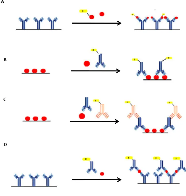
Most of the developed immunosensors are based either on a competitive or non-competitive assay, when applied to the detection of low and high molecular weight molecules, respectively (Ricci et al., 2007; Zhang et al., 2008). In a competitive immunoassay, sample analyte and labelled analyte compete for a limited number of antibody-binding sites. In electrochemical immunoassays, an enzyme label or an electroactive label is commonly used. The signal produced by the bound labelled analyte is usually inversely proportional to the amount of sample analyte. When immobilised antibodies react with the free antigens in competition with labelled antigens (Figure 4A) or, reversely, when immobilised antigens compete with free antigens for labelled free antibodies (Figure 4B), the immunoassay format is a defined as direct competitive. But when a secondary antibody is used as label after binding with the Fc region of the primary antibody, the format is denoted as indirect competitive immunoassay (Figure 4C) (Ricci et al., 2007; Ricci et al., 2012).
In a non-competitive immunoassay (also known as a two-site “sandwich” immunoassay), the sample analyte is captured by an excess of a capture antibody, separating it from the bulk sample (Figure 4D). After the interaction between immobilised antibodies and free antigens, an excess of secondary labelled antibodies, directed toward a second binding site of the antigen, is added. These labeled antibodies will only bind to the existing capture antibody–analyte complex. In an ideal non-competitive immunoassay, no signal would be produced in the absence of analyte because there are no appropriate binding sites available for the secondary labelled antibody. However, in practice, this is not the case due to non-specific interactions between the secondary antibody and other
15 components of the immunoassay. Therefore, it is always desirable to use a blocking agent to reduce these non-specific interactions. (Ricci et al., 2007; Zhang et al., 2008).
The ideal format to perform an immunoassay should be as simple as possible. Thus, a label-free direct competitive format, as shown in figure 4A, would be the ideal. However, besides simplicity other aspects, such as selectivity and sensitivity, have to be considered. Therefore, in some cases it is important to use a non-competitive assay with two antibodies, one of them monoclonal (usually the capture element), that react with the same antigen to ensure the selectivity of the assay.
Figure 4. Schematic representation of different immunoassays formats. (adapted from Ricci et al. 2007).
A
D C B
16
Cyclic voltammetry as a tool for development of electrochemical immunosensors
In electrochemical analysis involving voltammetry, a three-electrode cell configuration is used and is constituted of a working electrode (i.e. the electrode at which the reaction of interest occurs); a reference electrode (to maintain/ control the potential of the working electrode), and a counter or auxiliary electrode to carry the cell current (Cass 1990; Wang 2000). When the potential of the working electrode is maintained or shifted to a value where the oxidation or the reduction of species in solution occurs, a current flows between the solution and the electrode. With an appropriate control of the experimental conditions, the intensity of this current can be related to the analyte concentration and the applied potential can provide information about the identity of the species. Since the intensity of the measured current results from the oxidation (anodic current) or reduction (cathodic current) of chemical species in solution, voltammetric techniques can only be applied to electroactive species or non-electroactive species that are previously converted into electroactive species or coupled to faradaic processes.
The current resulting from an electrochemical reaction is called faradaic current, because it obeys Faraday's law (i.e. the reaction of one mole of electroactive substance involves a charge change of 96,487 coulombs). In non-faradaic processes no charge transfer occurs across the interface between the solution and the electrode. However, there are changes in the structure of this interface that may cause a transient flow of external currents. These two processes occur simultaneously, so the total current is the sum of the faradaic current and the non-faradaic (background) current. Thus, the most recent voltammetric techniques aim to minimize the contribution of non-faradaic processes in order to improve the signal to noise ratio (Wang 2000).
Cyclic voltammetry (CV) is a voltammetric technique widely used to generate the signal in EIs devices. CV involves sweeping the potential between two limits, E1 and E2, at a fixed scan rate. Once potential E2 is reached the scan direction is reversed whilst
maintaining the same scan rate (Figure 5 and 6). CV is a useful technique for the study of electrochemical characteristics and also for examining more complex electrochemical systems such as enzyme-coupled reactions. Bearing in mind that enzymes are common labels for immunoassays (e.g., alkaline phosphastase has been used extensively as label in immunoassays, including EIs) the employment of CV as detection technique is a good option to understand the mechanism of electron transfer at the electrode surface.
17
Figure 5. Potential-time excitation signal in a cyclic voltammetric experiment (Wang
2000).
18
Alkaline phosphatase as electrochemical enzymatic label
Alkaline phosphatase (AP) (orthophosphoric monoester phosphohydrolase) is widely used as enzyme label in immunoassays. AP has a molecular weight in the range of 80 kDa and exibiths optimum activity at alkaline pH values. Magnesium is also considered essential for maximum AP enzymatic activity. AP's role as lable in immunoassays is largely exploited because AP is easily conjugated to haptens, antibodies, and other proteins, and is the most active of all alkaline phosphatases; moreover it presents a high turnover number and broad substrate specificity (Fernández-Sánchez et al., 1998, Preechaworapun et al., 2008).
In EIs, AP is used to generate electroactive organic products, most of which can be detected and quantified. Electrochemical detectors are generally sensitive and rapid for the analysis of the product of the enzyme hydrolysis of an AP substrate. Several AP substrates, useful for electroanalysis, have been studied for the use in immunoassays, such as: catechol monophosphate (Szydlowska 2006), 3-indoyl phosphate (IP) (Martinez-Montequin et al., 2000; Gonzalez et al., 2002; Fanjul-Bolado et al., 2004; Diaz-Gonzalez et al,. 2005), hydroquinone diphosphate (HQDP) (Wilson and Rauh 2004), 4-nitrophenol phosphate (NPP) (Rosen and Rishpon 1989; Kim and Kwak 2005; Fanjul-Bolado et al., 2006), p-aminophenyl phosphate (APP) (Rosen and Rishpon 1989; Hart et al., 1997; Pemberton et al., 1999; Gyurcsanvi et al., 2002; Moore et al., 2003; Dong et al., 2006; Kwon et al., 2006), 1-naphthyl phosphate (NTP) (Pemberton et al., 1999; Chikae et al., 2007), phenyl phosphate (PheP) (Rosen and Rishpon 1989; Hart et al., 1997; Wilson and Rauh 2004; Sun et al., 2006), and 2-phospho-l-ascorbic acid (AAP) (Kokado et al., 2000; Gyurcsanyi et al,. 2002; Moore et al., 2003). During the enzymatic process, these substrates are converted to the electroactive species catechol, indigo carmine (IC), hydroquinone (HQ), 4-nitrophenol (NP), 4-aminophenol (AP), 1-naphthol (NT), phenol (Phe), and l-ascorbic acid (AA), respectively.
A new method in which AP catalyzes the deposition of metallic silver was developed by Fanjul-Bolado et al., (2007). In this method 3-indoxyl phosphate/silver (3-IP/Ag+) is used as the AP electrochemical substrate. 3-IP is basically constituted by an indolic ring substituted in position 3 by a phosphate group. In the presence of AP this substrate is hydrolyzed in position 3, giving an indoxyl intermediate. This indoxyl intermediate will oxidize, producing indigo blue, and reduce silver ions present in solution into a metallic deposit. Therefore, metallic silver is co-deposited with indigo blue. The deposited silver is electrochemically stripped into solution and the resulting current intensity is measured by anodic stripping voltammetry. The described mechanism is illustrated in Figure 7. Metallic silver will be deposited where the enzymatic AP label is attached, which is
19 especially important to avoid cross-contamination and to ensure that the measured signal correspondes to the immune-labelled reaction.
Figure 7. Mechanism of alkaline phosphatase-catalyzed silver deposition for electrochemical detection (adapted from Fanjul-Bolado et al., 2007).
Screen-printed electrodes
A wide variety of electrodes have been used as support to fabricate biosensor devices, including carbon paste electrodes (Fernández-Sánchez et al., 2000), glassy carbon electrodes (Dai et al., 2003), and gold electrodes (Ouerghi et al., 2001). Recently, several biosensor devices have been developed on screen-printed electrodes (Hernández-Santos et al., 2004; Díaz-González et al., 2005; Singh et al., 2005). The screen-printing microfabrication technology is nowadays well established for the production of thick-film electrochemical transducers (Tudorache and Bala 2007). This technology allows the mass production of reproducible, inexpensive, customized and mechanically robust solid-strip electrodes. Moreover, typical disadvantages of conventional electrodes such as memory-effects due to difficult, time-consuming and sometimes inefficient cleaning steps can be surpassed with these disposable SPEs. Another important feature that these electrodes exhibit is related to the miniaturisation of the corresponding device, along with their ease of handling and manipulation. Several SPE configurations based on different materials can be constructed. The process of production and design of SPEs consists of sequential layer-by-layer deposition of an ink onto an insulating support or substrate, which will define the geometry of the sensors (Bilitewski et al., 1992; Albareda-Sirvent et al., 2000; Mooring et al., 2005, Tudorache and Bala 2007). The substrates are commonly made of
Metalloenzymatic detection AP Ag+ Ag0 Voltammetric detection 3-IP Indigo blue


![Fig. 1 Proposed approach for the evaluation of patients with suspected celiac disease [39, 43]](https://thumb-eu.123doks.com/thumbv2/123dok_br/15717292.1070074/69.892.153.806.561.1077/fig-proposed-approach-evaluation-patients-suspected-celiac-disease.webp)
![Figure 1. Proposed algorithm of the process of clinical testing using central laboratory versus decentralized POC testing (from [1])](https://thumb-eu.123doks.com/thumbv2/123dok_br/15717292.1070074/94.892.166.685.168.630/figure-proposed-algorithm-process-clinical-testing-laboratory-decentralized.webp)