The Neuroradiology Journal 27: 85-90, 2014 - doi: 10.15274/NRJ-2014-10009 www.centauro.it
SUMMARY – Glioblastoma is a malignant infiltrative glial tumor occurring most often over 50 years of age, with diverse clinical presentations. We describe a case of temporal lobe glioblastoma with a rare presentation as an acute ischemic stroke, discussing the imaging and histopathological findings, and reviewing the literature. A 77-year-old woman had sudden onset of left hemiparesis and hemihypoesthesia. The neuroradiological studies revealed an acute ischemic lesion in the right lenticulostriate arteries territory and a right anterior temporal lobe tumor, enhancing heteroge-neously after contrast with enhancement of the right middle cerebral artery wall. Histopathological analysis of the resected temporal lesion revealed a glioblastoma multiforme with tumoral infiltra-tion of the vascular wall. Glioblastoma should be considered in the etiology of acute ischemic stroke, where neuroimaging plays an important diagnostic role, enabling a more immediate therapeutic approach, with a consequent impact on survival.
Acute Ischemic Stroke Secondary
to Glioblastoma
A Case Report
SOFIA PINA, ÂNGELO CARNEIRO, TIAGO RODRIGUES, RAQUEL SAMÕES, RICARDO TAIPA, MANUEL MELO-PIRES, CLÁUDIA PEREIRA
Serviço de Neurorradiologia, Hospital de Santo António - CHP; Porto, Portugal
Key words: glioblastoma, stroke, vasculopathy, computed tomography, magnetic resonance imaging
Introduction
There are only a few case reports 1-6 in the
literature describing acute ischemic stroke sec-ondary to brain tumors, which are not usually considered in the etiology of cerebral infarcts in clinical practice. We describe a histologi-cally proven case of vascular wall infiltration by glial tumoral cells, causing deep middle cer-ebral artery stroke.
Case Report
A 77-year-old woman, with a medical history of arterial pre-hypertension 7 and dyslipidemia
under medical control, had an acute onset of left hemiparesis and hemihypoesthesia. Ini-tially she had a brain computed tomography (CT) scan, an arterial angiographic CT study and a brain magnetic resonance (MR). The imaging studies revealed a focal right lenticu-locapsular lesion, with slight mass effect, hy-podense on CT, showing water molecules
re-stricted diffusion on MR (Figure 1). The lesion was suggestive of an acute ischemic stroke in the lenticulostriate arteries territory (perfo-rating branches of the right middle cerebral artery). There coexisted another lesion with pronounced mass effect in the ipsilateral an-terior temporal lobe (Figure 2), corticosubcor-tical, with heterogeneous enhancement, more prominent on the anterior lateral portion but extending into the mesial temporal lobe, and also enhancement of the right middle cerebral artery wall (Figure 3). The angiographic study showed a focal stenosis of the distal M1 portion of the right middle cerebral artery (MCA), as well as a reduced caliber of the right anterior cerebral artery (Figure 4). The histopathologi-cal study after anterior temporal partial lobec-tomy, performed 2.5 weeks after the clinical presentation, confirmed the suspected diagno-sis of glioblastoma with vascular wall infiltra-tion of small and medium-sized arteries, in-cluding the MCA, that were encompassed by a tumoral mass and also infiltration of leptome-ningeal vessels by glial tumoral cells (Figure
5). After surgery the patient remained with permanent left hemiplegia and immediate ad-juvant therapy was not implemented.
The follow-up brain CT obtained four weeks after surgery revealed recurrence of right
tem-poral glioblastoma, involution of lenticulocap-sular stroke to chronicity and more prominent left insular parenchymal tumoral infiltration. At this point, only palliative treatment was im-plemented.
C
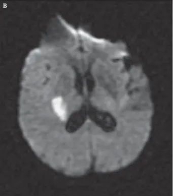
Figure 1 CT, diffusion-weighted imaging and ADC maps. Right lenticular focal hypodensity (A) with water molecules restricted diffusion (B,C).
www.centauro.it The Neuroradiology Journal 27: 85-90, 2014 - doi: 10.15274/NRJ-2014-10009
Discussion
The clinical presentation of a brain tumor as a sudden alteration of neurological status is frequently associated with intratumoral
hemor-rhage 8 or tumoral progression, and also
com-monly presents with seizure 1. Rarely the first
clinical presentation is due to an acute cerebral infarct secondary to direct vascular infiltration by the adjacent tumor.
A B
Figure 2 MR, Ax T2 TSE. Cyst-like lesion in the right lateral anterior temporal lobe, and expansion and slight hyperinten-sity on right temporal mesial structures.
Figure 3 MR, 3D T1 TFE post-gadolinium. A) Heterogene-ous enhancement in the right anterior temporal lobe (ring-like laterally and more diffuse on the mesial portion) and enhance-ment of the wall of the right middle cerebral artery. B) Focal superficial enhancement in the left insular region.
Glioblastoma is a malignant infiltrative glial cell tumor, grade IV in the World Health Or-ganization classification 9, comprising about
16% of primary brain tumors 10 and frequently
occurring in patients over 50 years 10. The
his-topathological study reveals densely compacted tumoral cells, typically with mitosis, vessels and necrosis. The immunohistochemical study shows strong reactivity to glial fibrillary acidic protein in malignant gliomas, with nearly 100% sensitivity as a glial differentiation marker 11.
Only seldom does a brain tumor lead to direct occlusion or vascular dissection causing acute cerebral infarct in the corresponding arterial territory. In these rare cases the underlying
pathophysiological mechanism is not completely clear, but various explanations include a proco-agulant effect mediated by tumor-secreting fac-tors, mechanical compression and/or tumoral cell infiltration causing occlusion or vascular dis-section 1,2. Leptomeningeal dissemination giving
rise to vasculopathy is also reported, with arte-rial vessels being encased by the tumor 12.
Clini-cally, patients present with an acute vascular syndrome and imaging studies show an acute ischemic lesion and a distinct tumoral mass that usually arises in the same brain hemisphere.
Our patient had an acute ischemic infarct in the perforating branches of the right middle cer-ebral artery territory (lenticulostriate arteries), Figure 4 Arterial angiographic CT study. Stenosis of right middle cerebral artery and narrowing of ipsilateral anterior cerebral artery.
Figure 5 Histopathology, after partial resection of right anterior temporal lobe (18th day) - glioblastoma multiforme (A-C: HE; D: GFAP). A) Highly cellular neoplasia with hyperchromatic pleomorphic nuclei and mitosis. B) Three tumoral vessels with promi-nent vascular wall proliferation. C) Tumor involving and infiltrating the leptomeningeal vessels. D) Tumor infiltrating an intrac-ortical vessel.
B D
www.centauro.it The Neuroradiology Journal 27: 85-90, 2014 - doi: 10.15274/NRJ-2014-10009
which were in close proximity to the anterior temporal lobe tumor. Brain MR with gadolin-ium disclosed enhancement of the right middle cerebral artery wall (Figure 3), and there are associated changes in the arterial angiographic CT study showing a focal stenosis of M1 seg-ment of the middle cerebral artery (Figure 4). During surgery, arterial vessels with apparent endoluminal thrombus were encased by the tu-moral mass, including the right MCA, and the histopathological findings confirmed tumoral cell infiltration of the wall of medium and small-sized arteries, as well as leptomeningeal vessels (Figure 5).
The cause of the ischemic stroke was in our case a thromboembolic effect of the tumoral cell infiltration of the vascular wall of MCA, and we do not exclude possible vascular compression by the tumoral mass. Leptomeningeal dissemina-tion is also an assumed concurrent etiological mechanism as there is a focal superficial en-hancement on the contralateral hemisphere (on the left insular region), also confirmed on the histopathological examination, and extension of glial tumors into the leptomeninges has been described before 2-4,11,12.
Chen et al. 3 described the most similar case
to ours, but they did not show the arterial wall imaging on a T1-weighted image after gadolin-ium injection, which in our case may be corre-lated with the histopathological finding of arte-rial wall infiltration by tumoral cells. This same arterial wall imaging feature was not present in the cases reported by Obeid et al. 1 and
Rojas-Marcos et al. 2, who show patients with
gliob-lastoma multiforme who developed infarcts sev-eral months after biopsy or tumoral resection and implementation of adjuvant radiotherapy/ chemotherapy. Hence, the etiology of the infarct was not explicitly related to the tumor, and ra-diotherapy is a main concurrent cause for arte-rial occlusion.
Aoki et al. 4 described a patient presenting
with a right MCA infarct and a parenchymal tu-mor in the corresponding area of the infarct ap-pearing after a three-month interval, suggest-ing the possibility of a malignant glial tumor growing in an infarcted area. There is a recog-nized association of gliosarcomas with infarcts, and it remains to be clarified if the tumoral cells first infiltrate the vessels causing infarct and then subsequent growth of tumor mass occurs 6.
The case reported by Hart et al. 5 disclosed
vascular invasion by tumoral cells in an anato-mopathological specimen but MR features were not shown.
We describe a case of glioblastoma whose initial manifestation was an acute ischemic syndrome, with imaging and histological proof of arterial wall invasion of the MCA and other smaller ves-sels, in a live patient, that would still fit the rec-ommendation criteria for treatment with radio-therapy or chemoradio-therapy, improving survival. Our assumption of the acute cerebral infarct being secondary to glioblastoma in this patient is supported by the fact that vascular risk fac-tors were under good medical control, there was no evidence of significant atherosclerosis in cervical and cerebral arteries, a cardioembolic source was lacking and the patient had not re-ceived previous radiotherapy.
The definite diagnosis is histopathological, but surgical macroscopic evaluation is also rel-evant. However, the neuroradiological studies (CT, arterial angiography and MR) disclosed the coexistence of distinct focal lesions, charac-terizing the enhancing patterns and giving the clue for suspicion of a tumoral infiltrative lesion underlying an acute cerebral infarct by direct influence on vascular structures in the vicinity. Imaging of the arterial wall with T1 post-gado-linium was an important resource.
Imaging follow-up after treatment provides information on the evolution to chronicity of the cerebral infarct, documenting the extension of tumoral resection, the progression or tumoral relapse and possible post-surgical complica-tions, contributing to the management of sur-vival expectations.
The treatment of choice is directed at the neo-plastic process and the most common therapeu-tic approaches are surgical resection, radiother-apy and chemotherradiother-apy. In these patients there is no need for administration of recombinant human tissue-type plasminogen activator, as usually indicated for strokes within three hours of the onset of symptoms 13, as it may promote
intratumoral hemorrhage.
The main concern of treatment in this case was directed to the leading cause of the symptoms, i.e., prompt treatment of the glioblastoma as soon as its diagnosis was recognized in a patient presenting with an acute ischemic syndrome.
The best treatment for glioblastomas is sur-gical resection of the tumor, combined with ra-diation therapy and chemotherapy that doubles the two-year survival rate of these patients, despite the overall prognosis remaining poor 14.
Current recommendations for the treatment of glioblastoma multiforme in elderly people ad-vise the use of hypofractionated radiotherapy or temozolomide only with deferred
radiother-apy (balancing the risk-benefit of radiotherradiother-apy towards the clinical status of the patient) 15,16.
The rarity of presentation of a glioblastoma as an acute ischemic stroke and the need for a histological examination led neurosurgeons in our case to opt for tumoral resection, delaying implementation of adjuvant therapy, ending in rapid tumor recurrence.
Another concern in this kind of patients is to be aware of the possibility of the malignant transformation of an infarct, thus requiring de-compressive craniectomy. For this, short-inter-val sequential imaging in an acute and suba-cute phase is important.
In conclusion, although rare, cerebral tumors should be considered in the etiology of acute cerebral infarct. Our case has exceptional imag-ing characterization of the infarct, tumor and imaging features pointing towards the cause of infarct, such as the arterial wall enhancement. We emphasize the relevance of MR imaging characteristics in elucidating the pathophysi-ological mechanism of stroke and tumor spread. Imaging also plays a pivotal role in the differen-tial diagnosis between stroke alone or coexist-ent stroke and tumor, allowing proper clinical management and an appropriate therapeutic approach.
References
1 Obeid M, Ulane C, Rosenfeld S. Pearls & Oy-sters: Large vessel ischemic stroke secondary to glioblas-toma multiforme. Neurology. 2010; 74 (13): e50-51. doi: 10.1212/WNL.0b013e3181d7d66a.
2 Rojas-Marcos I, Martin-Duverneuil N, Laigle-Donadey F, et al. Ischemic stroke in patients with glioblastoma multiforme. J Neurol. 2005; 252 (4): 488-489. doi: 10.1007/s00415-005-0665-7.
3 Chen H, Cebula H, Schott R, et al. Glioblastoma multi-forme presenting with ischemic stroke: case report and review of the literature. J Neuroradiol. 2011; 38 (5): 304-307. doi: 10.1016/j.neurad.2011.01.008.
4 Aoki N, Sakai T, Oikawa A, et al. Dissection of the middle cerebral artery caused by invasion of malig-nant glioma presenting as acute onset of hemiplegia. Acta Neurochir (Wien). 1999; 141 (9): 1005-1008. doi: 10.1007/s007010050408.
5 Hart MN, Byer JA. Rupture of middle cerebral artery branches by invasive astrocytoma. Neurology. 1974; 24: 1171-1174. doi: 10.1212/WNL.24.12.1171.
6 Züchner S, Kawohl W, Sellhaus B, et al. A case of gliosarcoma appearing as ischaemic stroke. J Neu-rol Neurosurg Psychiatry. 2003; 74 (3): 364-366. doi: 10.1136/jnnp.74.3.364.
7 Chobanian AV, Bakris GL, Black HR, et al. The Sev-enth Report of the Joint National Committee on Pre-vention, Detection, Evaluation, and Treatment of High Blood Pressure: the JNC 7 report. JAMA. 2003; 289 (19): 2560-2572. doi: 10.1001/jama.289.19.2560. 8 Kondziolka D, Berstein M, Resh L, et al. Significance
of hemorrhage into brain tumors: clinicopathological study. J Neurosurg. 1987; 67 (6): 852-857. doi: 10.3171/ jns.1987.67.6.0852.
9 Louis DN, Ohgaki H, Wiestler OD, et al. The 2007 WHO classification of tumours of the central nervous system. Acta Neuropathol. 2007; 114 (2): 97-109. doi: 10.1007/s00401-007-0243-4.
10 Dolecek TA, Propp JM, Stroup NE, et al. CBTRUS statistical report: primary brain and central nerv-ous system tumors diagnosed in the United States in 2005-2009. Neuro Oncol. 2012; 14 Suppl 5: v1-49. doi: 10.1093/neuonc/nos218.
11 Brat DJ, Prayson RA, Ryken TC, et al. Diagnosis of malignant glioma: role of neuropathology. J Neuroon-col. 2008; 89 (3): 287-311. doi: 10.1007/s11060-008-9618-1.
12 Herman C, Kupsky WJ, Rogers L, et al. Leptomenin-geal dissemination of malignant glioma simulating cerebral vasculitis. Case report with angiographic and
pathological studies. Stroke. 1995; 26 (12): 2366-2370. doi: 10.1161/01.STR.26.12.2366.
13 Tissue plasminogen activator for acute ischemic stroke. The National Institute of Neurological Disorders and Stroke rt-PA Stroke Study Group. 1995; 333 (24): 1581-1587. doi: 10.1056/NEJM199512143332401.
14 Omuro A, DeAngelis LM. Glioblastoma and other ma-lignant gliomas: a clinical review. JAMA. 2013; 310 (17): 1842-1850. doi: 10.1001/jama.2013.280319. 15 Mason WP, Maestro RD, Eisenstat D, et al. Canadian
recommendations for the treatment of glioblastoma multiforme. Curr Oncol. 2007; 14 (3): 110-117. doi: 10.3747/co.2007.119.
16 Chamberlain MC. Treatment of newly diagnosed ma-lignant glioma in the elderly people: new trials that impact therapy. Int J Clin Pract. 2013; 67 (12): 1225-1227. doi: 10.1111/ijcp.12258.
Sofia Pina, MD
Serviço de Neurorradiologia
Hospital de Santo António - Centro Hospitalar do Porto Largo Prof. Abel Salazar
4099-001 Porto, Portugal Tel.: +351962502494 E-mail: [email protected]