Analyzing the neuropsychological characteristics and
changes in serum markers of patients with chronic cerebral
circulation insufficiency
JIANHUA TANG1, YUQING ZHEN1, LING YU1, CUI LV1, JUAN ZHENG1, HUI LIANG1*
1Department of Neurology, Yantaishan Hospital, Yantai, China
SUMMARY
Study conducted at the Department of Neurology, Yantaishan Hospital,
Yantai, China
Article received: 5/23/2017
Accepted for publication: 6/16/2017
*Correspondence:
Department of Neurology, Yantaishan Hospital Address: No. 91 Jiefang Road Yantai, Shandong – China Postal code: 264401 lianghui_aikan@163.com
http://dx.doi.org/10.1590/1806-9282.64.01.41
Objective: To investigate the neuropsychological characteristics and changes in
CRP, S100B, MBP, HSP-7, and NSE in serum.
Method: Sixty-six (66) patients treated in our hospital as CCCI group were chosen
for our study, and 90 patients with depression were selected as the depression group. The patients in both groups were examined with CT perfusion, depression, anxiety and cognition evaluation. Their serum CRP, S100B, MBP, HSP-70 and NSE levels were detected. Neuropsychological and serum markers characteristics were compared.
Results: The CBF and CBV in bilateral basal ganglia, frontal lobes, greater oval
center, brain stem, and left and right regions of occipital lobes of the patients in CCCI group were significantly lower than in the depression group. The HAMD and HAMA scores of CCCI group patients were significantly lower than in the depression group; CCCI group performed better regarding attention, memory, abstract terms and delayed recall. CCCI also had significantly higher total scores than the depression group. Serum CRP, S100B, MBP, HSP-70 and NSE levels in CCCI group were significantly higher than in the depression group. The differences reach statistical significance (p<0.05).
Conclusion: CCCI patients who are accompanied by minor depressive disorder
have different degrees of cognitive impairment and experience a significant rise in serum CRP, S100B, MBP, HSP-70 and NSE.
Keywords: Neuropsychology. Biomarkers. Cerebrovascular Circulation.
I
NTRODUCTIONChronic cerebral circulation insufficiency (CCCI) is cere-bral vascular stenosis or hypoperfusion in patients induced by multiple factors. It can cause the cerebral blood flow to be incapable of satisfying the basic physiological de-mands.1-3 Its existence has been one of the most contro-versial issues in this field because this disease has no spe-cific clinical manifestations and there is no reliable diagnosis method for it.4-6 However, a large number of clinical experiences have demonstrated that such patients often experience the clinical manifestations of repeated dizziness and head heaviness, which are accompanied by emotional abnormalities such as anxiety-depression of varying degrees, with a very high possibility of acute stroke and cognitive impairment. During recent years, in order to find an accurate method to diagnose CCCI, the chang-es in the level of serum markers have drawn wide attention
from scholars.7,8 Therefore, our study reviewed the CCCI patients admitted in our hospital from April 2013 to April 2015 and analyzed their psychological characteristics as well as serum markers.
M
ETHODGeneral materials
with depression who sought our hospital during the same period were selected and included as a control group, with the clinical manifestations of similar dizziness and vertigo. The control group had 25 males and 20 females aged 61 to 78 years with a mean of 67.32±4.27 years.
Study methods
CT spiral perfusion examination: all patients underwent cerebral CT perfusion imaging examination within one week after their admissions. The examination was per-formed with a 64-layer spiral CT machine (Brilliance from Phillips Company). Using the canthomeatal line as the baseline, the conventional cross section scan with a layer thickness of 5 mm and a layer spacing of 5 mm was first performed, and then CT perfusion imaging was performed with basal ganglia plane as the center and a coverage area of 40 mm (layer thickness 5 mm × 8 layers). By means of high-pressure syringe, a bolus injection of 50 mL non-ion contrast medium (ultravist 300 mg/mL) was performed at the rate of 4 mm/s via the hand dorsum vein. In the meantime, dynamic scan (matrix 512 × 512, scan field 24 × 24 mm, tube voltage 120 kV, tube current 80 mA) was performed continuously for 50 seconds, the re-ac-quired 152 dynamic images were transmitted to the workstation, and Brain perfusion software was used for further processing. The planes at the basal ganglia and corona radiata areas of the CCCI patients were selected as the planes of interest, and a method of hand drawing of the region of interest (ROI) was adopted to observe the following regions: bilateral basal ganglia, frontal lobes, great-er oval centgreat-er, brain stem and occipital lobes. Parametgreat-ers such as cerebral blood flow, cerebral blood volume, mean transit time (MTT) and time-to-peak (TTP) were measured. Generally, each site was measured for three times to ensure that the size of ROI selected each time was reasonably consistent. Both qualitative and quantitative methods were adopted to evaluate the computed tomography per-fusion imaging (CTPI) parameters, i.e. a qualitative eval-uation on whether the left and right cerebral perfusion was symmetric on the TTP and MTT pseudocolor im-ages: with the measured values in the control group as the yardstick, if the absolute value of CTPI parameters of each ROI in CCCI group exceeded 95% confidence inter-val of the absolute inter-values in the control group, that was considered as perfusion abnormality.
In the next morning following admission, 5 mL of venous fasting blood were extracted as detection samples, then they were centrifuged, and finally the supernatant was saved and stored at -80oC. Radioimmune
turbidimet-(CRP) levels of the patients in the two groups after admis-sion and after the treatment, and the enzyme linked im-munosorbent assay was used to determine the S100 cal-cium-binding protein B (S100B), myelin basic protein (MBP), heat shock protein 70 (HSP-70) and neuron-spe-cific enolase (NSE) contents in plasma.
Grade indexes
Status evaluation: the Hamilton’s Depression Scale (HAMD) and Anxiety Scale (HAMA) were used to evaluate the patient’s depression and anxiety status after admis-sion and after treatment.
Cognitive function evaluation: the Montreal Cogni-tive Assessment (MoCA) scale was used to determine the patient’s cognitive functions after admission and after treatment, the scale is mainly used to explore eight aspects of cognitive functions, such as visual space, executive capacity, naming, ability to concentrate mentally and language. The test generally lasts 8 to 10 minutes. A high-er score means stronghigh-er cognitive abilities.
Statistical methods
SPSS 15 software was used for data processing, the enu-meration data were shown in the form of absolute numbers and frequencies, t and χ2 methods were used for the test. p<0.05 means the difference was of statistical significance.
R
ESULTSCT perfusion parameters in the left and right encephalic regions As is shown in Figure 1, the cerebral blood flow (CBF) and
cerebral blood volume (CBV) in bilateral basal ganglia, frontal lobes, greater oval center, brain stem, and left and right regions of occipital lobes of the patients in CCCI group were significantly lower than those seen in the depression group. The comparison generated a significant difference (p<0.05 or p<0.01), and the inter-group com-parison did not generate an obvious difference in MTT and TTP (p>0.05).
Depression and anxiety evaluation
FIGURE 1 Comparison between CCCI group and depression group in terms of the patient’s perfusion parameters of left and right encephalic regions when compared with the depression group. *p<0.05; #p<0.01.
CCCI: chronic cerebral circulation insufficiency; CBF: cerebral blood flow; CBV: cerebral blood volume; MTT: mean transit time; TTP: time-to-peak.
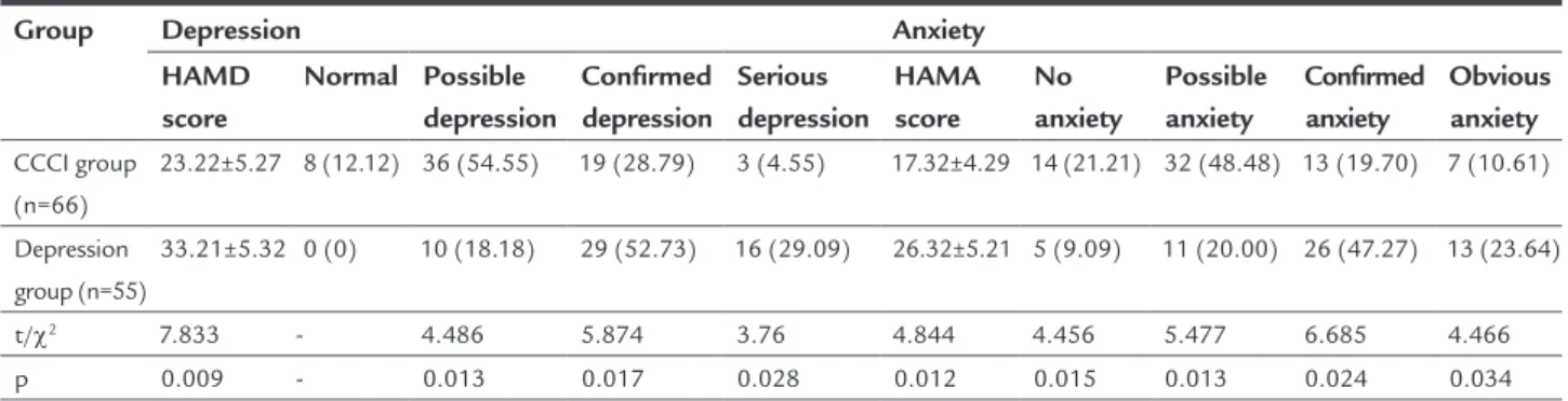
TABLE 1 Comparison between CCCI group and depression group in terms of anxiety-depression.
Group Depression Anxiety
HAMD score
Normal Possible depression
Confirmed depression
Serious depression
HAMA score
No anxiety
Possible anxiety
Confirmed anxiety
Obvious anxiety CCCI group
(n=66)
23.22±5.27 8 (12.12) 36 (54.55) 19 (28.79) 3 (4.55) 17.32±4.29 14 (21.21) 32 (48.48) 13 (19.70) 7 (10.61)
Depression group (n=55)
33.21±5.32 0 (0) 10 (18.18) 29 (52.73) 16 (29.09) 26.32±5.21 5 (9.09) 11 (20.00) 26 (47.27) 13 (23.64)
t/χ2 7.833 - 4.486 5.874 3.76 4.844 4.456 5.477 6.685 4.466 p 0.009 - 0.013 0.017 0.028 0.012 0.015 0.013 0.024 0.034
CCCI: chronic cerebral circulation insufficiency; HAMD: Hamilton’s Depression Scale; HAMA: Anxiety Scale (HAMA).
Frontal lobe
Frontal lobe
Frontal lobe
Frontal lobe Occipital
lobe 90
80 70 60 50 40 30 20 10 0
160
140
120
100
80
60
40
20
0 80 70 60 50 40 30 20 10 0
30
25
20
15
10
5
0
Occipital lobe
A B
C D
Occipital lobe
Occipital lobe Greater
oval center
Greater oval center
Greater oval center
Greater oval center Brain
stem
Brain stem
Brain stem CCCI group (left) CCCI group (right) Depression group (left) Depression group (right)
Brain stem Bilateral
basal ganglia
CBF
MTT TTP
C
BV
Bilateral basal ganglia
Bilateral basal ganglia
Bilateral basal ganglia #
#
#
# #
#
# # # # #
# #
#
# #
* *
Cognitive evaluation
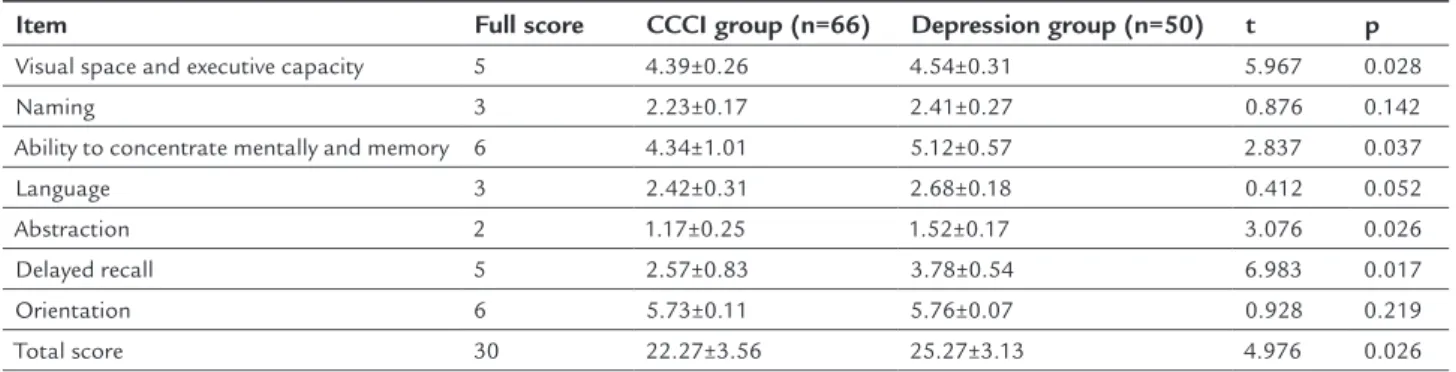
As shown in Table 2, the scores for visual space, executive capacity, ability to focus, memory, abstraction ability, de-layed recall and the total scores of the patients in the CCCI group were significantly lower than those in the depres-sion group, and the comparison generated a significant difference (p<0.05).
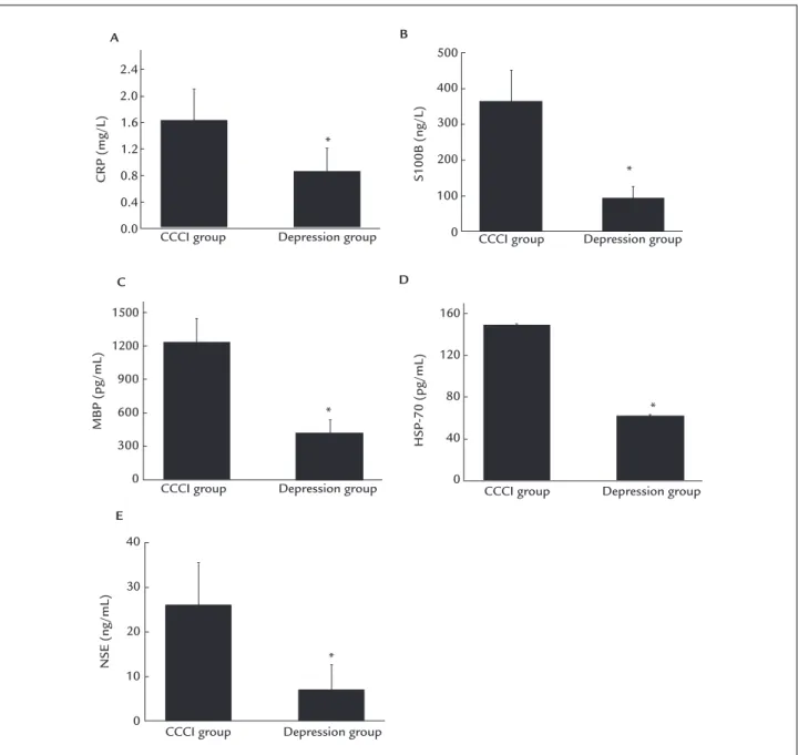
Plasma CRP, S100B, MBP, HSP-70 and NSE levels
As shown in Figure 2, the levels of CRP, S100B, MBP, HSP-70 and NSE in serum of the patients in the CCCI group were significantly higher than those of the depression group, and the comparison generated a significant dif-ference (p<0.05).
D
ISCUSSIONIn 1990, a scholar from Japan first proposed the concept of chronic cerebral circulation insufficiency, which refers to a phenomenon of overall blood flow decrease that oc-curs in the brain instead of the focal ischemic lesions.5 Studies have revealed that the primary reason for CCCI occurrence is as follows: atherosclerosis leads to the oc-currence of vascular plaques and stenosis, so that the cerebral blood flow decreases. When it decreases to a cer-tain threshold value, the local perfusion will also decrease, causing clinical manifestations such as dizziness and head heaviness, which are usually considered to be the early manifestations of cerebral infarction.9-12 As there is no specific manifestation for CCCI clinical symptoms and imaging characteristics, early accurate diagnosis is one of the difficulties in the treatment of this disease. Currently, domestic and foreign studies show that the early manifes-tations of many patients with chronic cerebral functional insufficiency are experiencing an increase in memory loss and emotional disorders, which is similar to neurosis such as depression and accompanied by mild cognitive
disor-der. Therefore, the studies on the neuropsychological characteristics of CCCI patients have drawn wide attention from scholars during the recent years.13
The results of our study revealed that 87.88% of the patients in the CCCI group have anxiety issues and 78.78% of the patients concomitantly suffer from the psycho-logical conditions of depression. Comparison between the CCCI group and the depression patients shows that the proportion of patients with possible depression and anx-iety in the CCCI group was significantly higher than that of the depression group, but the proportion of patients with confirmed depression and anxiety and obvious de-pression and anxiety was significantly lower than that of the depression group. It is also found that the HAMD and HAMA scores of the patients in the CCCI group were significantly lower than those seen in the depression group. The results indicated that the majority of the CCCI patients concomitantly suffer from depression, but the depression is mild and there is no specific report on the incidence rate of depression among CCCI patients. Secondly, in our study, the MoCA scale was used to evaluate the cognitive func-tions of the patients in the two groups. The results indi-cated that the scores for visual space, executive capacity, ability to focus attention, memory, abstraction and delayed recall and the total scores of the patients in the CCCI group were significantly lower than those in the depres-sion group. The result also indicated that the cognitive functions of the patients in the CCCI patients decreased significantly. The decrease in the cognitive functions of the patients in the CCCI patients has drawn wide atten-tion. Animal experiment studies showed that chronic cerebral ischemia will cause serious damages to the neurons of hippocampal CA1 area of rats while the hippocampal neurons are the key links that influence the cognitive abilities such as memory and learning, which can prop-erly explain the decrease in the cognitive functions of the
TABLE 2 Comparison between CCCI group and depression group in terms of MoCA score.
Item Full score CCCI group (n=66) Depression group (n=50) t p Visual space and executive capacity 5 4.39±0.26 4.54±0.31 5.967 0.028
Naming 3 2.23±0.17 2.41±0.27 0.876 0.142
Ability to concentrate mentally and memory 6 4.34±1.01 5.12±0.57 2.837 0.037
Language 3 2.42±0.31 2.68±0.18 0.412 0.052
Abstraction 2 1.17±0.25 1.52±0.17 3.076 0.026
Delayed recall 5 2.57±0.83 3.78±0.54 6.983 0.017
Orientation 6 5.73±0.11 5.76±0.07 0.928 0.219
Total score 30 22.27±3.56 25.27±3.13 4.976 0.026
FIGURE 2 Comparison the levels of CRP, S100B, MBP, HSP-70 and NSE between the CCCI group and depression group. Data are presented as mean + SD. *p<0.05 versus CCCI group.
CCCI: chronic cerebral circulation insufficiency; CRP: C-reactive protein; MBP: myelin basic protein; HSP-70: heat shock protein 70; NSE: neuron-specific enolase.
CCCI patients. Studies revealed that basal nuclei areas and greater oval centers are rich in a large number of neurons and fibers closely associated with cognitive functions such as learning and memory.14-17 By means of multi-layer spi-ral CT examination, our study found that the CBF and CBV in bilateral basal ganglia, frontal lobes, greater oval center, brain stem, and left and right regions of occipital lobes of the patients in CCCI group were significantly lower than those seen in the depression group, which leads to decrease in the cognitive functions of the CCCI patients.
Change in blood markers is also one of the most im-portant manifestations of chronic cerebral ischemic dis-eases.18,19 In our study, the research of all indices in blood indicated that CRP, S100B, MBP, HSP-70 and NSE in the blood of patients in the CCCI group had a significant increase compared with the depression group. CRP level is one of the commonest markers for body inflammatory reactions during the clinical application. A large number of studies revealed that increased levels of CRP in plasma of patients with chronic cerebral ischemia are closely
as-CCCI group
A B
C D
E
CCCI group
CCCI group
CCCI group 2.4
2.0 1.6 1.2 0.8 0.4 0.0
40
30
20
10
0 1500
1200
900
600
300
0
500
400
300
200
100
0
160
120
80
40
0
CCCI group Depression group
Depression group
Depression group
S100B (ng/L)
CRP (mg/L)
MBP (pg/mL)
HSP-70 (pg/mL)
NSE (ng/mL)
Depression group *
* *
*
Depression group
sociated with the degree of injuries in brain cells. And it can further aggravate the injuries of cerebral vascular endothelial cells and aggravate patient conditions.20 NSE is a type of specific enolase present in the cerebral neurons and endocrine cells and participates in the glycolysis process. Cerebral blood supply insufficiency will lead to functional disorder and structural injuries in neuron serous membrane. As a result, NSE will be released from the damaged cell membrane and enter the cerebrospinal fluid, passing through the disrupted blood-brain barrier and entering the blood circulation, further leading to an increase in the level of NSE in blood. S100B and MBP are present in the colloid and oligodendrocytes in the central nervous system. Their abundance can somewhat reflect the degree of damages of colloid and oligodendrocytes and is closely associated with the damages in cognitive functions.21 NSE, S100B and MBP are often used to eval-uate whether the damages occur to neurons, colloid and oligodendrocytes in the cerebral tissues. They can also be used to evaluate severity because they are important serum markers of cognitive function. Studies revealed that HSP-70 is a type of stress protein closely associated with patient cognitive functions, and HSP-70 can improve neurocyte tolerance and activate the anti-apoptosis route under the stress status so that the damaged neurocytes in the brain can be protected. Therefore, in the case of cerebral blood supply insufficiency in the human body, the HSP-20 level will be greatly increased to lower the neurocyte in-juries.17 As shown in the above results, in the case of the occurrence of CCCI, the CRP, S100B, MBP, HSP-70 and NSE will experience significant changes, and these can be used as important indices for the diagnosis of this disease. To sum up, CCCI patients often concomitantly suffer from mild depression and cognitive injuries of varying degrees, the CRP, S100B, MBP, HSP-70 and NSE in serum will increase significantly, and the diagnosis of CCCI can be made according to the neuropsychological character-istics and the changes in serum markers.
R
EFERENCES1. Wu C, Liao L, Yan X, Li M, Wu S, Wang J, et al.; Yangxue Qingnao Granule Chronic Cerebral Hypoperfusion Study Group. Effects of Yangxue Qingnao granules on chronic cerebral circulation insufficiency: a randomized, double-blind, double-dummy, controlled multicentre trial. Psychogeriatrics. 2013; 13(1):29-34.
2. Starosel’tseva NG. Neurophysiological studies of chronic cerebral ischemia. Neurosci Behav Physiol. 2009; 39(6):605-11.
3. Kufner A, Galinovic I, Ambrosi V, Nolte CH, Endres M, Fiebach JB, et al. Hyperintense vessels on FLAIR: hemodynamic correlates and response to thrombolysis. AJNR Am J Neuroradiol. 2015; 36(8):1426-30.
4. Pancucci G, Potts MB, Rodríguez-Hernández A, Andrade H, Guo L, Lawton MT. Rescue bypass for revascularization after ischemic complications in the treatment of giant or complex intracranial aneurysms. World Neurosurg. 2015; 83(6):912-20.
5. Starodubtsev VB, Bakharev AV, Stoliarov MS, Al’sov SA, Amelin ME, Vinogradova TE, et al. [Role of multispiral CT angiography in diagnosis and treatment of patients with chronic cerebral circulatory insufficiency]. Angiol Sosud Khir. 2008; 14(3):39-43.
6. Jung DK, Devuyst G, Maeder P, Bogousslavsky J. Atrial fibrillation with small subcortical infarcts. J Neurol Neurosurg Psychiatry. 2001; 70(3):344-9. 7. Zhang L, Dong W, Han J, Wang Z, Sun D, Ji X, et al. Montreal cognitive assessment
and analysis of related factors for cognitive impairment in patients with chronic cerebral circulation insufficiency. Int J Psychiatry Med. 2015; 50(3):257-70. 8. Tripathi M, Tripathi M, Sharma R, Jaimini A, Md’souza M, Saw S, et al.
Functional neuroimaging using F-18 FDG PET /CT in amnestic mild cognitive impairment: a preliminary study. Indian J Nucl Med. 2013; 28(3):129-33. 9. Gregg NM, Kim AE, Gurol ME, Lopez OL, Aizenstein HJ, Price JC, et al.
Incidental cerebral microbleeds and cerebral blood flow in elderly individuals. JAMA Neurol. 2015; 72(9):1021-8.
10. Medow MS, Sood S, Messer Z, Dzogbeta S, Terilli C, Stewart JM. Phenylephrine alteration of cerebral blood flow during orthostasis: effect on n-back performance in chronic fatigue syndrome. J Appl Physiol (1985). 2014; 117(10):1157-64.
11. Haratz S, Weinstein G, Molshazki N, Beeri MS, Ravona-Springer R, Marzeliak O, et al. Impaired cerebral hemodynamics and cognitive performance in patients with atherothrombotic disease. J Alzheimers Dis. 2015; 46(1):137-44. 12. Hartung EA, Laney N, Kim JY, Ruebner RL, Detre JA, Liu HS, et al. Design
and methods of the NiCK study: neurocognitive assessment and magnetic resonance imaging analysis of children and young adults with chronic kidney disease. BMC Nephrol. 2015; 16:66.
13. Lestou V, Lam JM, Humphreys K, Kourtzi Z, Humphreys GW. A dorsal visual route necessary for global form perception: evidence from neuropsychological fMRI. J Cogn Neurosci. 2014; 26(3):621-34.
14. Altamura C, Ventriglia M, Martini MG, Montesano D, Errante Y, Piscitelli F, et al. Elevation of plasma 2-arachidonoylglycerol levels in Alzheimer’s disease patients as a potential protective mechanism against neurodegenerative decline. J Alzheimers Dis. 2015; 46(2):497-506. 15. Isshiki R, Kobayashi S, Iwagami M, Tsutumi D, Mochida Y, Ishioka K, et al.
Cerebral blood flow in patients with peritoneal dialysis by an easy Z-score imaging system for brain perfusion single-photon emission tomography. Ther Apher Dial. 2014; 18(3):291-6.
16. Liang Y, Chu P, Wang X. Health-related quality of life of Chinese earthquake survivors: a case study of five hard-hit disaster counties in Sichuan. Social Indicators Research. 2014; 119(2):943-66.
17. Hermann DM, Kribben A, Bruck H. Cognitive impairment in chronic kidney disease: clinical findings, risk factors and consequences for patient care. J Neural Transm (Vienna). 2014; 121(6):627-32.
18. Liang Y, Guo M. Utilization of health services and health-related quality of life research of rural-to-urban migrants in China: a cross-sectional analysis. Social Indicators Research. 2015; 120(1):277-95.
19. Boyer L, Testart J, Michel P, Richieri R, Faget-Agius C, Vanoye V, et al. Neurophysiological correlates of metabolic syndrome and cognitive impairment in schizophrenia: a structural equation modeling approach. Psychoneuroendocrinology. 2014; 50:95-105.
20. Zhang L, Zhang J, Sun H, Zhu H, Liu H, Yang Y. An enriched environment elevates corticosteroid receptor levels in the hippocampus and restores cognitive function in a rat model of chronic cerebral hypoperfusion. Pharmacol Biochem Behav. 2013; 103(4):693-700.