OR
IGI
N
A
L
R
E
S
E
A
R
C
H
Corresponding address: Júlia Risso Parisi – Av. Jovino Fernandes Sales 2600, Alfenas, MG, Brazil – Zip Code: 37130-000 – Phone: (19) 98165-3991 – E-mail: [email protected] – Finance source: [Universidade Federal de Alfenas – UNIFAL-MG (PROBIC; Scientiic Initiation Scholarship Program fellowship LMSE); Coordenação de Aperfeiçoamento de Pessoal de Nível Superior (CAPES; Master’s Level Fellowship JRP) and by Fundação de Amparo à Pesquisa de Minas Gerais (FAPEMIG)] – Conlict of interests: nothing to declare. – Presentation:Aug. 3, 2016 – Accepted for publication: July 7, 2017 – Approved by the [Ethics Committee on Animal Use (CEUA protocol no. 591/2014)].
Study developed at the Universidade Federal de Alfenas (UNIFAL) – Alfenas (MG), Brazil. 1Physical Therapy Student, Universidade Federal de Alfenas – Alfenas (MG), Brazil.
2Physical therapist, Master of Science in Biosciences Applied to Health (PPGB), Universidade Federal de Alfenas – Alfenas (MG), Brazil. 3Physical therapist, PhD, professor in the Undergraduate Program in Physical Therapy and professor and advisor in the Master’s in Sciences and PhD in Biosciences Applied to Health (PPGB) Universidade Federal de Alfenas – Alfenas (MG), Brazil.
ABSTRACT | Although transcutaneous electrical nerve stimulation (TENS) has been proposed to modulate pain and the mechanisms underlying analgesia remain poorly understood, evidence of anti-inflammatory effect is more limited. The purpose of this study was to examine the opioidergic mechanisms of TENS effects in two different frequencies on pain and inflammatory edema in the ankle sprain model in rats. Threshold to mechanical stimulation was utilized to examine the changes produced by intraperitoneal injection of non-selective opioid antagonist naloxone on the antihyperalgesic effect induced by a 20-min period of 2Hz or 100Hz TENS in the ankle sprain model, produced by manually overextending the lateral ligaments. Ankle sprain induced a long-lasting reduction in paw withdrawn latency (PWL) after 30 minutes for up to 24 hours in sham TENS (SH-TENS) treated rats. The reduced PWL after the induction of ankle sprain was restored partially at 0,1,2,3 and 6, but not 24 hours, after the termination of 2 Hz-TENS (LF-Hz-TENS). In 100Hz (HF-Hz-TENS) the reduction in PWL was shorter than LF-TENS and both LF and HF effects were fully blocked in naloxone-treated rats. LF- and HF-TENS treated rats did not reach the elevation of edema and presented a progressive edema reduction for over 24 hours when compared to
288
SH-TENS group. Both effects were reduced by naloxone. TENS-induced antihyperalgesic and anti-edematous effects observed in ankle sprain model were mediated by the endogenous opioid system.
Keywords | Transcutaneous Electric Nerve Stimulation; Ankle Injuries; Pain, Inlammation.
RESUMO | Embora estimulação elétrica nervosa transcutânea (TENS) tem sido proposta para modular a dor e os mecanismos subjacentes a analgesia permanecem mal compreendidos, evidências do efeito anti-inflamatório são mais limitadas. O objetivo deste estudo foi examinar os mecanismos opioidérgicos de efeitos de TENS em duas frequências diferentes sobre dor e edema inflamatório no modelo de entorse de tornozelo em ratos. Limiar de estimulação mecânica foi utilizado para examinar as alterações produzidas pela injeção intraperitoneal de naloxona, um antagonista opioide não-seletivo, sobre o efeito anti-hiperalgésico induzido por um período de 20 min de 2Hz ou 100Hz de TENS no modelo de entorse de tornozelo, produzido ultrapassando manualmente os ligamentos laterais. Entorse de tornozelo induziu uma redução duradoura na latência de retirada da pata (PWL) depois de 30 minutos até 24 horas em ratos tratados para TENS “simulada” (SH-TENS). A PWL reduzida após a indução
Opioidergic efects of transcutaneous electrical
nerve stimulation on pain and inlammatory edema
in a rat model of ankle sprain
Efeitos de opioidérgico da estimulação elétrica nervosa transcutânea sobre a dor e edema
inlamatório em um modelo de entorse de tornozelo em ratos
Efectos opioidergicos de la estimulación nerviosa eléctrica transcutánea sobre el dolor y el
edema inlamatorio en un modelo de rata con esguince de tobillo
de entorse de tornozelo foi restaurada parcialmente em 0,1,2,3 e 6, mas não em 24 horas, após o término do 2 Hz-TENS (LF-TENS). Em 100Hz (HF-TENS) a redução de PWL foi menor do que LF-TENS e tanto os efeitos HF e LF foram totalmente bloqueados em ratos tratados com naloxona. Ratos tratados com LF- e HF-TENS não alcançou a elevação do edema e apresentaram uma redução progressiva do edema por mais de 24 horas, quando comparado ao grupo SH-TENS. Ambos os efeitos foram reduzidos pela naloxona. Efeitos anti-hiperalgésicos induzidos por TENS e efeitos antiedematosos observados no modelo de entorse de tornozelo foram mediados pelo sistema de opioides endógenos.
Descritores | Estimulação Elétrica Nervosa Transcutânea; Traumatismos do Tornozelo; Dor, Inlamação.
RESUMEN | Aunque la estimulación nerviosa eléctrica transcutánea (TENS) ha sido propuesta para modular el dolor y los mecanismos subyacentes a la analgesia sigue siendo mal entendida, la evidencia del efecto antiinflamatorio es limitada. El propósito de este estudio fue examinar los mecanismos opioidérgicos de los efectos de la TENS en dos frecuencias diferentes sobre el dolor y el edema inflamatorio en un modelo de ratas con esguince de tobillo. Se utilizó el umbral a la estimulación mecánica para examinar los cambios producidos
por inyección intraperitoneal del antagonista opiáceo no selectivo naloxona sobre el efecto antihiperalgésico inducido por un período de 20 minutos de 2Hz o 100Hz TENS en el modelo con esguince de tobillo, producido por sobrecarga manual de los ligamentos laterales. El esguince de tobillo indujo una reducción de larga duración en latencia de la pata retraída (PWL) después de 30 minutos por hasta 24 horas en simulación de la TENS (SH-TENS) para las ratas tratadas. El PWL reducido después de la inducción del esguince de tobillo fue restaurado parcialmente en 0,1,2,3 y 6, pero no por 24 horas, después de la terminación de 2 Hz-TENS (LF-TENS). La reducción en PWL fue menor que LF-TENS en 100Hz (HF-TENS) y tanto los efectos de LF como de HF fueron completamente bloqueados en ratas tratadas con naloxona. Las ratas tratadas con LF- y HF-TENS no alcanzaron la elevación del edema y presentaron una reducción progresiva del edema durante más de 24 horas en comparación con el grupo SH-TENS. Ambos efectos fueron reducidos por la naloxona. Efectos antihiperalgésicos y antiedematosos TENS-inducidos observados en el modelo con esguince de tobillo fueron mediados por el sistema opioide endógeno.
Palabras clave | Estimulación Eléctrica Transcutánea del Nervio; Traumatismos del Tobillo; Dolor; Inlamación.
INTRODUCTION
Transcutaneous electrical nerve stimulation (TENS)
is a noninvasive treatment commonly used to manage
pain. While strongly supporting an analgesic efect on
pain thresholds
1-5, evidence of anti-inlammatory efect
of TENS is more limited.
Two diferent theories have been proposed to explain
TENS-induced analgesia. First, the gate control theory
of pain
6,7proposed that the stimulation of large-diameter
aferent ibers inhibits second order neurons in the
dorsal horn and prevents pain impulses carried by
small-diameter ibers from reaching higher brain centers.
Second, TENS activates pain inhibitory pathways
stimulating the release of endogenous opioids
8and
serotonin
9. Endogenous opioid peptides such as
beta-endorphin activate opioid receptors both at the level of
the spinal cord
10,11and on peripheral sensory neurons at
the site of inlammation
12,13. In inlammatory pain model
in rats serotonin also contributes to TENS-induced
analgesia via spinal 5-HT
2Aand 5-HT
3,but not
5-HT
1Areceptors and 5-HT
3receptors involves
GABAergic, enkephalinergic, and other classes of spinal
intrinsic neurons related to gate control and descendent
pain inhibitory pathways
14.
he spinal cord is also involved in the modulation
of peripheral inlammatory edema
15,16. he spinal
segmental modulation of dorsal root relex generation
seems to be the main responsible for peripheral control
of the neurogenic component of inlammation
17.
Recently, it was demonstrated that TENS could
suppress the spinal release of substance P and
proinlammatory cytokines
18. Although, TENS has
proved to be an efective therapy against several pain
conditions, few studies have shown that TENS can
modulate or suppress the inlammatory edema.
METHODOLOGY
Animals
he experiments were conducted using male Wistar
rats (200-250g) from the main animal house of the
Universidade Federal de Alfenas (Unifal-MG). Animals
were housed at a controlled temperature (24±2°C) and
on a 12-hour light-dark cycle (dark cycle beginning at
7 am), and they had free access to food and water. he
experiments were approved by Ethics Committee (CEUA –
Unifal-MG, protocol 591/2014).
Procedure for ankle sprain
he rats were anesthetized with isolurane vaporized in
air (3% for induction and 2.0% for maintenance). Ankle
sprain was produced by manually overextending the lateral
ligaments by the same person, without breaking them, to
imitate a lateral ankle sprain in a human
19,20. Anesthesia
was discontinued and the rats recovered from anesthesia
within 5-10 minutes. A single operator performed this
procedure to guarantee the same strength of the procedures.
Inlammatory edema
Paw edema was measured with a plethysmometer
(Model 7140, Ugo Basile, Rome, Italy). Briely, the paw is
inserted into water, contained in a special water cell whose
resistance is changed due to the immersion of the animals’
paws. his resistance change is calibrated in ml and shown
on the electronic monitor
21. After determination of the
basal volume, the animals (n=6
per
group) were divided into
experimental groups in such a way that the mean volumes
of the diferent groups were similar. he paw volume was
measured 0, 1, 2, 3, 6 and 24 h after TENS or sham treatment.
Paw mechanical sensitivity
Mechanical sensitivity was measured using an
electronic von Frey device (Insight Equipamentos,
Ribeirão Preto, SP, Brazil). Briely, a pressure-meter which
consisted of a hand-held force transducer itted with a
0.5mm
2polypropylene tip was applied perpendicularly
to the central area of the hindpaw with a gradual increase
in pressure
22. he corresponding force was recorded (in
grams). he smaller the force applied for inducing paw
withdrawal, the more sensitive the animals were to the
nociception stimulus.
Drug treatment
To evaluate the possible involvement of opioidergic
mechanisms in antinociceptive efect of TENS, naloxone
hydrochloride (Sigma, St. Louis, MO, USA), an opioid
receptor antagonist, was dissolved in saline and injected
intraperitoneally 10 minutes before the stimulation (NAL
groups). Saline (1ml) was used as control (SAL groups).
TENS Treatment
Rats were lightly anesthetized with isoflurane
(2%, 20 minutes) and TENS (Neurodyn TENS unit,
IBRAMED, SP, Brazil) was applied to the ankle joint.
One-inch round pregelled electrodes were placed on the
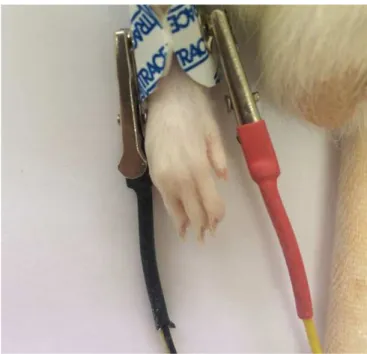
medial and lateral aspects of the ankle joint (Figure 1).
Sensory-intensity TENS was determined by increasing
the intensity until a palpable muscle contraction was
elicited and then reducing the intensity to just below
that point. Rats received either 1) low-frequency TENS
to the inlamed ankle joint at sensory intensity
(LF-TENS, 10Hz; 20 minutes); 2) high-frequency TENS at
sensory intensity (HF-TENS, 100Hz; 20 minutes); or
3) Sham TENS (SH-TENS, 0Hz; 20 minutes) during
which the animal was placed in the same apparatus,
had electrode placed but no current was applied. he
pulse duration is constant at 100μs and intensities were
constant at sensory-level intensity and are based on
those used clinically
8.
Experimental Protocol
Each rat was initially taken to determine its baseline
PWL (baseline). he animal was then anesthetized with
isolurane and submitted to the ankle sprain procedure.
PWL were again determined 30 minutes later (sprain).
Saline (SAL groups, 1 ml) or naloxone (NAL groups,
10mg/kg, 1ml, i.p.) was injected and 10 minutes later
HF-, LF- or SH-TENS were applied for 20 minutes.
PWL were measured ive minutes (T0), one (T1), two
(T2), three (T3), six (T6) and twenty-four hours (T24)
after the period of stimulation.
Statistics
Data were analyzed using the GraphPad software
program Version 5.0 and expressed as the mean ±
S.E.M. Statistically signiicant diferences between
the groups were calculated using two-way analysis
of variance (Anova) followed by the Newman–Keuls
post-hoc test. P-values less than 0.05 were considered
signiicant.
RESULTS
he PWL (Figure 2) and edema (Figure 3) were
measured before and after the ankle sprain. he PWL
and edema baselines in each group were not diferent in
all the experiments in the present study.
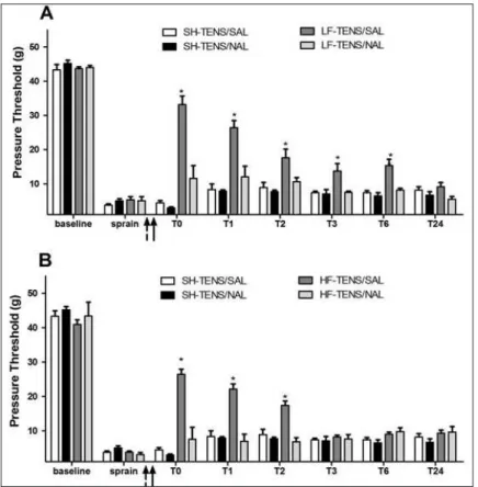
Ankle sprain induced a long-lasting reduction in PWL
for up to 24 hours in SH-TENS/SAL rats (Figure 2). In
this group, naloxone (SH-TENS/NAL) or saline-treated
(SH-TENS/SAL) rats were not diferent throughout
the period of observation. After the LF-TENS/SAL,
the PWL was increased at 0, 1, 2, 3 and 6 hours, but
were not diferent from SH-TENS/SAL after 24 hours
(Figure 2A). his antinociceptive efect was fully blocked
in naloxone-treated rats (LF-TENS/NAL) at all evaluated
times. Similar results were obtained with
HF-TENS-treated rats (Figure 2B). After the HF-TENS/SAL, the
PWL was increased at 0, 1 and 2 hours, but were not
diferent from SH-TENS/SAL after 3, 6 or 24 hours.
his antinociceptive efect was shorter than LF-TENS/
SAL, but also fully blocked in naloxone-treated rats
(HF-TENS/NAL) at all times.
Figure 2. Efect of LF-TENS (A) or HF-TENS (B) application on sprain-induced hyperalgesia. The experiment was conducted before (baseline) and after ankle sprain (sprain) and the mechanical threshold was measured 5 min after TENS (T0) and at diferent times T1, T2, T3, T6 and T24 hours. The animals were pretreated (dashed arrow) with saline (SAL) or naloxone (NAL) and then 10 minutes later were treated with TENS (black arrow). Points are means ± SD of 6 rats per group. p < 0.05 compared with SH-TENS/SAL (*).
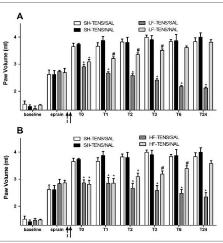
Figure 3. Efect of LF-TENS (A) or HF-TENS (B) application on sprain-induced edema. The experiment was conducted before (baseline) and after ankle sprain (sprain) and the mechanical threshold was measured 5 min after TENS (T0) and at diferent times T1, T2, T3, T6 and T24 hours. The animals were pretreated (dashed arrow) with saline (SAL) or naloxone (NAL) and then 10 minutes later were treated with TENS (black arrow). Points are means ± SD of 6 rats per group. p < 0.05 compared with SH-TENS/SAL (*) or LF- or HF-TENS/SAL groups (#). The data in A were signiicantly diferent regarding treatments (F = 25,89; P < 0.001), time (F = 127,11; P < 0.001) and had signiicant treatment x time interaction (F = 8,71; P < 0.001). The data in B were signiicantly diferent regarding treatments (F = 56,04; P < 0.001) and were not signiicantly diferent regarding time (F = 207,55; P < 0.001) or treatment x time interaction (F = 13,85; P < 0.001).
Before the induction of ankle sprain, the average
volume of the foot was just below 2 ml (Figure 3). he
ankle swelled rapidly after the sprain induction, almost
doubling its volume in after SH-TENS treatment.
For this group, naloxone (SH-TENS/NAL) or
saline-treated (SH-TENS/SAL) rats were not diferent from
SH-TENS rats throughout the period of observation.
LF-TENS/SAL-treated rats had a decreased edema
less intense than SH-TENS/SAL-treated rats at all
times after ankle sprain (Figure 3A). his decrease in
edema after LF-TENS was blocked after pre-treatment
with naloxone (LF-TENS/NAL) 1 hour after the ankle
sprain and maintained for 24 hours.
HF-TENS/SAL-treated rats had a decreased edema
less intense than SH-TENS/SAL-treated rats at all
times after ankle sprain (Figure 3B). his decrease in
edema after LF-TENS was blocked after pre-treatment
with naloxone (HF-TENS/NAL) 3 hours after the
ankle sprain and maintained for 24 hours.
DISCUSSION
TENS has been extensively used for diferent
purposes mainly for pain relief. However, the mechanism
involved in TENS analgesic efect is not completely
understood
23. In the present study, we extend these
observations showing that a single session of LF-TENS
or HF-TENS (10 or 100Hz) produced long-lasting
reduction in mechanical inlammatory hyperalgesia and
edema induced by ankle sprain in rat hind paw.
TENS efects depend on activation of opioid
receptors by endogenously released opioids
8. In fact,
and dynorphin, has been extensively described
24.
TENS-stimulated opioids can reduce the activity of nociceptive
neurons and the release of important neurotransmitters,
such as substance P, which are involved in the
transmission of nociceptive information
18.
In the present study, we show that endogenous
opioids are involved in the antinociceptive efect of
TENS against inlammatory pain evoked by ankle
sprain. he opioids released can counteract the
migration of neutrophils to the site of inlammation
and this efect might account for the efect of TENS
against hyperalgesia and edema. he migration of
neutrophils to the inlammatory site is a crucial step in
the development of inlammatory hyperalgesia
22.
An important question point emerging from these
results is the local from where opioids are released by
TENS stimulation. here is evidence that opioids can
be released through the entire nociceptive system,
including peripherally, in the spinal cord and
supra-spinal sites
25.
In the periphery, two diferent mechanisms may result
in a decreased nociception. First, opiate agonists may act
directly on opioid receptors of primary aferent neurons
26,27.
It has been shown that peripheral opioid receptors of
primary aferents may be physiologically important
because they may represent targets for endogenous opioids
released during inlammatory process
26.
Second, opioids can reduce inlammation through
actions on leukocytes
28. Numerous leukocytes,
including lymphocytes, macrophages, monocytes, and
polymorphonuclear cells such as neutrophils have been
reported to express opioid receptors
29. Direct activation
of opioid receptor reduced neutrophil migration toward
the inlammatory site
30. In TENS efect opioid might be
afecting several steps involved in neutrophil migration
such as neutrophil adhesion by reducing adhesion
molecules expression, chemokines-induced neutrophil
chemotaxis and edema
31.
TENS can also stimulate the release of opioids in the
spinal dorsal horn where they exhibit inhibitory actions
against excitatory transmission
24. he mechanisms
activated by TENS difer according to the frequency of
stimulation. Low-frequency TENS (2–10Hz) increases
the spinal release of met-enkephalin, endomorphin
and beta-endorphins, whereas high-frequency TENS
(50-100Hz) increases the spinal release of dynorphin
and both activate pain inhibitory pathways
24,32that
are represented by serotonergic and noradrenergic
ibers from rostral ventromedial medulla
33. his central
mechanism might be involved in the TENS efect
against inlammatory edema.
In summary, using the ankle sprain model, an
efect of TENS was demonstrated on hyperalgesia
and edema. he efect lasted for several hours and is
diferent when compared LF-TENS higher than
HF-TENS. For the irst time, an efect of TENS against
inlammatory edema was demonstrated. Furthermore,
the TENS-induced analgesia observed was mediated by
the endogenous opioid system and opioids are involved
in efect of TENS against inlammatory edema. hese
results suggest that the antihyperalgesic action of TENS
might be triggered by a peripheral and/or central-acting
opioid mechanism.
ACKNOWLEDGEMENTS
We are grateful for the excellent technical support of
Luciana Costa Teodoro and Zélia de Fátima Fernandes.
We would also like to acknowledge the help of Prof. Dr.
Wiliam Alves do Prado.
REFERENCES
1. Chesterton LS, Foster NE, Wright CC, Baxter DG, Barlas P. Efects of TENS frequency, intensity and stimulation site parameter manipulation on pressure pain thresholds in healthy human subjects. Pain. 2003;106(1-2):73-80. doi: 10.1016/S0304-3959(03)00292-6.
2. Ellrich J, Lamp S. Peripheral nerve stimulation inhibits nociceptive processing: an electrophysiological study in healthy volunteers. Neuromodulation. 2005;8(4):225-32. doi: 10.1111/j.1525-1403.2005.00029.x.
3. Krabbenbos IP, Brandsma D, van Swol CF, Boezeman EH, Tromp SC, Nijhuis HJ, et al. Inhibition of cortical laser-evoked potentials by transcutaneous electrical nerve stimulation. Neuromodulation. 2009;12(2):141-5. doi: 10.1111/j.1525-1403.2009.00204.x.
4. Ristić D, Spangenberg P, Ellrich J. Analgesic and antinociceptive efects of peripheral nerve neurostimulation in an advanced human experimental model. Eur J Pain. 2008;12(4):480-90. doi: 10.1016/j.ejpain.2007.07.013.
5. Vassal F, Créac’h C, Convers P, Laurent B, Garcia-Larrea L, Peyron R. Modulation of laser-evoked potentials and pain perception by Transcutaneous Electrical Nerve Stimulation (TENS): a placebo-controlled study in healthy volunteers. Clin Neurophysiol. 2013;124(9):1861-7. doi: 10.1016/j. clinph.2013.04.001.
activity in the cat. Neuromodulation. 2002;5(4):231-7. doi: 10.1046/j.1525-1403.2002.02036.x.
7. Melzack R, Wall PD. Pain mechanisms: a new theory. Science. 1965;150(3699):971-9.
8. Sluka KA, Bailey K, Bogush J, Olson R, Ricketts A. Treatment with either high or low frequency TENS reduces the secondary hyperalgesia observed after injection of kaolin and carrageenan into the knee joint. Pain. 1998;77(1):97-102. doi: 10.1016/S0304-3959(98)00090-6.
9. Radhakrishnan R, King EW, Dickman JK, Herold CA, Johnston NF, Spurgin ML, et al. Spinal 5-HT2 and 5-HT3 receptors mediate low, but not high, frequency TENS-induced antihyperalgesia in rats. Pain. 2003;105(1-2):205-13. doi: 10.1016/S0304-3959(03)00207-0.
10. Zhang RX, Lao L, Wang L, Liu B, Wang X, Ren K, et al. Involvement of opioid receptors in electroacupuncture-produced anti-hyperalgesia in rats with peripheral inlammation. Brain Res. 2004;1020(1-2):12-7. doi: 10.1016/j. brainres.2004.05.067.
11. Zhang RX, Wang L, Liu B, Qiao JT, Ren K, Berman BM, et al. Mu opioid receptor-containing neurons mediate electroacupuncture-produced anti-hyperalgesia in rats with hind paw inlammation. Brain Res. 2005;1048(1-2):235-40. doi: 10.1016/j.brainres.2005.05.008.
12. Taguchi R, Taguchi T, Kitakoji H. Involvement of peripheral opioid receptors in electroacupuncture analgesia for carrageenan-induced hyperalgesia. Brain Res. 2010;1355:97-103. doi: 10.1016/j.brainres.2010.08.014.
13. Wang Y, Hackel D, Peng F, Rittner HL. Long-term antinociception by electroacupuncture is mediated via peripheral opioid receptors in free-moving rats with inlammatory hyperalgesia. Eur J Pain. 2013;17(10):1447-57. doi: 10.1002/j.1532-2149.2013.00325.x.
14. Woolf CJ, Mitchell D, Barrett GD. Antinociceptive efect of peripheral segmental electrical stimulation in the rat. Pain. 1980;8(2):237-52. doi: 10.1016/0304-3959(88)90011-5. 15. Daher JB, de Melo MD, Tonussi CR. Evidence for a spinal
serotonergic control of the peripheral inlammation in the rat. Life Sci. 2005;76(20):2349-59. doi: 10.1016/j.lfs.2004.11.012. 16. Daher JB, Tonussi CR. A spinal mechanism for the peripheral
anti-inlammatory action of indomethacin. Brain Res. 2003;962(1-2):207-12. doi: 10.1016/S0006-8993(02)04056-8. 17. Willis WD Jr. Dorsal root potentials and dorsal root relexes: a
double-edged sword. Exp Brain Res. 1999;124(4):395-421. 18. Chen YW, Tzeng JI, Lin MF, Hung CH, Wang JJ. Transcutaneous
electrical nerve stimulation attenuates postsurgical allodynia and suppresses spinal substance P and proinlammatory cytokine release in rats. Phys Ther. 2015;95(1):76-85. doi: 10.2522/ptj.20130306.
19. Kim HY, Koo ST, Kim JH, An K, Chung K, Chung JM. Electroacupuncture analgesia in rat ankle sprain pain model:
neural mechanisms. Neurol Res. 2010;32(Suppl 1):10-7. doi: 10.1179/016164109X12537002793689.
20. Koo ST, Park YI, Lim KS, Chung K, Chung JM. Acupuncture analgesia in a new rat model of ankle sprain pain. Pain. 2002;99(3):423-31. doi: 10.1016/S0304-3959(02)00164-1. 21. Brock SC, Tonussi CR. Intrathecally injected morphine
inhibits inlammatory paw edema: the involvement of nitric oxide and cyclic-guanosine monophosphate. Anesth Analg. 2008;106(3):965-71. doi: 10.1213/ane.0b013e318162cebf. 22. Cunha TM, Verri WA Jr, Valerio DA, Guerrero AT, Nogueira
LG, Vieira SM, et al. Role of cytokines in mediating mechanical hypernociception in a model of delayed-type hypersensitivity in mice. Eur J Pain. 2008;12(8):1059-68. doi: 10.1016/j.ejpain.2008.02.003.
23. Bennett MI, Hughes N, Johnson MI. Methodological quality in randomised controlled trials of transcutaneous electric nerve stimulation for pain: low idelity may explain negative indings. Pain. 2011;152(6):1226-32. doi: 10.1016/j.pain.2010.12.009. 24. Sluka KA, Walsh D. Transcutaneous electrical nerve
stimulation: basic science mechanisms and clinical efectiveness. J Pain. 2003;4(3):109-21. doi: 10.1054/ jpai.2003.434.
25. Millan MJ. Descending control of pain. Prog Neurobiol. 2002;66(6):355-474. doi: 10.1016/S0301-0082(02)00009-6. 26. Cunha TM, Roman-Campos D, Lotufo CM, Duarte HL,
Souza GR, Verri WA Jr, et al. Morphine peripheral analgesia depends on activation of the PI3Kgamma/AKT/nNOS/ NO/KATP signaling pathway. Proc Natl Acad Sci U S A. 2010;107(9):4442-7. doi: 10.1073/pnas.0914733107.
27. Fields HL, Emson PC, Leigh BK, Gilbert RF, Iversen LL. Multiple opiate receptor sites on primary aferent ibres. Nature. 1980;284(5754):351-3. doi: 10.1038/284351a0. 28. Wybran J. Enkephalins and endorphins as modiiers of the
immune system: present and future. Fed Proc. 1985;44(1 Pt 1):92-4.
29. Carr DJ, DeCosta BR, Kim CH, Jacobson AE, Guarcello V, Rice KC, et al. Opioid receptors on cells of the immune system: evidence for delta- and kappa-classes. J Endocrinol. 1989;122(1):161-8.
30. Kapitzke D, Vetter I, Cabot PJ. Endogenous opioid analgesia in peripheral tissues and the clinical implications for pain control. Ther Clin Risk Manag. 2005;1(4):279-97.
31. Kulkarni-Narla A, Walcheck B, Brown DR. Opioid receptors on bone marrow neutrophils modulate chemotaxis and CD11b/ CD18 expression. Eur J Pharmacol. 2001;414(2-3):289-94. 32. Toda K. Response of raphe magnus neurons after acupuncture
stimulation in rat. Brain Res. 1982;242(2):350-3.