Bracken (
Pteridium aquilinum
)-Induced DNA Adducts
in Mouse Tissues Are Different from the Adduct
Induced by the Activated Form of the Bracken
Carcinogen Ptaquiloside
R. N. Freitas,*
,† P. J. O’Connor,* A. S. Prakash,‡ M. Shahin,‡ and A. C. Povey*
,§
,1*Cancer Research Campaign Carcinogenesis Group, Paterson Institute for Cancer Research, Manchester, M20 9BX United Kingdom;†School of Nutrition, Federal University of Ouro Preto, Ouro Preto, Minas Gerais, 35400-000, Brazil;‡National Research Centre for Environmental Toxicology, 39 Kessels Road, Coopers Plains, Queensland 4108, Australia; and §School of Epidemiology and Health Sciences, University of Manchester, Manchester M13 9PT, United Kingdom
Received January 22, 2001
Following treatment with bracken fern (Pteridium
aquilinum) extract and bracken spores a number of
DNA adducts were detected by32
P-postlabeling. Three of these adducts have been described previously (Poveyet al., Br. J. Cancer(1996) 74, 1342–1348) and in this study, using a slightly different protocol, four new adducts, with higher chromatographic mobility, were detected at levels ranging from 50 to 230% of those previously described. When DNA was treatedin vitro
with activated ptaquiloside (APT) and analysed by bu-tanol extraction or nuclease P1 treatment, only one adduct was detected by 32
P-postlabeling. This adduct was not present in the DNA from mice treated with bracken fern or spores, suggesting either that bracken contains genotoxins other than ptaquiloside or that the metabolism of ptaquiloside produces genotoxins not reflected by activated ptaquiloside. However, as the ATP-derived adduct has been detected previously in ileal DNA of bracken-fed calves, species-specific dif-ferences in the metabolism of bracken genotoxins may exist, thereby leading to differences in their biological outcomes. © 2001 Academic Press
Key Words:bracken fern; ptaquiloside; DNA adducts;
32
P-postlabelling.
Bracken fern (genus
Pteridium
) has been described
as one of the most common plants on the planet
(Tay-lor, 1990) and it is the only plant that is known to cause
tumours naturally in animals (Shahin
et al.,
1999). The
toxicity and carcinogenicity of bracken fern to domestic
and experimental animals has been extensively
de-scribed (Evans and Mason, 1965; Evans, 1984; IARC,
1986; Pamacku and Price, 1969). This plant contains a
number of toxic components, including flavonoid
anti-oxidants such as quercertin and an unstable glycoside,
ptaquiloside (PT), which is thought to be the principal
carcinogenic compound present in bracken (Hirono
et
al.,
1984a, 1987). Recent work indicates that other
toxic compounds related to ptaquiloside are also
present in bracken fern (Castillo
et al.,
1998) but in
lower concentrations.
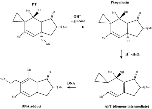
PT is stable at room temperatures but decomposes in
aqueous solutions. Under acidic conditions it
under-goes aromatisation by elimination of the glucose to give
pterosin B, whereas under alkaline conditions it is
activated to give a conjugated dienone (activated
ptaquiloside: APT) upon the liberation of the sugar
group (Fig. 1; Ojika
et al.,
1987)). APT has been shown
to alkylate DNA via a reactive cyclopropyl ring to form
a number of different DNA adducts (Ojika
et al.,
1987,
1989). The main labile adducts occur at the N-3 of
adenine, and to a minor extent with N-7 of guanine
(Smith
et al.,
1994b; Kushida
et al.,
1994). This
N3-adenine selectivity has also been observed in
anti-carcinogenic alkylating agents possessing a
cyclo-propyl ring intermediate (Hurley
et al.,
1984; Shalder
et al.,
1999). Following nuclease P1 enrichment and
32
P-postlabelling, a single adduct, as yet
uncharacter-ised, was found in DNA from the ileum of bracken fed
calves (Prakash
et al.,
1996) or APT dosed rats (Shahin
et al.,
1998), and in DNA treated
in vitro
with APT
(Smith
et al.,
1994b). As ptaquiloside-N3 adenine
ad-ducts are labile and depurinate within 24 h (Prakash
et
al.,
1996) they may not be readily detectable in the
postlabeling assay. Previously, it has been
demon-strated that administration of either extract or spores
of bracken fern collected in the UK (
Pteridium
aquili-1To whom correspondence should be addressed at School of Epi-demiology and Health Sciences, University of Manchester, M13 9PT UK. Fax: 61 275 5595. E-mail: [email protected].
Biochemical and Biophysical Research Communications281,589 –594 (2001) doi:10.1006/bbrc.2001.4388, available online at http://www.idealibrary.com on
589 0006-291X/01 $35.00
num
subsp.
aquilinum
) induced DNA adducts
detect-able in the upper gastrointestinal tract of BDF1 mice
by using
32P-postlabeling after butanol extraction for
DNA adducts (Povey
et al.,
1996). However, the adduct
enrichment method used in the former studies (Smith
et al.,
1994a; Prakash
et al.,
1996; Shahin
et al.,
1998)
differed from that used in the UK study (Povey
et al.,
1996) and as these enrichment methods may extract
chemically different adducts (Gupta and Early, 1988) it
was not immediately apparent whether or not
APT-induced DNA adducts were present in the DNA
iso-lated from BDF1 mice.
We thus carried out the present work, using both
butanol and nuclease P1 enrichment procedures, to
determine whether the uncharacterised adducts found
in gastrointestinal tissue from BDF1 mice were the
same or different from the one formed by APT
in vitro.
MATERIALS AND METHODS
32
P-g-ATP (specific activity 7000 Ci/mmol) was purchased from
ICN Pharmaceuticals, Inc. (ICN Biomedicals, Kingston-upon-Thames, UK). T4 polynucleotide kinase (T4-PNK) and calf spleen phosphodiesterase (CSPE) were both obtained from Boehringer-Mannheim (Germany). Micrococcal nuclease (MN) and alkaline phosphatase were obtained from Sigma (Poole, UK). MN and CSPE were also purchased from Worthington (Lorne Laboratories, Read-ing, UK). Plastic PEI-cellulose plates were supplied by Schleicher-Schuell (Anderman and Co., Kingston-upon-Thames, UK).
Preparation of Bracken Samples
An extract of fresh bracken fern (Pteridium aquilinum subsp.
aquilinum) collected in Bellech, Anglesey, UK was prepared (Povey
et al., 1996). Ptaquiloside was isolated and purified as described
elsewhere (Oelrichs et al., 1995) from bracken collected from in Southeast Queensland.
Treatment of Animals
Treatment of animals with bracken fern extracts and spores from the UK has been described previously (Poveyet al.,1996) and the DNA obtained in these experiments was used here.
In Vitro Modification of DNA with Activated
Ptaquiloside
Ptaquiloside was activated by incubation with NaOH and usedin vitroto modify calf thymus DNA (Smithet al.,1994a).
Analysis of DNA Adducts
DNA samples from thein vivoexperiments were extracted using a standard phenol-chloroform method after treatment of the tissue with RNase A and proteinase K (Poveyet al.,1996). Both butanol and nuclease P1 enrichment procedures were used to analyse for DNA adducts by32P-postlabelling.
DNA digestion to nucleotides. 10 –25mg of each DNA sample was
digested for 4 h at 37°C using CSPE (0.432 mU/mg DNA) and MN
(0.495 U/mg DNA) obtained from Worthington, with 10 mM Tris–
HCl, pH 7.4, 5 mM CaCl2 and 1mM deoxycorfomycin in a final
volume of 2.5ml/mg DNA.
Butanol extraction. Aliquots containing 4mg of the DNA digest
were extracted with 1-butanol (Poveyet al.,1996) but using 3 rather than 2 back washes with 1-butanol saturated water.
Nuclease P1 digestion. Aliquots containing 7.89mg of the initial
DNA digest was further incubated with sodium acetate pH 5, zinc chloride, and nuclease P1 at final concentrations of 40 mM, 0.2 mM, and 0.31mg/ml, respectively. The reaction was allowed to proceed for
30 min at 37°C, then stopped by adding 1ml of 0.92 mM Tris and the
samples were driedin vacuo.
32P-postlabelling and TLC.
The adducted nucleotides were 32 P-postlabelled for 30 min at 37°C in 30 mM Tris-HCl (pH 9.5) contain-ing 10 mM magnesium chloride, 10 mM DTT, 1 mM spermidine, 3.0
mM ATP (total concentration) using 2.5 units T4PNK and 170mCi
32
P-g-ATP in a total volume of 10ml. Labelled samples were spotted
on PEI-cellulose TLC plates and chromatographed as previously described (Poveyet al.,1996) but with the plates only run once in D3 and D4. Solvents used for development of the thin layer plates were as follows: D1, 1.0 M sodium phosphate, pH 6.5; D3, 3.5 M lithium formate, 8.5 M urea, pH 3.5; D4, 1.2 M lithium chloride, 0.5 M Tris, 8.5 M urea, pH 8; and D5, 1.7 M sodium phosphate, pH 6.
Normal nucleotides and adduct quantitation. The plates were dried and exposed to a phosphorimager screen for up to 72 h. The samples were visualised using a Molecular Dynamic Storm 860 image analysis system with ImageQuant software. Adducts were quantified by comparing the signal with that of a known amount of the labelling mix spotted onto a TLC plate and exposed together on the same phosphor screen. To determine the normal levels of nucle-otides released, 10mg aliquots of the same DNA digest were treated
with alkaline phosphatase to give nucleosides that were quantified by HPLC (Cooperet al.,1992).
RESULTS
Upper gastrointestinal DNA samples (oesophagus,
stomach and ileum) from BDF1 mice treated
intragas-trically with bracken extract or spores contained
sev-eral adducts (Figs. 2B and 2C) that were not found in
the DNA samples from untreated control animals (Fig.
2A), or in untreated calf thymus DNA used as a
nega-tive control (data not shown). The adducts located near
to the origin (Nos. 1, 2, and 3 in Fig. 2C) were described
previously (Povey
et al.,
1996). In addition to these
adducts, 4 other spots with higher chromatographic
mobility were detected (Nos. 4 –7 in Fig. 2C). Assuming
quantitative recovery, the levels of adducts 4 and 5
were 50 and 90% of adduct 2 respectively whereas
levels of adducts 6 and 7 were, respectively, 160 and
230% higher than the level of adduct 2. Adducts
recov-ered using butanol extraction were similar to those
recovered using nuclease P1 digestion, except for
ad-duct 1, which was poorly recovered after nuclease P1
digestion (Fig. 3).
Only one adduct was detected in calf thymus DNA
reacted
in vitro
with APT when the sample was treated
with either nuclease P1 APT (Fig. 4) or extracted with
butanol (data not shown). This APT derived adduct
had a chromatographic mobility that was similar to
adducts 6 and 7 but subsequent cochromatography
experiments indicated a chemically different identity
for the APT-derived adduct. Hence, we conclude that
the APT induced DNA adduct was not present in
de-tectable amounts in the DNA obtained from BDF1 mice
treated with bracken fern extract or bracken spores.
DISCUSSION
Previously, the presence of DNA adducts in upper
gastrointestinal tissue of BDF1 mice treated with a
single dose of bracken extracts or spores has been
described (Povey
et al.,
1996). Using butanol extraction
to enrich the adducted nucleotides in these samples,
three major (Nos. 1, 2, and 3: Fig. 2C) adducts were
found that were not present in untreated animals.
These adducts were not characterised, but their
chro-matographic mobility was similar to adducts formed in
the same tissue from mice dosed with synthesised
com-pounds containing a cyclopropyl ring (Povey
et al.,
1996).
Using a slightly different protocol, not only have the
same spots in these samples been detected, but also at
least 4 other adducts (Nos. 4, 5, 6, and 7) with higher
chromatography mobility (Figs. 2B and 2C) were found
that were not present in the control samples (Fig. 2A).
Procedures that result in the preferential enrichment
of different adduct classes (Reddy and Randerath,
1986; Gupta and Early, 1988) were used to further
characterise these adducts. The DNA samples were
digested to nucleotides and then either extracted with
FIG. 2. Phosphorimages of butanol-extractable DNA adducts detected in the upper gastrointestinal tract of mice following treatment with water (A), bracken fern extract (B), and bracken spores (C).1-butanol (Gupta, 1985) or treated with nuclease P1
(Reddy and Randerath, 1986). Most of the adducts
(Nos. 2–7, Fig. 2C) were readily detected by both
tech-niques indicating that these were not sensitive to
nu-clease P1 digestion unlike certain adducts, notably
those derived from aromatic amines or simple
alkylat-ing agents and some cyclic nucleotide adducts (Reddy
et al.,
1984; Hemmink
et al.,
1991; Szyfter
et al.,
1991;
Vaca
et al.,
1992).
In order to know if any of the adducts detected in
mouse DNA were identical to that formed by APT
in
vitro,
a cochromatography experiment was performed
(Fig. 4). This indicated that the principal DNA adduct
formed by APT
in vitro
was not found in digests of the
upper gastrointestinal tissue DNA of mice treated with
bracken fern extracts or spores. The amount of PT in
the bracken fern extract and bracken spores used in
the mouse experiments was not quantified, but it was
previously calculated that approximately 8 mg of
ptaquiloside was administered to each animal (Povey
et al.,
1996) based on a published bracken fern content
(Hirono, 1986). However, as PT levels can depend on a
number of factors including bracken species, freshness,
season and drying conditions (Oelrichs
et al.,
1995;
Smith
et al.,
1994b), the amount of PT in these samples
may have been too low to permit the detection of
PT-derived DNA adducts. In a previous work, we (Shahin
et al.,
1998) could detect in ileum tissue a PT derived
DNA adduct from rats dosed intravenously with 3 mg
ptaquiloside weekly for 10 weeks. Although, the
ab-sence of PT derived DNA adducts may potentially be
ascribed to insufficient PT in the initial bracken
ex-tract, the presence of DNA adducts as detected by
32
P-postlabelling, clearly indicates that the bracken
from APT-DNA (Prakash
et al.,
1996), was found in the
ileum from a calf fed with bracken fern, species specific
differences in PT metabolism may exist. In this regard,
it is of interest to note that in calves, the most
impor-tant site of tumour occurrence is the ileum, followed by
urinary bladder and mammary gland whereas in mice,
leukaemic, stomach and lung tumours are common but
with ileal tumours being a relatively rare occurrence
(Evans, 1984; IARC, 1985).
In summary, DNA adducts derived from APT were
not detected in tissues from mice treated with
Pteridium aquilinum.
The possibility that this is due to
low PT levels in the bracken samples used to treat the
mice cannot be ruled out. However as other DNA
ad-ducts
were
detected
following
treatment
with
Pteridium aquilinum
in mice, it is evident that there
is more than one genotoxic substance is present
in
Pteridium aquilinum.
Whether these genotoxins
are related to the new PT-like compounds such as
Ptaquiloside Z (Castillo
et al.,
1998) that are now being
identified remains to be determined.
ACKNOWLEDGMENTS
The authors are grateful to Dr. Barry Smith, AgResearch, NZ, for his helpful comments; to the CNPq and CAPES of the Brazilian government and from FAPEMIG from Minas Gerais State govern-ment, Brazil, for grants to support R.N.F.; the Cancer Research
FIG. 4. Phosphorimages of high mobility DNA adducts detected in calf thymus DNA modifiedin vitrowith APT (A) and the upper gastrointestinal tract of mice following treatment with bracken fern extract (B) or bracken spores (C). D and E are respectively 32 P-postlabelled samples of DNA adducts induced by bracken fern extract (as in B) and bracken spores (as in C) cochromatographed with the adduct from calf thymus DNA modifiedin vitrowith APT (as in A). Circles in D and E locate the APT-derived adduct.
of aromatic carcinogen:DNA adducts. Cancer Res.45, 5656 – 5662.
Gupta, R. C., and Early, K. (1988)32P-adduct assay: Comparative recoveries of structurally diverse DNA adducts in the various enhancement procedures.Carcinogenesis9,1687–1693. Hemminki, K., Szyfter, K., and Kadublar, F. F. (1991) Quantitation
of the32P-postlabeling reaction using cyclic N1,N2and C8 mod-ified deoxyguanosine-3-monophosphates as substrates. Chem-Biol. Interact.77,51– 61.
Hirono, I. (1986) Carcinogenicity of plant constituents: Pyrrolizidine alkaloids, bracken fern. In Genetic Toxicology of the Diet (Knudsen, I., Ed.), pp. 45–53. Alan Liss, New York.
Hirono, I., Yamada, K., Niwa, N., Shizuri, M., Ojika, M., Hasaka, S., Yamaji, T., Wakamatsu, K., Kigashi, H., Nuyama, K., and Uosakki, Y. (1984a) Separation of carcinogenic fraction of bracken fern.Cancer Lett.21,239 –246.
Hirono, I., Aiso, S., Yamaji, T., Mori, H., Yamada, K., Niwa, H., Ojika, M., Wakamatsu, K., Kigoshi, H., Niiyama, K., and Uosaki, Y. (1984b) Carcinogenicity in rats of ptaquiloside iso-lated from bracken.Gann75,833– 836.
Hirono, I., Ojino, H., Fujimoto, M., Yamada, K., Yoshida, Y., Igawa, M., and Okumura, M. (1987) Induction of tumours in ACI rats given a diet containing ptaquiloside, a bracken carcinogen.
J. Natl. Cancer Inst.79,1143–1149.
Hurley, L. H., Reynolds, V. L., Swenson, D. H., Petzold, G. L., and Scahill, T. A. (1984) Reaction of the antitumor antibiotic CC-1065 with DNA: Structure of a DNA adduct with DNA sequence specificity.Science226,843– 844.
IARC (1986) Bracken fern (Pteridium aquilinum) and some of its constituents.InMonographs on the Evaluation of Carcinogenic Risk to Humans. No. 40. pp. 47– 65. WHO, IARC, Lyon. Kushida, T., Uesugi, M., Sugiura, Y., Kigoshi, H., Tanaka, H.,
Hiro-kawa, J., Ojika, M., and Yamada, K. (1994) DNA damage by ptaquiloside, a potent bracken carcinogen: Detection of selective strand breaks and identification of DNA cleavage products.
J. Am. Chem. Soc.116,479 – 486.
Oelrichs, P. B., Ng, J., and Bartley, J. (1995) Purification of ptaquiloside, a carcinogen fromPteridium aquilinum. Phyto-chemistry40(1), 53–56.
Ojika, M., Wakamatsu, K., Niwa, H., and Yamada, K. (1987) Ptaquiloside, a potent carcinogen isolated from bracken fern
Pteridium aquilinum var. laticulum. Structure elucidation
analysis of DNA adducts formed in the upper gastrointestinal tissue of mice fed bracken extract or bracken spores. Br. J. Cancer74,1342–1348.
Reddy, M. V., and Randerath, K. (1986) Nuclease P1-mediated en-hancement of sensitivity of32P-postlabeling test for structurally diverse DNA adducts.Carcinogenesis7,1543–1551.
Reddy, M. V., Gupta, R. C., Randerath, E., and Randerath, K. (1984) 32
P-postlabeling test for covalent DNA binding of chemicalsin vivo:Application to a variety of aromatic carcinogens and meth-ylating agents.Carcinogenesis5,231–243.
Shahin, M., Smith, B. L., Worral, S., Moore, M. R., Seawright, A. A., and Prakash, A. S. (1998) Bracken fern carcinogenesis: Multiple intravenous doses of activated ptaquiloside induce DNA ad-ducts, monocytosis, increased TNFalevels and mammary gland
carcinoma in rats.Biochem. Biophys. Res. Commun.244,192– 197.
Shahin, M., Snith, B. L., and Prakash, A. S. (1999) Bracken carcin-ogens in the human diet.Mutat. Res.443,69 –79.
Shalders, R. L., Blanch, G., Brown, C. L., Young, D. J., and Prakash, A. S. (1999) Stability and DNA alkylation rates of simplest functional analogs of CC-1065,p-hydroxy and p-amino phen-ethyl bromides.Chem.-Biol. Interact.117,83–94.
Smith, B. L., Shaw, G., Prakash, A. S., and Seawright, A. A. (1994a) Studies on DNA adduct formation by ptaquiloside, the carcino-gen of bracken ferns (Pteridiumspp.).InPlant Associated Tox-ins (Colgate, S. M., and Dorling, P. R., Eds.), Chap. 31, pp. 167–172. CAB International, Wallingford, UK.
Smith, B. L., Seawright, A. A., Ng, J. C., Hertle, A. T., Thomson, J. A., and Bostock, P. D. (1994b) Concentration of ptaquiloside, a major carcinogen in bracken fern (Pteridiumspp.), from east-ern Australia and from a cultivated worldwide collection held in Sydney, Australia.Nat. Toxins2,347–353.
Szyfter, K., Hemminki, K., Crane, A. E., and Watson, W. P. (1991) Quantitative and kinetic examination of 32P-postlabeling of etheno-substituted nucleotides. Chem.-Biol. Interact. 80, 99 – 107.
Taylor, J. A. (1990) The bracken problem: A global perspective. AIAS Occasional Publ.40,3–19.