Polyamine Ligand-Mediated Self-Assembly of Gold and
Silver Nanoparticles into Chainlike Structures in Aqueous
Solution: Towards New Nanostructured Chemosensors
Adrin Fernndez-Lodeiro,
[a]Javier Fernndez-Lodeiro,
[a]Cristina NfflÇez,*
[a, b]Rufina Bastida,
[c]Jos Luis Capelo,
[a]and Carlos Lodeiro*
[a]Introduction
Noble metal nanoparticles (MNPs) have been intensively pur-sued in recent years, not only for their fundamental scientific interest[1]but also for their technological applications, ranging
from analytical sensors to catalysis and fuel cells.[2] Recently,
the attention paid to 1D nanomaterials has been increasing significantly, because of the need to fabricate alternative func-tional 1D nanostructures for applications in the fields of nano-electronics and nanobiotechnology,[3]due to the fact that they
can act as interconnects between functional nanoscale compo-nents.[4]
Several experimental routes have been recently proposed to efficiently self-assemble preformed MNPs into 1D chains: methods involving hard,[5] polymeric[6] or surfactant-based[7]
templates, molecular recognition,[8]specific functionalization,[9]
and surface- or solvent-induced phase separation[10]have been
successfully demonstrated.[11] Chains of MNPs also have been
prepared by using linear macromolecular templates, such as, DNA,[12, 13] peptide,[14, 15] insulin fibrils,[16] protein fibrils[17, 18] or
carbon nanotubes.[19]
The growth mechanism of self-assembled metal nanostruc-tures using (macro)molecular ligands has been also reported to exhibit common features with molecular step-growth poly-merization.[20] Similar to functional monomers, metal
nano-structures assemble to form chains. The assembly was per-formed by small molecules (<2 nm), called molecular linkers, that contain at least two reactive ending groups, capable of at-taching to a solid surface by chemisorption (thiol, amine) or in-teracting electrostatically with other functional groups (hy-droxy, carboxyl, amine) present on the surface of nanoparticles (NPs).[20] The governing factor in linker-mediated assembly of
MNPs is the equilibrium between the attractive and repulsive forces.[21]In particular, fabrication of anisotropic 1D noble MNP
chains to obtain integrated optics operating below the diffrac-tion limit of light has attracted much attendiffrac-tion.[22]
Stellaci and co-workers[10a]have introduced anisotropic
prop-erties on ligand-stabilized AuNPs. Face-centered cubic (fcc) metallic NPs exhibit no intrinsic electric dipole, however, heter-ogeneities in surface charge and polarity, associated for exam-ple with the non-uniform spatial distribution of capping li-gands on different crystal faces,[23]or nanophase separation in
mixed-ligand stabilization layers,[24] are possible driving forces
for anisotropic self-assembly.[25] In the case of spherical NPs,
Polyamine ligands are very versatile compounds due to their water solubility and flexibility. In the present work, we have ex-ploited the binding ability of a polyamine molecular linker (L2) bearing different functional groups, which favors the self-assembling of silver nanoparticles (AgNPs) and gold nanoparti-cles (AuNPs) into 1D nanochains in aqueous solution. The chainlike assemblies of AuNPs and AgNPs were structurally stable for a long period of time, during which their characteris-tic opcharacteris-tical properties remained unchanged. The mechanism of
AuNPs and AgNPs chain assembly associated with the induc-tion of electric dipole–dipole interacinduc-tions arising from the par-tial ligand exchange of surface-adsorbed citrate ions by (L2) was investigated. UV/Vis spectrophotometry and transmission electron microscopy (TEM) were used to determine time-dependent structural changes associated with formation of the 1D nanoparticle structures. Finally, the sensing of Hg2+
in aqueous solution using AgNPs@(L)2 and AuNPs@(L)2 assem-blies was also carried out in aqueous solution.
[a]A. Fernndez-Lodeiro, Dr. J. Fernndez-Lodeiro, Dr. C. NfflÇez, Dr. J. L. Capelo, Dr. C. Lodeiro
BIOSCOPE Group, REQUIMTE-CQFB, Chemistry Department Faculty of Science and Technology, University NOVA of Lisbon 2829-516, Monte da Caparica (Portugal)
E-mail: [email protected] [email protected]
[b]Dr. C. NfflÇez
Ecology Research Group, Department of Geographical and Life Sciences Canterbury Christ Church University
CT1 1QU, Canterbury (UK)
[c] Prof. R. Bastida
Inorganic Chemistry Department, Faculty of Chemistry University of Santiago de Compostela
15782 Santiago de Compostela (Spain)
Supporting information for this article is available on the WWW under http://dx.doi.org/10.1002/open.201300023.
controlling the surface chemistry of the fabricated NPs allows the creation of an anisotropic ligand organization.[26] Enthalpy
minimization, is obtained by promoting dipole alignment and reducing interdipole distances through the formation of linear chains of single NPs. This facilitates the orientation of specific interactions in one direction, which helps directing the self-assembly into 1D arrays. The self-self-assembly of the NPs into a well-defined 1D array is also
in-fluenced by interparticle chemi-cal bonding, hydrogen bonding, van der Waals interactions, elec-trostatic forces, or any combina-tion of these forces. In addicombina-tion, entropy can be maximized at finite temperature by introduc-ing some disorder in the linear
chain, which corresponds to the incorporation of branching junctions and to chain reticulation that should be favored at elevated temperature.
Aggregation of NPs induces variations in absorption spectra accompanied by significant color changes of solutions.[27]
Simi-lar color changes can be observed upon the addition of an an-alyte, which initiates the aggregation of noble MNPs, and this feature can be used for permitting their industrial application in biosensing, immunological, and biochemical investiga-tions.[28, 29] In the particular case of AgNPs, the geometrical
shape also plays an important role in determining plasmon res-onance properties.[30]For example, triangular, pentagonal, and
spherical silver particles are colored red, green, and blue, re-spectively. Consequently, it is important to develop approaches that can manipulate NPs into different shapes and dimensions. While many studies have tackled the synthesis and charac-terization of gold dimers and networks with peculiar plasmon resonance behavior,[31] organization of AgNPs in 2D
superlatti-ces is less common.[32, 33] For example, Chang et al. reported
a variety of 1D- and 2D-nanostructured assemblies formed from AgNPs by variations in pressure, temperature, and time in supercritical water (SCW) without the need for any external linking agents.[34]
To follow our interest in new emissive materials, and func-tionalized NPs and to explore their applications,[35]herein, we
investigate the mechanism of AuNP and AgNP chain assembly associated with the induction of electric dipole–dipole interac-tions. The nanoassembly capacity arises from the partial ligand exchange of surface-adsorbed negatively charged citrate ions, by covalently bound neutral molecular ligand L to produce a final mixed-ligand surface layer.
We show that exchange of surface adsorbed citrate withL, results in the formation of chain-like superstructures with topo-logical features that are dependent on the extent of surface-ligand substitution. We determine the time-dependent struc-tural changes associated with the formation of 1D NP super-structures. Morphological and optical characteristics of various nanostructures were investigated by TEM and UV/Vis.
Results and Discussion
Formation of chainlike structures from AgNPs and AuNPs
1D metallic silver or gold nanostructures, can be obtained by exploiting the binding ability of the linear polyamine molecular probe L in water (see chemical structure in Scheme 1). The
donor atoms presented in the structure of compoundLcould be responsible for the formation of NP chainlike aggregates and the partial removal of the citrate ion from the starting metal nanoparticle (MNP) surface. This method is similar to that reported by Zhang et al.[36, 37]and it is different to that in
which the assembly was induced by electrostatic interactions and the disassembly was labile in the presence of stronger chelating agents.[38]
In that case, firstly, we assumed that the level of ionization (protonation-deprotonation of functional groups) of potential modifying compounds, might play a role in the process of MNP functionalization, and consequently, could affect the sta-bility of their dispersions. Therefore, the pH of the mixture was modified with a NaOH solution, to an approximate value of pH 12, which leads to the change in the linker ionization degree. After the addition of base to a solution ofL, this com-pound was dissociated bearing negative charges L2 because of deprotonation of the amine groups. Second, we assumed that the chemisorption of the linker on the AuNP surface could take place through the sulfur atom and/or by deprotonated amine groups, leading to the modification of the zpotential from z0 (initial potential) to z (potential that determines the
equilibrium between attractive and repulsive forces).
Finally, it is well known that changing the medium surround-ing the nanoparticles (NPs) for another medium, havsurround-ing a mark-edly different refractive index, strongly alters the surface plas-mon resonance (SPR) band of the NPs in the UV/Vis spectrum. The position, intensity, and shape of SPR band strongly de-pends on the dielectric constant of surrounding medium, the size and shape of NPs as well as the electronic interaction be-tween the stabilizing ligand and NP.[39] Therefore, UV/Vis
ab-sorption spectroscopy is an important analytical tool to probe the stability, surface chemistry, and aggregation behavior of AgNPs and AuNPs.
Formation of chainlike structures from AgNPs
Spherical shape
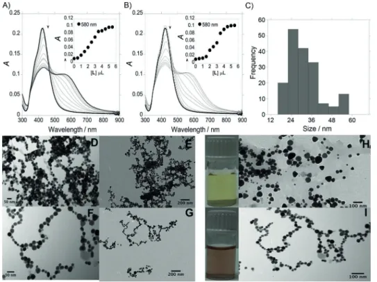
Initial characterization of spherical AgNPs prepared by citrate reduction of a silver nitrate solution revealed an absorption
maximum (lmax) of the SPR peak for single particles at 420 nm (transverse SPR band; Figure 1 A,B). Analyses of TEM imaging shows that the prepared citrate-capped silver NPs (AgNPs@citrate) are nearly monodisperse spheres with an aver-age size of about (253) nm. The AgNP concentration was es-timated to be 7.8 1010in terms of molar concentration. This
value was obtained taking into account that the entire mass of silver in AgNPs employed for colloidal dispersion preparation was fully transformed into NPs.
Figure 1 A and 1 B show the absorption titration of spherical AgNPs@citrate with compoundLandL2, respectively. As men-tioned above, because AgNPs@citrate are highly dispersed in solution, the spectrum is characterized by a single SPR band at
420 nm (transverse SPR band). AgNPs@L and AgNPs@(L)2 exhibit two absorption bands, at 420 nm and a new band at
580 nm, respectively. In both cases, the appearance of the second band (longitudinal SPR band) is a clear evidence of the assembly of AgNPs in solution, and a color change from yellow (Figure 1 H) to deep red (Figure 1 I) was also observed.
Chainlike structure formations of AgNPs in aqueous solution were induced in both cases byLandL2, with the contact be-tween AgNPs being easier at higher concentrations of L and L2. Thezpotential distribution plays the major role in linker-mediated self-assembly of MNPs, and the development of linear chains and branched chain network is directed by the fact that the electrostatic double layer is rearranged around the dimers and becomes anisotropic.
The replacement of citrate ions most probably takes place after the addition of the nega-tively charged ligand L2. In that case, the amount of negative charge on the surface of NPs does not decrease significantly, and as a result, a slight change of the zpotential was observed from z0 33.5 mV cm1 toz0 27.8 mV cm1. The formations
of chain-like superstructures with topological features are de-pendent on the extent of sur-face-ligand substitution. The re-placement of citrate ions of the AgNPs surface by L2 probably induces less electric dipole– dipole interactions, observing that the 1D NP assembly takes place less prominently (Fig-ure 1 F,G) compared with that observed withL(Figure 1 D,E).
In that case, IR spectra were recorded to demonstrate the re-placement of citrate ions of the AgNP surface by polyamine ligand L. The IR spectrum of compound L shows peaks char-acteristic of the carbonyl group at 1676 cm1 and the n˜(C=C)
stretching mode at 1436 cm1(see Figure S1, Supporting
Infor-mation). In addition, a peak at 3262 cm1 is observed due to
then˜(NH) stretching mode. A decrease in the intensity of the
n˜(C=O) band was observed in the IR spectra of AgNPs@L in comparison withL. The most interesting part of the spectrum is the region from 3000 to 3200 cm1due ton˜(NH) stretching
modes. The IR spectrum for AgNPs@Lis very different from the spectrum ofLin the same region. These results suggest the in-teraction ofLwith the MNP surface through the carbonyl and amine groups, confirming the replacement of the citrate ions from the NP surface.
Triangular shape
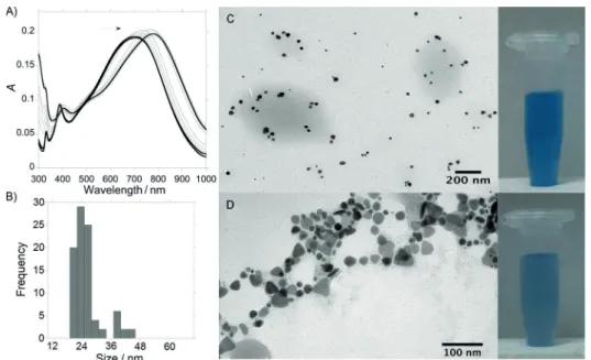
Because better results were obtained after titration of spherical AgNPs@citrate with the deprotonated ligandL2, we used the same method with the triangular AgNPs@citrate. The forma-tion of triangular AgNPs@citrate in aqueous soluforma-tion with sizes in the range of (6410) nm (see Figure S2, Supporting Infor-mation) was confirmed by the presence of the blue SPR band at 700 nm in the UV/Vis spectrum (Figure 2 A). As shown in Figure 2 C, the triangular AgNPs@citrate were well dispersed in milli-Q water before addingL2, and they remained well sepa-rated on the TEM grid. After the addition of 5mL of L2 (1.103m) to a solution of triangular AgNPs@citrate in aqueous
solution, a redshift in the absorption band was observed to
780 nm, due to the formation of chains in which L2 ions
played the role of connectors between the AgNPs (Figure 2 D). As shown in Figure 2 C,D, a slight color change from intense to pale blue was also observed.
Formation of chainlike structures from AuNPs
As a model experiment, we also used citrate-capped spherical gold NPs (AuNP@citrate) with a hydrodynamic diameter of (205) nm (see Figure S3, Supporting Information). The self-assembly was performed using a AuNP@citrate solution with a CNP=3.7 109m (2.2 1015particle L1) and a concentration
of 1.6 106
mforL2(
pH 12) (CL/CNP4.5 102).
As we mentioned above, the assembly of AuNPs has a signifi-cant effect on the optical prop-erties of the NPs, reflected by a dramatic change of the UV/Vis extinction value of the SPR band. As shown in Figure 3 A, single spherical AuNPs were characterized by an extinction transverse plasmon band at l1 520 nm.[40] After addition of
the linker L2, a second low-energy longitudinal surface plas-mon band (l2) appears at higher
wavelengths (630–710 nm), which is a result of the plasmon-ic coupling of linearly assembled NPs. The position of this longitu-dinal surface plasmon band (l2)
could be modified in function of the topological distortions in the chains (Y-junction, zigzag
de-fects, loop domains) in place of a strictly linear assembled super-structure. The second band shifts with time toward higher wavelengths and its intensity (Il2) increases. As the interparticle
spacing decreases, the first peak becomes weaker, while the second peak intensifies and shifts to longer wavelengths. The wide range of different chain morphologies observed in the extended networks accounts for the broadness of the emerg-ing longitudinal plasmon band and the absence of isosbestic points in the time-dependent spectra.[15a]
The spectral change associat-ed with the progressive aggre-gation of the NPs into chains and branched networks was also demonstrated by TEM (Fig-ure 3 C,D). Consequently, the number of single NPs progres-sively decreases, leading to a decrease in the intensity of the first peak (Il1).
The zpotential of the original AuNPs@citrate solution was
z0 33.5 mV cm1, which is high enough (in absolute value)
to keep the NPs electrostatically stable, and avoid aggregation due to repulsive forces between the negatively charged citrate ions. The use of citrate permits a controlled ligand exchange of the ions adsorbed on the surface. This spontaneous assem-bly is attributed to the electric dipole formed by the anisotrop-ic organization of the ligands on the surface of the NP.[41]After
the addition of L2, a destabilization of the system was
ob-Figure 2.A) UV/Vis absorption spectra of triangular citrate@AgNPs during titration to AgNPs@(L)2and B) size dis-tribution diagram for AgNPs@(L)2. Visual color changes and TEM images obtained C) before and D) after the addi-tion ofL2to an aqueous solution of triangular AgNPs@citrate.
tained with a value of potential z 27.0 mV cm1.
The effect of amine functionality on binding to the surface of AuNPs has been investigated,[42] and
re-cently Chegel et al. reported experimental and theo-retically studies about the cooperative functionalities of amine and thiol groups for aggregation of AuNPs.[43] However, some authors have claimed that
one type of amine group can readily bind to Au col-loids, whereas others cannot.[44] In that case, an
effi-cient surface displacement of citrate ions on the presence ofL2could take place due to a cooperative functionality of the deprotonated amine groups, and the sulfur atom capable of forming stable chemical bonds with gold atoms.
In the absence of linker molecules, the aggregation of gold nanospheres could be mainly caused by van der Waals attraction, yielding random and irregular geometries and usually leading to rapid precipitation. The presence of a functional linker asL2in the sur-face of NPs minimizes the effect of van der Waals at-tractions by significantly increasing long-range inter-actions represented by electrostatic forces.[45]
Experi-mentally speaking, thezpotential distribution seems to play the major role in linker-mediated self-assem-bly of AuNPs. As shown in Figure 3 C,D, a color change from intense red to blue was also observed. In this particular case, the chainlike-structured AuNPs@(L)2 were observed after no longer reaction time (45 min) due to connection between one parti-cles with other NPs from solution. These superstruc-tures remained unchanged even for prolonged incu-bation times such as two weeks.
Interaction of AgNPs@(L)2and AuNPs@(L)2with Hg2+
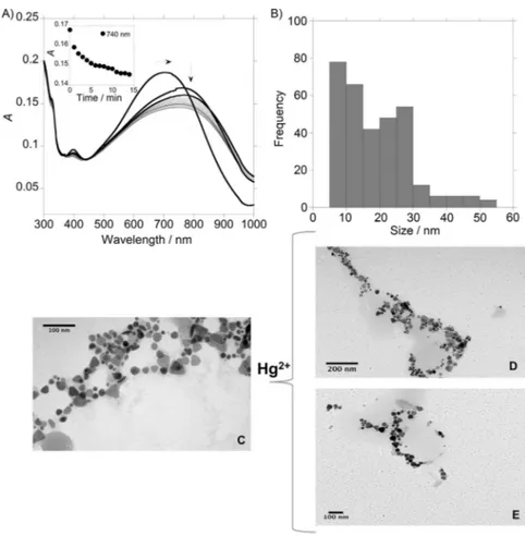
Trying to apply the obtained nanostructures as optical effec-tive nanochemosensors, the sensing of Hg2+
using the colloi-dal systems AgNPs@L2 and AuNPs@L2 was carried out in aqueous solution. Changes in the absorption spectra of the colloidal systems AgNPs@(L)2with spherical (Figure 4 A,B) and triangular (see Figure 5) shapes were observed after the addi-tion of increasing amounts of Hg(NO3)2. In both cases, a
blue-shift of the SPR bands in the UV/Vis spectra suggest a continu-ous deformation of the chains, that is confirmed by TEM mi-croscopy (Figure 4 D,E and Figure 5 D,E). Thezpotentials of the spherical and triangular AgNPs@(L)2 solutions were z
spherical 27.8 mV cm1 and z
triangular 19.1 mV cm1, respectively. In
both cases, after the addition of Hg2+
, a destabilization of the systems was observed, and the value of zpotentials changes
tozspherical 22.5 mV cm1andztriangular 17.0 mV cm1.
In view of these results, we can conclude that the destabili-zation of the spherical and triangular AgNPs@(L)2systems and the loss of the assembly, could be due to the more favorable interaction of L2 with Hg2+
to that observed for L2 with AgNPs. The exchange ofL2adsorbed on the surface produces a decrease of the negative net charge in the NP surface, which causes an increase of thezpotentials.
On the other hand, different results were obtained for linker L2 adsorbed on the AuNPs surface because a more stable bond is formed compared with that obtained forL2and Hg2+ , explaining that the AuNPs@(L)2 assembly remains un-changed after the interaction with this metal ion.
The sensitivity of this nanoassembly system AgNPs@(L)2 (spherical shape) toward Hg2+
was found to be comparable to the small fluorescent molecular system L. The value for the limit of detection (LOD) shows that the best candidate for the detection of this metal ion is system AgNPs@(L)2, with the minimum amount of Hg2+
detectable being 8.3 ppm, whereas the LOD value with compoundLwas 19.0 ppm. The selectively of systemLtowards Hg2+
was also explored as shown in Fig-ure S4 and S5.
Conclusions
This study of 1D self-assembly of silver nanoparticles (AgNPs) and gold nanoparticles (AuNPs) demonstrates direct evidence of the cooperative interaction between the metal nanoparticles (MNPs) and the deprotonated polyamine compoundL2at the nanoscale in aqueous solution.
The unexpected symmetry breaking that occurs whenL2is added to an aqueous suspension of citrate-capped isotropic AgNPs and AuNPs was attributed to the ligand-mediated in-duction of a surface electrical dipole. At least three features of
Figure 4.A) Spectrophotometric titration of spherical AgNPs@(L)2with the addition of increasing amounts of HgCl2in aqueous solution. B) Modification with time in the ab-sorption spectra of AgNPs@(L)2with the addition of 6mL of HgCl
potentially modifying L2 could influence citrate-stabilized AgNPs and AuNPs: (1) the presence of a sulfur atom, which can form covalent bonds with silver and gold atoms; (2) the presence of ionizable functional groups (amine); and (3) the charge (+/) of ionizable functional groups. Electrostatic re-pulsion between AgNPs and AuNPs was progressively reduced, and the stability of the electric dipole associated with charge separation on the nanocrystal surface was potentially en-hanced by spatial partitioning of L2 and citrate-capping li-gands. As a consequence, highly extended 1D NP assemblies in the form of discrete chains, bifurcated and looped chains, or interconnected chain networks are assembled spontaneously as the concentration of surface-adsorbed L2 molecules in-creases.
Monoanionic compound L2 causes shifts to the initial ab-sorption spectra of AgNPs and AuNPs, and can be used for de-velopment of a surface plasmon resonance (SPR) chemical and biomolecular sensing platform because the interaction of cit-rate-stabilized AgNPs and AuNPs with the aforementioned compound is a very sensitive easy-to-visualize process.
The sensing of Hg2+
in aqueous solution using AgNPs@(L)2 and AuNPs@(L)2 was carried out. A destabilization of the system AgNPs@(L)2 (spherical and triangular shape) and the loss of the assembly were observed due to the interaction of
L2 with Hg2+
is more favorable to that observed for L2 with AgNPs. LinkerL2binds to AuNP surfaces and forms a more stable bond compared with that obtained betweenL2and Hg2+
, explaining that the AuNPs@(L)2 assembly remains unchanged after the interaction with this metal ion.
Experimental Section
General
Instrumentation: Elemental analy-ses were carried out with Fisons In-struments EA1108 microanalyzer (Ipswich, UK) at the University of Vigo (CACTI), Spain. Infrared spec-tra were recorded in KBr windows using a JASCO FT/IR-410 spectro-photometer (Spain). 1
H and 13
C NMR were carried out on a Bruker Avance III400 at an oper-ating frequency of 400 MHz for 1
H NMR and 100.6 MHz for 13
C NMR using the solvent peak as an internal reference at 258C. MALDI-MS analyses were per-formed with a MALDI-TOF/TOF MS model Ultraflex II (Bruker, Germa-ny) equipped with nitrogen from the BIOSCOPE group. Each spec-trum represents accumulations of 5 50 laser shots. The reflection mode was used, and the ion source and flight tube pressure were less than 1.80 107
and 5.60 108
Torr, respectively. The MALDI mass spectra of the soluble samples (1 or 2mgmL1
) were recorded using the conventional sample preparation method for MALDI MS. One microliter was put on the sample holder on which the ligand had been previously spotted. The sample holder was inserted in the ion source. UV/Vis absorption spectra (220–900 nm) were per-formed using a JASCO-650 UV/Vis spectrophotometer (Oklahoma City, OK, USA) and fluorescence spectra on a HORIBA JOVIN-IBON Spectramax 4 (Irvine, CA, USA). All measurements were performed at 298 K.
Limit of detection: The limit of detection (LOD) for Hg2+
with the small fluorescent molecular systems L and the nanoassembly system AgNPs@(L)2 (spherical) were performed having in mind
their use for real ion detection and for analytical applications. The LOD was obtained using Equation 1:
ydl¼yblankþ3std ð1Þ
where ydl=signal detection limit and std=standard deviation.
Concentration determination: Assuming a spherical shape and uniform face centered cubic (fcc) structure, the molar concentra-tions of the silver nanoparticle (AgNP) and gold nanoparticle (AuNP) solutions was calculated using Equations 2 and 3.[46]
N¼p
6
1D3
M ð2Þ
where Nis the number of atoms per AgNP or AuNP,1[g cm3 ] is the density of face centered cubic (fcc) silver (10.5 g cm3
) of gold (19.3 g cm3
), and M[g mol1
] is the atomic mass of sliver (107.86 g mol1
) or gold (196.97 g mol1 ).
C¼ NT NVNA
ð3Þ
where Cis the molar concentration of AgNPs or AuNPs,NTis the total number of silver atoms added as AgNO3or gold atoms added as HAuCl4,Nis the number of NPs,Vis the volume of the reaction solution in L, NA is Avogadro’s constant (number of atoms per mole).
Characterization of the assemblies of AgNPs and AuNPs: We characterized the AgNPs, AuNPs and the chainlike assemblies of AgNPs and AuNPs using a number of optical tools including, trans-mission electron microscopy (TEM) and dynamic light scattering (DLS). To collect TEM images, the samples were prepared dropping 1mL of the colloidal suspension onto a copper grid coated with a continuous carbon film and allowing the solvent to evaporate. TEM images were obtained using a JEOL JEM 1010F TEM operating at 200 kV. To perform the Fourier transformations, we used the Dig-ital Micrograph (Gatan) software.[47]
The NP size distributions were measured using the DLS system, Malvern Nano ZS instrument (Worcestershire, UK) with a 633 nm laser diode. We investigated the optical properties of these structures using a JASCO-650 UV/Vis spectrophotometer.
Synthesis
2,2’-((Thiobis(ethane-2,1-diyl))bis(azanediyl))bis(N -(naphthalen-1-yl)acetamide) (L): A solution of 20 % NaOH (3.4 g, 0.085 mol) was added to a stirred solution of 1-naphthylamine (9.34 g, 0.051 mol) in CH2Cl2(30 mL). The mixture was cooled to 08C and chloroacetyl chloride (9.29 g, 0.083 mol) was added dropwise for 45 min. After stirring at 08C for 100 min, the mixture was allowed to warm to RT. The aqueous layer was separated and extracted with CH2Cl2 (2 25 mL). The combined organic phases were washed with HCl (5 % v/v), NaHCO3(5 %v/v) and H2O, dried over MgSO4and filtered to obtain a white solid. The crude product was purified by silica column chromatography and characterized as 2-chloro-N-(1-naph-thyl)acetamide (74 %).
A solution of 2-chloro-N-(1-naphthyl)acetamide (439.34 mg, 2 mmol) in tetrahydrofuran (THF; 25 mL) was added dropwise to a solution of 2,2’-thiobis(ethylamine) (120 mg, 1 mmol) and tri-ethylamine (202.24 mg, 2 mmol) dissolved in THF (50 mL) over 1 h in an ice bath. After the addition was completed, the reaction mix-ture was kept at reflux for 4 h. The solvent was removed in vacuo, and the residue was washed with H2O/CHCl3 (1:3v/v; 4 20 mL). The resulting organic phase was dried in vacuo to giveLas a pink powder (407.05 mg, 84 %): 1H NMR (500 MHz, CDCl
3): d=2.81 (t, 4 H), 3.12 (t, 4 H), 3.72 (m, 4 H), 5.84 (s, 2 H), 7.41–7.59 (m, 2 H), 7.72 (m, 2 H), 7.80 (m, 2 H), 7.92 (m, 4 H), 8.10 (m, 4 H), 10.35 ppm (s, 2 H); 13C NMR (500 MHz, CDCl
3) d=30.32, 54.85, 58.37, 120.69, 124.80, 125.73, 125.90, 127.20, 128.05, 128.19, 133.03, 133.56, 169.83 ppm; IR (KBr):n˜=1436 ((C=C)ar), 1676 (C=O), 3262 cm1(N H); MALDI-TOF MS: m/z 487.21 [M+H]+
; Anal. calcd for
C28H30N4O2S: C 69.1, N 11.5, S 6.6, H 6.2, found: C 69.3, N 11.2, 6.4, H 6.6.
Preparation of AgNPs: Citrate-stabilized AgNPs of different shapes (spherical and triangular) were synthesized in aqueous solution fol-lowing the Frank methodology.[48]
For the synthesis of AgNPs with spherical shape, sodium citrate (2.0 mL, 1.25 102
m), AgNO3 (5.0 mL, 3.75 104
m), and H2O2(5.0 mL, 5.0 10 2
m) were. After that, freshly prepared NaBH4 (2.5 mL, 5.0 10
3
m) was added. To obtain AgNPs with triangular shape, before the addition of NaBH4, KBr (40mL, 1.0 103
m) was added to the solution. Once all re-agents were combined, the resulting solutions were carefully swirled to fully mix the reactants. Almost immediately, the progres-sion of the reaction becomes evident through the visual changes consistent with the growth of silver nanoprisms. Yellow and blue colors were observed for the spherical and triangular AgNPs, re-spectively (see Figure 1S, Supporting Information). Using Equa-tions 2 and 3,N=30.70D3
, and the resulting spherical AgNPs solu-tion had a concentrasolu-tion of CAgNP=7.8 10
10
m with AgNPs of (253) nm size.
Preparation of AuNPs: Preparation of AuNPs was performed fol-lowing the Turkevish method[49]
through reduction of tetrachlor-oaurate ions (AuCl4) by boiling in aq sodium citrate solution. HAuCl4·3 H2O (49.5 mg, 0.125 mmol) dissolved in nanopure H2O (125 mL; 18.2 MWcm) was added to a preheated solution of sodium citrate (12.5 mL, 1 wt %). The resulting solution was heated to 1008C for 60 min and turned colorless before changing to violet and finally to ruby red. AuNPs obtained using this method appear as almost monodispersed globular structures with a size of (20
5) nm, which are stabilized by weakly bound citrate anions. Using Equations 2 and 3, N=30.896D3
for AuNPs and the resulting AuNPs solution was found to have a ofCAuNPs=3.7 10
9 m.
Chainlike assemblies of AgNPs and AuNPs: We used the poly-amine molecular probe L2 to investigate the effect of the
pres-ence of different donor atoms in the AuNP assemblies and their optical properties. The formation of chainlike assemblies of AuNPs was controlled and modulated observing that the total formations were obtained by adding an acetonitrile solution ofL2(5mL, 1
103
m) into a suspension of AuNPs and AgNPs (circular and trian-gular) in nanopure H2O (10
8 –109
min 3 mL).
Acknowledgements
We are grateful to the Scientific Association ProteoMass (Portu-gal) for financial support. C.N. thanks Xunta de Galicia (Spain) for her postdoctoral contract (I2C program).
Keywords: 1D nanochains · self-assembly · gold · mercury · nanoparticles·silver
[1] a) M. C. Daniel, D. Astruc, Chem. Rev. 2004, 104, 293; b) T. Sau, A. Rogach,Adv. Mater.2010,22, 1781 – 1804.
[2] a) G. J. Hutchings, M. Brust, H. Schmidbaur,Chem. Soc. Rev.2008,37, 1759; b) S. Guo, E. Wang,Nano Today2011,6, 240 – 264.
[3] a) M. A. Kumar M., S. Jung, T. Ji,Sensors2011,11, 5087 – 5111; b) F. Ratto, P. Matteini, S. Centi, F. Rossi, R. Pini,J. Biophotonics2011,4, 64 – 73; c) J. Stone, S. Jackson, D. Wright,Wiley Interdiscip. Rev. Nanomed. Nanobio-technol. Nanomed. NanobioNanobio-technol.2011,3, 100 – 109; d) F. S.-J. Kim, G.-Q. Ren, S. A. Jenekhe,Chem. Mater.2011,23, 682 – 732.
[5] T. Teranishi,C. R. Chim.2003,6, 979 – 987.
[6] D. F. Zhang, L. Y. Niu, L. Jiang, P. G. Yin, L. D. Sun, H. Zhang, R. Zhang, L. Guo, C. H. Yan,J. Phys. Chem. C2008,112, 16011 – 16016.
[7] Y. Yang, S. Matsubara, M. Nogami, J. L. Shi, W. M. Huang, Nanotechnolo-gy2006,17, 2821 – 2827.
[8] a) C. L. Chen, P. J. Zhang, N. L. Rosi,J. Am. Chem. Soc.2008,130, 13555 – 13557; b) J. Y. Chang, H. M. Wu, H. Chen, Y. C. Ling, W. H. Tan,Chem. Commun.2005, 1092 – 1094; c) J. Zhang, M. Riskin, R. Freeman, R. Tel-Vered, D. Balogh, H. Tian, I. Willner,ACS Nano2011,5, 5936 – 5944. [9] K. G. Thomas, S. Barazzouk, B. I. Ipe, S. T. S. Joseph, P. V. Kamat,J. Phys.
Chem. B2004,108, 13066 – 13068.
[10] a) G. A. DeVries, M. Brunnbauer, Y. Hu, A. M. Jackson, B. Long, B. T. Nelt-ner, O. Uzun, B. H. Wunsch, F. Stellacci, Science 2007, 315, 358 – 361; b) Z. H. Nie, D. Fava, E. Kumacheva, S. Zou, G. C. Walker, M. Rubinstein,
Nat. Mater.2007,6, 609 – 614.
[11] M. Li, S. Johnson, H. Guo, E. Dujardin, S. Mann,Adv. Funct. Mater.2011,
21, 851 – 859.
[12] a) B. Ding, Z. Deng, H. Yan, S. Cabrini, R. N. Zuckermann, J. Bokor,J. Am. Chem. Soc.2010, 132, 3248 – 3249; b) H. J. Kim, Y. Roh, S. K. Kim, B. Hong,J. Appl. Phys.2009,105, 074302 – 074304.
[13] a) A. Ongaro, F. Griffin, P. Beecher, L. Nagle, D. Iacopino, A. Quinn, G. Redmond, D. Fitzmaurice,Chem. Mater.2005,17, 1959 – 1964; b) G. Wei, L. Wang, L. Sun, Y. Song, Y. Sun, C. Guo, T. Yang, Z. Li,J. Phys. Chem. A J. Phys. Chem. B J. Phys. Chem. A## B##2007,111, 1976 – 1982.
[14] a) M. Higuchi, K. Ushiba, M. Kawaguchi, J. Colloid Interface Sci.2007,
308, 356; b) S. Si, T. K. Mandal,Langmuir2006,22, 190 – 195.
[15] a) Z. Zhong, S. Patskovskyy, P. Bouvrette, J. H. T. Luong, A. Gedanken,J. Phys. Chem. A J. Phys. Chem. B J. Phys. Chem. B.2004,108, 4046 – 4052; b) Z. Zhong, J. Luo, L. T. Ang, J. Highfield, J. Lin, A. Gedanken,J. Phys. Chem. A J. Phys. Chem. B.2004,108, 18119 – 18123; c) P. R. Selvakannan, S. Mandal, S. Phadtare, A. Gole, R. Pasricha, S. D. Adyanthaya, M. Sastry,
J. Colloid Interface Sci.2004,269, 97 – 102; d) Y. Shao, Y. Jin, S. Dong,
Chem. Commun.2004, 1104 – 1105; e) Z. Zhong, A. S. Subramanian, J. Highfield, K. Carpenter, A. Gedanken, Chem. Eur. J. 2005, 11, 1473 – 1478; f) L. Polavarapu, Q.-H. Xu,Nanotechnology 2008, 19, 075601 – 075606.
[16] S. Hsieh, C.-W. Hsieh,Chem. Commun.2010,46, 7355 – 7357.
[17] D. Lee, Y.-J. Choe, Y. S. Choi, G. Bhak, J. Lee, S. R. Paik,Angew. Chem.
2011,123, 1368 – 1373;Angew. Chem. Int. Ed.2011,50, 1332 – 1337. [18] L.-X. Qin, Y. Li, D.-W. Li, C. Jing, B.-Q. Chen, W. Ma, A. Heyman, O.
Sho-seyov, I. Willner, H. Tian, Y.-T. Long,Angew. Chem. Int. Ed.2012,51, 140 – 144.
[19] M. A. Correa-Duarte, L. M. Liz-Marzn,J. Mater. Chem.2006,16, 22 – 25. [20] K. Liu, Z. Nie, N. Zhao, W. Li, M. Rubinstein, E. Kumacheva,Science2010,
329, 197 – 200.
[21] A. E. Nel, L. Madler, D. Velegol, T. Xia, E. M. V. Hoek, P. Somasundaran, F. Klaessig, V. Castranova, M. Thompson,Nat. Mater.2009,8, 543 – 557. [22] a) N. Sharma, A. Top, K. L. Kiick, D. J. Pochan,Angew. Chem.2009,121,
7212 – 7216;Angew. Chem. Int. Ed.2009,48, 7078 – 7082 ; b) M. L. Bron-gersma, J. W. Hartman, H. A. Atwater,Phys. Rev. B 2000, 62, R16356 – R16359; c) M. Quinten, A. Leitner, J. R. Krenn, F. R. Aussenegg,Opt. Lett.
1998,23, 1331 – 1333.
[23] a) A. N. Shipway, M. Lahav, R. Gabai, I. Willner,Langmuir2000,16, 8789 – 8795; b) B. Nikoobakht, M. A. El-Sayed,Langmuir2001,17, 6368 – 6374; c) C. J. Johnson, E. Dujardin, S. A. Davis, C. J. Murphy, S. Mann,J. Mater. Chem.2002,12, 1765 – 1770.
[24] A. M. Jackson, J. W. Myerson, F. Stellacci,Nat. Mater.2004,3, 330 – 336. [25] S. C. Glotzer, M. J. Solomon,Nat. Mater.2007,6, 557 – 562.
[26] Z. Tang, N. A. Kotov, M. Giersig,Science2002,297, 237 – 240. [27] M. Faraday,Philos. Trans. R. Soc. London1857,147, 145 – 181.
[28] a) C. S. Thaxton, D. G. Georganopoulou, C. A. Mirkin, Clin. Chim. Acta
2006,363, 120 – 126; b) E. H. Witlicki, C. Johnsen, S. W. Hansen, D. W. Sil-verstein, V. J. Bottomley, J. O. Jeppesen, E. W. Wong, L. Jensen, A. H. Flood,J. Am. Chem. Soc.2011,133, 7288 – 7291; c) N. Nerambourg, R.
Praho, M. H. V. Werts, D. Thomas, M. B. Desce,Int. J. Nanotechnol.2008,
5, 722 – 740; d) X. Wu, S. Chang, X. Sun, Z. Guo, Y. Li, J. Tang, Y. Shen, J. Shi, H. Tian, W. Zhu,Chem. Sci.2013,4, 1221 – 1228.
[29] a) P. K. Jain, K. S. Lee, I. H. El-Sayed, M. A. El-Sayed, J. Phys. Chem. B
2006,110, 7238 – 7248 ; b) C. A. Mirkin, R. L. Letsinger, R. C. Mucic, J. J. Storhoff, Nature 1996, 382, 607 – 609; c) A. de la Escosura-MuÇiz, C. Parolo, A. MerkoÅi,Mater. Today2010,13, 24 – 34; d) F. Wei, R. Lam, S. Cheng, S. Lu, D. Ho, N. Li,Appl. Phys. Lett.2010,96, 133702 (3)e) W. J. Qi, D. Wu, J. Ling, C. Z. Huang,Chem. Commun.2010,46, 4893 – 4895. [30] J. J. Mock, M. Barbic, D. R. Smith, D. A. Schultz, S. Schultz,J. Chem. Phys.
2002,116, 6755 – 6762.
[31] a) Y. Cheng, M. Wang, G. Borghs, H. Chen,Langmuir2011,27, 7884 – 7891; b) E. Charrault, M. He, P. Muller, M. Maaloum, C. Petit, P. Petit,
Langmuir2009,25, 11285 – 11288.
[32] Y. Xia, Y. Xiong, B. Lim, S. E. Skrabalak,Angew. Chem.2009,121, 62 – 108;
Angew. Chem. Int. Ed.2009,48, 60 – 103.
[33] a) A. Taleb, C. Petit, M. P. Pileni,Chem. Mater. 1997,9, 950 – 959; b) R. Matassa, I. Fratoddi, M. Rossi, C. Battocchio, R. Caminiti, M. V. Russo,J. Phys. Chem. C2012,116, 15795 – 15800.
[34] J.-Y. Chang, J.-J. Chang, B. Lo, S.-H. Tzing, Y.-C. Ling,Chem. Phys. Lett.
2003,379, 261 – 267.
[35] a) C. Lodeiro, J. L. Capelo, J. C. Mejuto, E. Oliveira, H. M. Santos, B. Pedras, C. NfflÇez,Chem. Soc. Rev.2010,39, 2948 – 2976 ; b) E. Oliveira, C. Nfflnez, B. Rodrguez-Gonzlez, J. L. Capelo, C. Lodeiro, Inorg. Chem.
2011,50, 8797 – 8807; c) E. Oliveira, D. Genovese, R. Juris, N. Zaccheroni, J. L. Capelo, M. M. M. Raposo, S. P. G. Costa, L. Prodi, C. Lodeiro, Inorg. Chem.2011, 50, 8834 – 8849; d) E. Oliveira, J. D. Nunes-Miranda, H. M. Santos,Inorg. Chim. Acta2012,380, 22 – 30; e) J. Fernndez-Lodeiro, C. NfflÇez, O. Nieto Faza, J. L. Capelo, C. Lodeiro, J. S. Seixas de Melo, C. S. Lpez,Inorg. Chim. Acta2012,381, 218 – 228; f) R. Lpez-Corts, E. Oli-veira, C. NfflÇez, C. Lodeiro, M. Pez de La Cadena, F. Fdez-Riverola, H. Lpez-Fernndez, M. Reboiro-Jato, D. Glez-PeÇa, J. L. Capelo, H. M. Santos,Talanta2012,100, 239 – 245.
[36] S. Zhang, X. Kou, Z. Yang, Q. Shi, G. D. Stucky, L. Sun, J. Wang, C. Yan,
Chem. Commun.2007, 1816 – 1818.
[37] X. Kou, Z. Sun, Z. Yang, H. Chen, J. Wang,Langmuir2009, 25, 1692 – 1698.
[38] T. S. Sreeprasad, T. Pradeep,Langmuir2011,27, 3381 – 3390. [39] A. Moores, F. Goettmann,New J. Chem.2006,30, 1121 – 1132. [40] A. S. de Dios, M. E. Daz-Garca,Anal. Chim. Acta2010,666, 1 – 22. [41] H. Zhang, K.-H. Fung, J. Hartmann, C. T. Chan, D. Wang,J. Phys. Chem. C
2008,112, 16830 – 16839.
[42] S. Mandal, S. Phadtare, M. Sastry,Curr. Appl. Phys.2005,5, 118 – 127. [43] V. Chegel, O. Rachkov, A. Lopatynskyi, S. Ishihara, I. Yanchuk, Y. Nemoto,
J. P. Hill, K. Ariga,J. Phys. Chem. C2012,116, 2683 – 2690.
[44] P. R. Selvakannan, S. Mandal, S. Phadtare, R. Pasricha, M. Sastry, Lang-muir2003,19, 3545 – 3549.
[45] K. J. M. Bishop, C. E. Wilmer, S. Soh, B. A. Grzybowski,Small 2009, 5, 1600 – 1630.
[46] X. Liu, M. Atwater, J. Wang, Q. Huo,Colloids Surf. B Biointerfaces2007,
58, 3 – 7.
[47] A. Snchez-Iglesias, I. Pastoriza-Santos, J. Prez-Juste, B. Rodrguez-Gon-zlez, F. Garca de Abajo, L. Liz-Marzn, Adv. Mater. 2006, 18, 2529 – 2534.
[48] A. J. Frank, N. Cathcart, K. E. Maly, V. Kitaev,J. Chem. Educ.2010,87, 1098 – 1101.
[49] a) J. Turkevich, P. C. Stevenson, J. Hillier,Discuss. Faraday Soc.1951,11, 55 – 75; b) B. V. En st n, J. Turkevich,J. Am. Chem. Soc.1963,85, 3317 – 3328; c) J. Turkevich,Gold Bull. 1985,18, 86 – 91; d) J. Turkevich,Gold Bull.1985,18, 125 – 131.
Received: May 10, 2013