UNIVERSIDADE DE LISBOA
Faculdade de Medicina Veterinária
DEVELOPMENT AND OPTIMISATION OF A GROUP-SPECIFIC REAL-TIME RT-PCR ASSAY FOR THE BROAD DETECTION OF THE SIMBU SEROGROUP
ORTHOBUNYAVIRUSES
ANTÓNIO ALEXANDRE RIACHOS CAMARÃO
CONSTITUIÇÃO DO JÚRI ORIENTADOR Doutor Virgílio da Silva Almeida Doutor Melvyn Quan Doutor Fernando Jorge Silvano Boinas CO-ORIENTADOR Doutora Ana Isabel Simões Pereira Duarte Doutor Fernando Jorge Silvano Boinas
2018 LISBOA
UNIVERSIDADE DE LISBOA
Faculdade de Medicina Veterinária
DEVELOPMENT AND OPTIMISATION OF A GROUP-SPECIFIC REAL-TIME RT-PCR ASSAY FOR THE BROAD DETECTION OF THE SIMBU SEROGROUP
ORTHOBUNYAVIRUSES
ANTÓNIO ALEXANDRE RIACHOS CAMARÃO
DISSERTAÇÃO DE MESTRADO INTEGRADO EM MEDICINA VETERINÁRIA
CONSTITUIÇÃO DO JÚRI ORIENTADOR Doutor Virgílio da Silva Almeida Doutor Melvyn Quan Doutor Fernando Jorge Silvano Boinas CO-ORIENTADOR Doutora Ana Isabel Simões Pereira Duarte Doutor Fernando Jorge Silvano Boinas
2018 LISBOA
i
Acknowledgements
As long as we are people before we become professionals, it makes absolute sense to start expressing my eternal gratitude to my parents, sister and grandparents, an example since ever, basis of my education, for having given me all the basic values and necessary experiences for me to be the person I am today.
On what concerns to the professional level, generally speaking, I am deeply grateful to everyone that contributed to my veterinary knowledge and experience during these academic years. But particularly, I am indebted to my co-supervisor and infectious diseases professor, Prof. Fernando Boinas, which contributed to my special interest for the infectious diseases, and opened me “the doors of Africa”, for the helpful guidance and for the trust placed in me. I wish to express my sincere thanks to my supervisor, virologist and expert in molecular diagnostics, Prof. Melvyn Quan, not only for all the knowledge transmitted, in the most varied aspects, but also for the friendship. I want to record my gratitude to the Department of Veterinary Tropical Dieases, University of Pretoria, which made everything possible funding this project, and to some of the staff, for the confidence, help and for the “corridor conversations”, a thanks to Prof. Tshepo Matjila (Head of Department), Faith Nkosi, Elise van der Heijden, Donald Mukolwe, Paida Hove, Herman Geyer, Prof. Luís Neves, Dr. Jan Van Wyk, as well as to Karen Ebersohn for the help with the cell cultures and to Carien van den Bergh for the mosquito samples. I am deeply grateful to Prof. Robert Swanepoel, respectful virologist, for the lessons in arbovirology and for the help with the literature review. I owe a debt of gratitude to my microbiology and immunology professor, virologist and expert in molecular diagnostics, Prof. Ana Duarte, who introduced me to the “molecular world” applied to the diagnosis of infectious diseases. I would like to express my thanks to another respectful virologist, Prof. Carlos Martins, and to Dr. Alexandre Leitão, for the valuable comments on this manuscript. I also want to thanks to my “officemate” while I was writing, Sara Madeira, and to my friend, Miguel Amado, for the help with the graphic design of some of the figures.
And last but not least, to all my friends, “non-vets” and “vets”, as well as to my colleagues, that have been shaping my character, personality and professionalism.
ii
Abstract
The Simbu serogroup within the genus Orthobunyavirus belongs to the family Peribunyaviridae and comprises 32 recognised three-segmented negative-sense single-stranded RNA viruses, divided into two phylogenetic clades. Some members of this group of arthropod-borne viruses, cosmopolitan distributed, cause neurologic disease in humans as well as reproductive and neurologic disease in domestic animals, however, definitive diagnosis always requires laboratorial confirmation. Few real-time RT-PCR assays have been developed for the molecular diagnosis of Simbu serogroup orthobunyaviruses. There are two published methods with broad detection capacity, utilising either a SYBR Green based chemistry able to recognise viruses from both clades, which is not absolutely specific, or a TaqMan based chemistry that recognises only clade B viruses. A novel group-specific TaqMan-based real-time RT-PCR assay was developed, optimised and laboratory validatedfor the broad detection of the Simbu serogroup orthobunyaviruses. The published genomic data of the Simbu serogroup members were evaluated, and a conserved region, situated in the segment encoding the nucleocapsid protein, was selected to design a universal primer set and a pair of differently labelled hydrolysis probes, which allowed for the distinction between the two phylogenetic clades of the Simbu serogroup. Seven prototype Simbu serogroup isolates were used for the development of the assay, namely Akabane orthobunyavirus, Simbu orthobunyavirus, Shuni orthobunyavirus, Sathuperi orthobunyavirus, Shamonda orthobunyavirus, Ingwavuma virus and Sabo virus. The primer and probe concentrations in the reaction were optimised. Amplification efficiency was determined for each one of the viruses: AKAV (99%), SIMV (96%), SHUV (96%), SATV (97%), SHAV (84%), INGV (93%) and SABOV (110%). A panel constituted of genetically related, causative agents of abortion in ruminants and arthropod-borne viruses was selected for in vitro specificity analysis, and in silico analysis was also performed. The assay was shown to be specific, as no cross-reactions were observed either in vitro or in silico, and sensitive, with a 95% limit of detection ranging from 100,39 to 10-3,61 TCID50/reaction, for the detection of Simbu serogroup viruses. The repeatability of the assay was evaluated for both probes detection, using the intra- and inter-run standard deviations and coefficient of variation. This work resulted in a manuscript in submission process to a peer-reviewed journal. In addition, a comprehensive review of the viruses of the Simbu serogroup was carried out, including sites of viral isolation and seroconversion, and a map was generated using a geographic information system tool.
Keywords: Peribunyaviridae, Molecular diagnosis, TaqMan assay, Wide spectrum,
iii
Resumo
O serogrupo Simbu pertence ao género Orthobunyavirus, família Peribunyaviridae, e é constituído por 32 vírus de RNA tri-segmentado de cadeia simples e polaridade negativa, divididos em duas clades filogenéticas. Alguns membros deste grupo de vírus transmitidos por artrópodes, com distribuição cosmopolita, causam doença neurológica em humanos bem como doença reprodutiva e neurológica em animais domésticos, no entanto, o diagnóstico definitivo requer sempre confirmação laboratorial. Poucos ensaios de RT-PCR em tempo real têm sido desenvolvidos para o diagnóstico molecular destes vírus, ainda assim, existem dois que se destacam pela capacidade de detecção em largo espectro. Um deles, com sistema de detecção de fluorescência baseado na utilização de SYBR Green, é capaz de detectar vírus das duas clades, mas não é absolutamente específico, e o outro, baseando-se na utilização de sondas TaqMan, só detecta vírus de uma das clades. Um ensaio de RT-PCR em tempo real grupo-específico, inédito, com sondas TaqMan, foi desenvolvido, optimizado e caracterizado em termos laboratoriais, para a detecção em largo espectro dos orthobunyavirus do serogrupo Simbu. Os dados publicados referentes ao genoma dos membros do serogrupo foram analisados, e uma região conservada, situada no segmento codificante da proteína da nucleocápdise, foi eleita para o desenho de um par de primers específicos universal, bem como de duas sondas de hidrólise específicas, distintamente marcadas, e que, portanto, permitem a diferenciação entre as clades filogenéticas. Sete isolados de referência foram utilizados no desenvolvimento do ensaio, nomeadamente Akabane orthobunyavirus, Simbu orthobunyavirus, Shuni orthobunyavirus, Sathuperi orthobunyavirus, Shamonda orthobunyavirus, Ingwavuma virus e Sabo virus e, consequentemente, as concentrações dos primers e sondas na reação foram optimizadas. A eficiência de amplificação foi determinada para cada um dos vírus: AKAV (99%), SIMV (96%), SHUV (96%), SATV (97%), SHAV (84%), INGV (93%) e SABOV (110%). Um painel constituído por vírus geneticamente relacionados, agentes causais de aborto em ruminantes e transmitidos por artrópodes foi seleccionado no sentido de avaliar a especificidade do ensaio in vitro, tendo sido também efectuada uma análise in silico. O ensaio é específico, visto que não foram observadas reacções cruzadas quer in vitro quer in silico, e sensível, com um limite de detecção de 95% entre 100,39 a 10-3,61 TCID50/reacção, para a detecção de vírus do serogrupo Simbu. A repetibilidade foi avaliada para a detecção com ambas as sondas, pelo cálculo do desvio padrão intra- e inter-corridas bem como do coeficiente de variação. Este trabalho originou um manuscrito em processo de submissão para uma revista cientifíca. Além disso, foi levada a cabo uma revisão bibliográfica do serogrupo Simbu, incluindo locais de isolamento viral e seroconversão, e um mapa inédito desta distribuição foi gerado.
Palavras-chave: Peribunyaviridae, Diagnóstico molecular, Ensaio TaqMan, Largo espectro,
iv
List of Contents
1. Introductory Note ... 1
1.1. The Internship ... 1
1.2. The Research Project ... 1
2. Introduction ... 2
3. Literature Review ... 4
3.1. The Simbu serogroup, Genus Orthobunyavirus, Family Peribunyaviridae, Order Bunyavirales ... 4
3.1.1. General characteristics ... 4
3.1.2. Genome organisation ... 15
3.2. Molecular diagnosis using real-time polymerase chain reaction (qPCR)... 17
3.2.1. General review ... 17
3.2.2. Simbu molecular diagnostic assays based on real-time RT-PCR ... 18
4. Aims and Objectives ... 21
4.1. Aim ... 21
4.2. Objectives ... 21
5. Materials and Methods ... 22
5.1. Simbu serogroup viral isolates ... 22
5.1.1. Infectivity ... 22
5.2. Virus distribution ... 23
5.3. Bioinformatics ... 23
5.3.1. Evaluation of genome variation ... 23
5.3.2. Primer and probe design ... 24
5.3.2.1. Sequencing primers ... 24
5.3.2.2. Real-time RT-PCR primers and probes ... 24
5.4. Nucleic acid purification ... 24
5.5. Sequencing ... 25
5.6. The group-specific RT-qPCR assay ... 25
5.6.1. Development ... 26
5.6.1.1. Pilot run ... 26
5.6.1.2. Comparison with a pan-Simbu assay (Fischer et al., 2013) ... 26
5.6.2. Optimisation ... 27
5.6.2.1. Primer concentration ... 27
5.6.2.2. Simbu_CladeAP probe concentration ... 27
5.6.2.3. Simbu_CladeBP probe concentration ... 27
5.6.3. Efficiency ... 27
5.6.4. Sensitivity ... 27
5.6.5. Specificity ... 28
5.6.6. Repeatability ... 28
5.6.7. Testing of field samples ... 28
5.6.7.1. Abortion products from Namibia... 28
5.6.7.2. Calf with neurological signs ... 29
5.6.7.3. Mosquitoes from Bumbe lake ... 29
6. Results and Discussion ... 31
6.1. Infectivity ... 31
v
6.3. Bioinformatics ... 33
6.3.1. Evaluation of the genome variation ... 33
6.3.2. Primer and Probe Design ... 36
6.3.2.1. Sequencing primers ... 36
6.3.2.2. Real-time RT-PCR primers and probes ... 37
6.4. Sequencing ... 39
6.5. The group-specific RT-qPCR assay ... 39
6.5.1. Development ... 40
6.5.1.1. Pilot run ... 40
6.5.1.2. Comparison with a pan-Simbu assay (Fischer et al., 2013) ... 41
6.5.2. Optimisation of primer and probe concentrations ... 42
6.5.3. Efficiency ... 43
6.5.4. Sensitivity ... 43
6.5.5. Specificity ... 45
6.5.6. Repeatability ... 45
6.5.7. Testing of field samples ... 47
6.5.7.1. Abortion products from Namibia ... 47
6.5.7.2. Calf with neurological signs ... 47
6.5.7.3. Mosquitoes from Bumbe lake ... 48
7. Conclusion ... 49
8. Bibliography ... 50
vi
List of Figures
Figure 3.1. General characteristics of virions of the Order Bunyavirales (Original). ... 4
Figure 3.2. Genome organisation of Orthobunyavirus (Original). ... 15
Figure 6.1. Effect of Simbu viral infection in Vero cells (400). ... 31
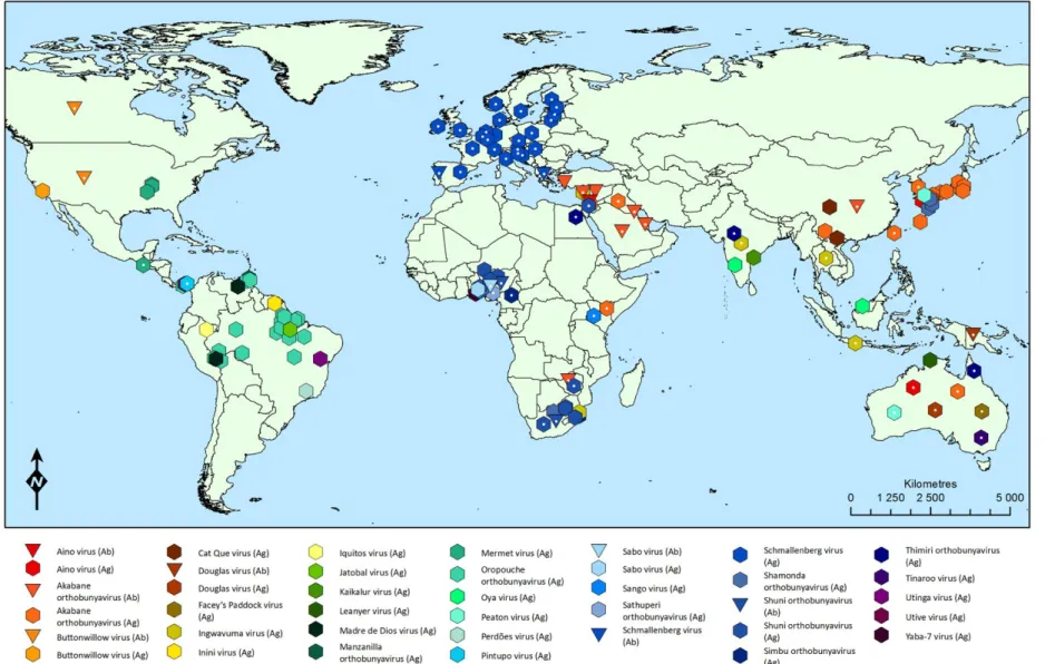
Figure 6.2. Map of the known distribution of the Simbu serogroup orthobunyaviruses ... 32
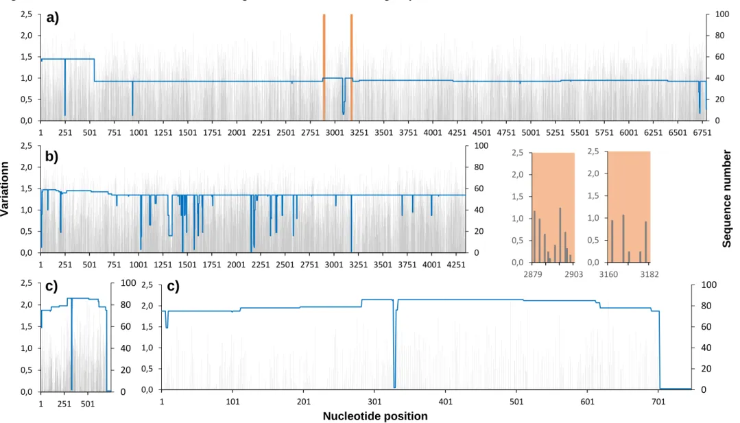
Figure 6.3. Plot of nucleotide variation in the genome of the Simbu serogroup viruses ... 34
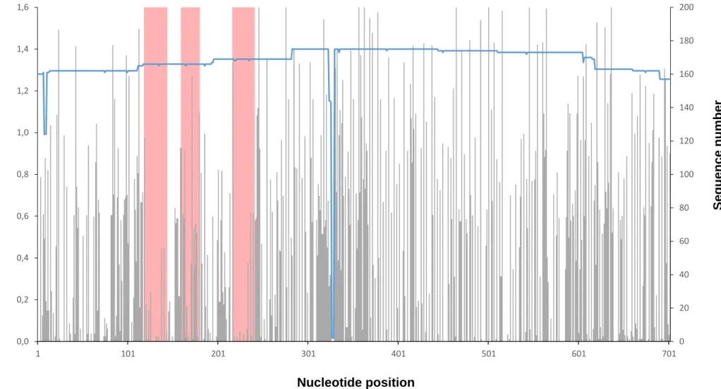
Figure 6.4. Plot of nucleotide variation in the S segment showing primer and probe position ... 35
Figure 6.5. Agarose gel electrophoresis ... 37
Figure 6.6. Simbu serogroup sequence alignment ... 38
Figure 6.7. Pilot run amplification plot ... 40
Figure 6.8. DVTD’s isolates sequence alignment ... 41
Figure 6.9. Amplification plot obtained for primer optimisation... 42
Figure 6.10. Amplification plots obtained for probe optimisation ... 42
Figure 6.11. Linear regression analysis for efficiency calculation ... 44
Figure 6.12. 95% limit of detection of the assay ... 46
Figure 6.13. Amplification plot of the Namibian samples ... 47
Figure 6.14. Amplification plot of the calf samples ... 48
vii
List of Tables
Table 3.1. Known and potential members of the Simbu serogroup orthobunyaviruses, showing
the origins of the prototype isolates (Swanepoel & Camarão, 2017). ... 6
Table 3.2. Phylogeny of members of the Simbu serogroup orthobunyaviruses (Ladner et al., 2014; ICTV, 2015). ... 7
Table 5.1. Simbu serogroup orthobunyavirus isolates stored in the Department of Veterinary Tropical Diseases (DVTD), Faculty of Veterinary Sciences, University of Pretoria. ... 22
Table 6.1. Log TCID50/ml values ... 31
Table 6.2. Sequencing primers ... 36
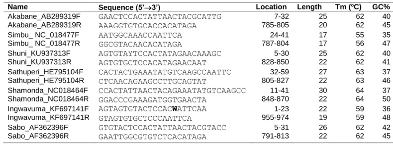
Table 6.3. Group-specific real-time RT-PCR primers and probes... 37
Table 6.4. Pilot run Cq values ... 40
Table 6.5. Comparison with the pan-Simbu ... 41
Table 6.6. Amplification efficiency of a group-specific Simbu serogroup Orthobunyavirus assay. ... 43
Table 6.7. The 95% limit of detection of a group-specific Simbu serogroup Orthobunyavirus assay. ... 43
Table 6.8. Intra and Inter-run variation for probe Simbu_CladeAP detection ... 45
viii
List of Abbreviations
AG/HE – Arthrogryposis and hydranencephaly AINOV – Aino virus
AKAV – Akabane orthobunyavirus BEFV – Bovine fever ephemerovirus BHK – Baby hamster kidney
BLAST – Basic Local Alignment Search Tool BoHV-1 – Bovine alphaherpesvirus 1
BTV – Bluetongue virus BVDV-1 – Pestivirus A CF – Complement fixation CNS – Central nervous system CPE – Cytophatic effect
Cq – Quantification cycle CQV – Cat Que virus
CV – Coefficient of Variation
dNTP – Deoxyribonucleotide phosphate DOUV – Douglas virus
dsDNA – Double-stranded deoxyribonucleic acid DVTD – Departement of Veterinary Tropical Diseases ELISA – Enzyme-linked immunosorbent assay
FBS – Foetal bovine serum
GIS – Geographic Information System HI – Hemagglutination inhibition
ICTV – International Committee on Taxonomy of Viruses INGV – Ingwavuma virus
IQTV – Iquitos virus JATV – Jatobal virus KZN – KwaZulu-Natal LOD – Limit of Detection MDDV – Madre de Dios virus MEM – Minimum essential medium MGB – Minor groove binder
MM – Master mix
mRNA – Messenger ribonucleic acid N – Nucleocpasid
NFQ – Non-fluorescent quencher NS – Non-structural
ORF – Open reading frame
ix OYAV – Oya virus
PALV – Palyam virus
PBS – Phosphate buffered saline PCR – Polymerase chain reaction
qPCR – Quantitative polymerase chain reaction RdRp – RNA-dependent RNA polymerase RNA – Ribonucleic acid
RSA – Republic of South Africa
RT-PCR – Reverse transcripton polymerase chain reaction RVFV – Rift Valley fever phlebovirus
SABOV – Sabo virus
SATV – Sathuperi orthobunyavirus SBV – Schamallenberg virus
SHAV – Shamonda orthobunyavirus SHUV – Shuni orthobunyavirus SIMV – Simbu orthobunyavirus SNT – Serum neutralisation test TAE – Tris-acetate-EDTA
TCID50 – 50% Tissue Culture Infectious Dose TM – Melting temperature
UTR – Untranslated region WSLV – Wesselsbron virus
1
1. Introductory Note
1.1. The Internship
The final mandatory internship for the acquisition of the Integrated Masters in Veterinary Medicine (MIMV) degree by the Faculty of Veterinary Medicine, University of Lisbon, took place at Onderstepoort, Republic of South Africa, in the internationally accredited Faculty of Veterinary Sciences of the University of Pretoria.
The internship with a duration of six months had a rotational structure and a broad spectrum on what concerns to the diverse fields of activity of the Veterinary Sciences. The first rotation, programmed by the Department of Paraclinical Sciences, Section of Public Health, included training in Food Safety, applied to the Udder Health and Milk Hygiene concepts, with visits to dairy farms, as well as to the Meat Safety concept with visits to a private abattoir and a 150 000 head of cattle feedlot. The second rotation, also in the Department of Paraclinical Sciences, took place in the Section of Pathology, and included a practical approach to the anatomopathological diagnosis in domestic and wildlife species, as well as to the discussion of cases which required histopathology for a definitive diagnosis. Posteriorly, and one of my most remarkable veterinary experiences, was a rotation in the Hluvukani Animal Clinic, located in a rural village in Mpumalanga Province with an interface with the Kruger National Park, which is accommodated in a Community Programme that aims to provide primary public and animal health care, in a One Health concept approach, and where the veterinary services are reinvented due to the lack of resources. The forth rotation was in the Clinical Section of the Department of Production Animal Studies, that provides medical and surgical services to livestock and wildlife in the Onderstepoort Veterinary Academic Hospital and surrounding communities. Alongside side these, the internship’s main component was the work developed in the Department of Veterinary Tropical Diseases (DVTD), which was carried out for almost the totality of time in Onderstepoort, either in part time in conjunction with the other activities, and mainly in full time. This dissertation is based on the research project developed in the DVTD.
1.2. The Research Project
The research project, basis of this dissertation was carried out in the DVTD, which funded this study, providing all the facilities, laboratory equipment and reagents used during its development, optimisation and validation.
All the work described furtherly was done entirely by me, under the guidelines and supervision of the DVTD’s Associate Professor Melvyn Quan, who is my supervisor for this dissertation.
2
2. Introduction
The Simbu serogroup within the genus Orthobunyavirus belongs to the family Peribunyaviridae (Adams, Lefkowitz, King, Harrach, Harrison, Knowles, Kropinski, Krupovic, Kuhn, Mushegian, Nibert, Sabanadzovic, Sanfaçon, Siddell, Simmonds, Varsani, Zerbini, Gorbalenya, & Davison, 2017) and comprises 32 recognised three-segmented negative-sense single-stranded RNA viruses (Saeed, Li, Wang, Weaver, & Barrett, 2001a; Elliott & Blakqori, 2011; Ladner, Savji, Lofts, Travassos da Rosa, Wiley, Gestole, Rosen, Guzman, Vasconcelos, Nunes, T, Lipkin, Tesh, & Palacios, 2014; Tilston-Lunel, Hughes, Acrani, da Silva, Azevedo, Rodrigues, Vasconcelos, Nunes, & Elliott, 2015). This group of arthropod-borne viruses (arboviruses) is known to cause central nervous system (CNS) disease in humans (Anderson, Spence, Downs, & Aitken, 1961; Aguilar, Barrett, Saeed, Watts, Russell, Guevara, Ampuero, Suarez, Cespedes, Montgomery, Halsey, & Kochel, 2011; Ladner et al., 2014) and reproductive system and CNS disease in domestic and livestock species (Coverdale, Cybinski, & St George, 1978; Charles, 1994; Coetzer & Howell, 1998; Hoffmann, Scheuch, Hoper, Jungblut, Holsteg, Schirrmeier, Eschbaumer, Goller, Wernike, Fischer, Breithaupt, Mettenleiter, & Beer, 2012; van Eeden, Williams, Gerdes, van Wilpe, Viljoen, Swanepoel, & Venter, 2012; Hirashima, Kitahara, Kato, Shirafuji, Tanaka, & Yanase, 2017). These agents have been isolated from a wide range of wild mammals and birds (Anderson, Spence, Downs, & Aitken, 1960; McIntosh, McGillivray, & Dickinson, 1965; Calisher, Kokernot, De Moore, Boyd, Hayes, & Chappell, 1969; Reeves, Scrivani, Hardy, Roberts, & Nelson, 1970; Carey, Reuben, George, Shope, & Myers, 1971; Pajot, 1980; Seymour, Peralta, & Montgomery, 1983; Navarro, Giambalvo, Hernandez, Auguste, Tesh, Weaver, Montanez, Liria, Lima, Travassos da Rosa, da Silva, Vasconcelos, Oliveira, Vianez, & Nunes, 2016). The viruses in the serogroup have a cosmopolitan distribution and have been divided phylogenetically into two different clades, designated A, those associated with clinical disease in humans, and B, associated with abortion and teratology in ruminants, as well as neurologic disease in horses (Ladner et al., 2014).
Rapid viral detection and large population screening capability provide important epidemiological data, enhance the surveillance and control of emerging, reemerging or novel diseases and consequently optimise the management in terms of public and animal health (Richman, Cleveland, Redfield, Oxman, & Wahl, 1984).
Culture-based and serologic methods play a pivotal role in the diagnostic of viral infections but can be time-consuming and technically demanding, thus limiting its usefulness when rapid diagnosis is needed, further requiring skilled and experienced personnel as well as adequate facilities to manipulate cell lines or pathogenic viruses (Souf, 2016). The use of molecular technologies based on nucleic acid amplification, such as conventional or real-time PCR, has increased to the point where it is now considered as the gold standard for viral diagnostics (Mackay, Arden, & Nitsche, 2002). In comparison with the conventional technique, although the cost of equipment and reagents are substantially higher, real-time PCR has greater
3
analytical sensitivity and specificity, is quicker to perform, has multiplex capacitiy and allow viral genome quantification, being the most widely used method for direct virus detection (Mackay et al., 2002; Wernike & Beer, 2017).
A few laboratory techniques for the broad detection of Simbu serogroup viruses have been described, based on serology, which in fact led to its original designation as serogroup (Kinney & Calisher, 1981), or on molecular techniques. Real-time PCR assays with broad detection capacity have been described, and utilise an intercalated dye chemistry for the detection of both phylogenetic clades (Fischer, Schirrmeier, Wernike, Wegelt, Beer, & Hoffmann, 2013) or Taqman chemistry for the detection of clade B viruses (Shirafuji, Yazaki, Shuto, Yanase, Kato, Ishikura, Sakaguchi, Suzuki, & Yamakawa, 2015).
In this dissertation, a novel real-time RT-PCR assay is described, utilising Taqman based chemistry for the broad detection of Simbu serogroup viruses, which allows for the distinction between both clades, based on fluorescence emission spectral discrimination. The development of the real-time RT-PCR assay from the evaluation of all Simbu serogroup viruses’s published genomic data, in order to design specific primers and probes, until the testing of field samples, including the laboratory characterisation in terms of efficiency, sensitivity, specificity and repeatability, is described.
This work resulted in a research article that will be submitted (June 2018) for publication to the Journal of Virological Methods, peer-reviewed scientific journal.
4
3. Literature Review
3.1. The Simbu serogroup, Genus Orthobunyavirus, Family
Peribunyaviridae, Order Bunyavirales
3.1.1. General characteristics
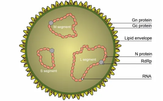
The virions are spherical, enveloped, approximately 100 nm in diameter and have a three-segmented genome, composed of negative-sense single-stranded RNA (Elliott & Blakqori, 2011) (Figure 3.1).
Figure 3.1. General characteristics of virions of the Order Bunyavirales (Original).
The genus Orthobunyavirus currently belongs to the family Peribunyaviridae (Adams et al., 2017) comprising 49 species of virus represented by approximately 170 examplar isolates, the exact number varies with sporadic discovery of new isolates or taxonomic revision of existing viruses. Historically, the viruses were classified into 18 serogroups according to their antigenic relationships in hemagglutination inhibition (HI), complement fixation (CF) and serum neutralisation tests (SNT) (Kinney & Calisher, 1981; Calisher, 1983; Travassos da Rosa, Tesh, Pinheiro, Travassos da Rosa, & Peterson, 1983; Elliott & Blakqori, 2011; Chowdhary, Street, Travassos da Rosa, Nunes, Tee, Hutchison, Vasconcelos, Tesh, Lipkin, & Briese, 2012; Gauci, McAllister, Mitchell, Boyle, Bulach, Weir, Melville, & Gubala, 2015). The Simbu serogroup is one of the largest of these serogroups and is named after the prototype virus (Saeed et al., 2001a). The concept of serogroup played an important role in the development of viral taxonomy (Calisher & Karabatsos, 1988), especially for the classification of the arboviruses (Casals, 1957) although the term is no longer used by the International Committee on Taxonomy of Viruses (ICTV), as the comparative analysis of nucleic acid and protein
5
sequences has become the primary method for determining virus relationships (Saeed et al., 2001a). However, due to the lack of genetic data for many named arboviruses, most current taxonomic assingments are still based on serology (Nichol, Beaty, Elliott, Goldbach, Plyusnin, Schmaljohn, & Tesh, 2005; Plyusnin, Beaty, Elliott, Goldbach, Kormelink, Lundkvist, Schmaljohn, & Tesh, 2012). In addition, antigenic relationships remain important in establishing serodiagnosis or in conducting serosurveys.
The Simbu serogroup consists of a highly diverse group of related arboviruses that infect not only humans, but also economically important livestock species and an extensive number of wildlife animals (Ladner et al., 2014; Zhang, Wang, Wang, Fu, Li, Zhao, Zhu, Wang, & Liang, 2015). The members of the group were identified in a widely geographically distributed range of vertebrate and invertebrate hosts, from dry deserts to tropical forests (Costa Mendes, 1984). In 1981, Kinney & Calisher published the most comprehensive comparison of the antigenic relationships among all recognised Simbu serogroup viruses. Even though the serogroup members were distinguishable by serum neutralization assays, the viruses showed extensive cross-reactions in the CF assays. On the basis of CF cross reactivity and subsequent difficulty to distinguish viruses from each other, the authors proposed the allocation of the 24 viruses into five complexes named after the first isolated member of each complex: the Simbu complex (Simbu, Akabane, Yaba-7, Shamonda, Sabo, Tinaroo, Sango, Peaton, Aino and Sathuperi viruses), the Manzanilla complex (Manzanilla, Ingwavuma, Mermet, Inini and Buttonwillow viruses) and the Oropouche complex (Oropouche, Utinga, Utive and Facey's Paddock viruses), plus Thimiri and Nola viruses which were distinct in the CF test and therefore designated as the sole members of their complexes (Kinney & Calisher, 1981).
Almost thirty years later, the Simbu serogroup included 25 antigenically related viruses, by then classified into seven different complexes, including Simbu, Manzanilla, Oropouche, Akabane, Sathuperi, Shamonda and Shuni (Yanase, Kato, Aizawa, Shuto, Shirafuji, Yamakawa, & Tsuda, 2012).
At present, the Simbu serogroup has 32 recognised members, of which 28 have been subjected to full genome sequencing (Table 3.1) (Saeed et al., 2001a; Ladner et al., 2014; Tilston-Lunel et al., 2015; Zhang et al., 2015). The viruses fall into two phylogenetic clades, designated A and B. Clade A comprise mainly viruses that occur in the New World (but including Facey’s Paddock virus from Australia, Cat Que virus from Asia, and Ingwavuma virus that occurs in Africa and Asia) and clade B members are distributed widely in Africa, Asia and Oceania, with a few individual viruses occurring in all three regions. The recent discovery of Schmallenberg virus (SBV) extends the known distribution of the clade into Europe (Table 3.2). Leanyer virus (LEAV), of Australia, does not fall into either of the two clades. The 31 viruses of clades A and B have recently been assigned to 8 species on the basis of close antigenic relationships and the existence of less than 10% differences in amino acid sequences of the nucleocapsid protein gene between the members of each species (Table 3.2) (ICTV, 2015).
6
Table 3.1. Known and potential members of the Simbu serogroup orthobunyaviruses, showing the origins of the prototype isolates (Swanepoel & Camarão, 2017).
Virus ICTV
Acronym
Prototype isolate
Year Country Source Putative vectors Known
distribution
1 Manzanilla MANV TRVL 3587 1954 Trinidad Howler monkey (Alouatta insularis) Mosquitoes, midges Central America
2 Simbu SIMV SA Ar 53 1955 South Africa Mosquitoes Mosquitoes, midges Africa
3 Oropouche OROV TRVL 9760 1955 Trinidad Human Mosquitoes, midges Central/South
America
4 Sathuperi SATV IG 10310 1957 India Mosquitoes Mosquitoes Asia, Africa
5
Ingwavuma INGV SA An 4165 1959 South Africa Spectacled Weaver (Hyphanturgus
ocularius) Mosquitoes, midges Africa, Asia
6
Akabane AKAV Ja Gar 39 1959 Japan Mosquitoes Midges Asia, Africa,
Australia
7 Buttonwillow BUTV A 7956 1962 USA Cottontail rabbit (Sylvilagus auduboni) Mosquitoes, midges North America
8 Yaba-7 Y7V Y-7 1963 Nigeria Culicoides midges Africa
9 Thimiri THIV VRC 66414 1963 India Indian Pond Heron (Ardeola grayii) Asia
10
Aino AINOV JaNAr 28 1964 Japan Mosquitoes Mosquitoes, midges Asia, Australia
11 Mermet MERV AV-782 1964 USA Purple martin (Progne subis) Mosquitoes North America
12 Utinga UTIV BeAn 84785 1965 Brazil Brown-throated sloth (Bradypus trydactylus) South America
13 Sango SANV An 5077 1965 Nigeria Bovine Mosquitoes, midges Africa
14 Shamonda SHAV An 5550 1965 Nigeria Bovine Midges Africa
15 Shuni SHUV An 10107 1966 Nigeria Bovine Mosquitoes, midges Africa
16
Sabo SABOV AN 9398 1966 Nigeria Caprine Midges Africa
17 Kaikalur KAIV VRC7 13423-2 1971 India Mosquitoes Mosquitoes Asia
18 Inini INIV CaAn-128d 1973 French Guiana Aracari bird (Pteroglossus aracari) South America
19 Facey's
Paddock FPV Aus Ch 16129 1974 Australia Mosquitoes Australia
20 Leanyer LEAV NT 16701 1974 Australia Mosquitoes Mosquitoes Australia
21 Utive UVV Pan An 48878 1975 Panama Pale-throated sloth (Bradypus variegatus) Central America
22 Pintupo Pending PanAr 517 1976? Panama Culicoides midges Midges Central America
23 Peaton PEAV CSIRO 110 1976 Australia Culicoides midges Midges Australia
24
Douglas DOUV CSIRO 150 1978 Australia Bovine Midges Australia, New
Guinea
25 Tinaroo TINV CSIRO 153 1978 Australia Culicoides midges Midges Australia
26 Jatobal JATV BeAn 423380 1985 Brazil Coati (Nasua nasua) Mosquitoes, midges South America
27 Iquitos IQTV IQT9924 1999 Peru Human Midges South America
28 Oya OYAV 1999 Malaysia Pig Asia
29 Cat Que Pending VN 04-2108 2004 Vietnam Mosquitoes Mosquitoes Asia
30 Madre de Dios MDDV FMD 1303 2007 Peru Human Mosquitoes, midges? South America
31 Schmallenberg SBV BH80/11-4 2011 Germany Bovine Midges Europe
7
Table 3.2. Phylogeny of members of the Simbu serogroup orthobunyaviruses (Ladner et al., 2014; ICTV, 2015).
Species Virus Distribution
CLADE A
Oropouche orthobunyavirus Oropouche Central/South America
Facey’s Paddock Australia
Iquitos South America
Jatobal South America
Madre de Dios South America
Perdões South America
Pintupo Central America
Utinga South America
Utive Central America
Manzanilla orthobunyavirus Manzanilla Central America
Buttonwillow North America
Oya Asia
Cat Que Asia
Ingwavuma Africa, Asia
Inini South America
Mermet North America
CLADE B
Akabane orthobunyavirus Akabane Asia, Africa, Australia
Tinaroo Australia
Sabo Africa
Yaba-7 Africa
Sathuperi orthobunyavirus Sathuperi Asia, Africa
Douglas Australia, New Guinea
Schmallenberg Europe
Shamonda orthobunyavirus Shamonda Africa
Sango Africa
Peaton Australia
Simbu orthobunyavirus Simbu Africa
Shuni orthobunyavirus Shuni Africa
Aino Asia, Australia
Kaikalur Asia
Thimiri orthobunyavirus Thimiri Asia
UNGROUPED
Leanyer orthobunyavirus? Leanyer Australia
Manzanilla orthobunyavirus (MANV), the first to be mentioned in the literature, was recovered in Trinidad and Tobago, in 1954, from the blood of a howler monkey (Alouatta seniculus insularis) (Anderson et al., 1960). Recently, MANV was isolated from pools of Culex tritaeniorhynchus in Yunnan Province, People’s Replubic of China (Feng, Fu, Yang, Zhang, He, Tu, Liang, & Zhang, 2015).
Simbu orthobunyavirus (SIMV) was isolated originally in the Republic of South Africa (RSA) from Aedes circumluteolus mosquitoes caught between 1955 and 1957, at Lake Simbu, Northern KwaZulu-Natal (KZN) (Weinbren, Heymann, Kokernot, & Paterson, 1957; Brooke Worth, Paterson, & De Meillon, 1961). SIMV was isolated subsequently in Cameroon, in 1966, from a pool of Eretmapodites chrysogaster mosquitoes (Salaun, Rickenbach, Bres, Brottes, Germain, Eouzan, & Ferrara, 1969). Human seroconversion was recorded in RSA and Botswana (Fraenkel-Conrat & Wagner, 1979).
8
Oropouche orthobunyavirus (OROV) was first isolated from the blood of a febrile forest worker in a village called Vega de Oropouche in 1955, and then from a pool of Coquillettidia venezuelensis mosquitoes in the Bush Bush Forest in Trinidad and Tobago, in 1960 (Anderson et al., 1961). In Brazil, OROV has been isolated from vertebrates such as humans, the three-toed sloth (Bradypus tridactylus), and the marmoset (Callithrix sp.), as well as from arthropods such as Aedes (Ochlerotatus) serratus, Culex fatigans, C. pipiens quinquefasciatus mosquitoes and Culicoides paraensis midges (Pinheiro, Pinheiro, Bensabath, Causey, & Shope, 1962; Pinheiro, Bensabath, Andrade, & Woodal 1968; Pinheiro, Travassos da Rosa, Travassos da Rosa, & Bensabath, 1976; Pinheiro, Hoch, Gomes, & Roberts, 1981; Nunes, Martins, Rodrigues, Chiang, Azevedo Rdo, da Rosa, & Vasconcelos, 2005). In 1989, the presence of OROV was reported in the village of Bejuco in Panama (Pinheiro, Travassos da Rosa, & Vasconcelos, 2004) and between 1992 and 1994, the pathogen was isolated in the Amazon region of Peru, in the cities of Iquitos, Puerto Maldonado e Madre de Dios (Watts, Lavera, Callahan, Rossi, Oberste, Roehrig, Cropp, Karabatsos, Smith, Gubler, Wooster, Nelson, & Hayes, 1997; Baisley, Watts, Munstermann, & Wilson, 1998). OROV exists as three different genotypes, namely I (Trinidad and Brazil), II (Brazil and Peru), and III (Panama and Brazil) (Saeed, Wang, Nunes, Vasconcelos, Weaver, Shope, Watts, Tesh, & Barrett, 2000). The true public health implication of OROV infection in South America remains unclear, due to the clinical resemblance of OROV infection to other endemic arboviral diseases such as Dengue, Mayaro, Chikungunya or Zika fevers, and the paucity of cases confirmed by laboratorial diagnosis (Navarro et al., 2016). Since the first isolation of the virus in 1955, it has been estimated to have caused more than 30 outbreaks and affected more than half a million people, which would make it one of the most common arboviruses affecting humans in tropical America (Travassos da Rosa, de Souza, Pinheiro, Figueiredo, Cardoso, Acrani, & Nunes, 2017). The clinical presentation of an affected human is an acute febrile illness, usually accompanied by headache, myalgia, arthralgia, anorexia, dizziness, chills, photophobia, nausea, vomiting, diarrhoea, conjunctive congestion, epigastric pain, retro-orbital pain and other systemic manifestations (Pinheiro, Travassos da Rosa, Travassos da Rosa, Ishak, Freitas, Gomes, LeDuc, & Oliva, 1981).
Sathuperi orthobunyavirus (SATV), recovered originally in India from pools of Culex vishnui mosquitoes (Dandawate, Rajagopalan, Pavri, & Work, 1969) was identified thereafter in Nigeria in dairy cattle and pools of Culicoides spp. (Lee, 1979). In 1999, two viruses closely related to the ones described previously were isolated from sentinel cattle in the Okayama Prefecture of Japan (Yanase, Fukutomi, Yoshida, Kato, Ohashi, Yamakawa, & Tsuda, 2004). Ingwavuma virus (INGV), isolated originally in South Africa, in 1959, from the organs of a spectacled weaver (Hyphanturgus ocularius) and from a pool of Culex univittatus mosquitoes (McIntosh et al., 1965), was a posteriori recovered from the whole blood of an Indian pond heron (Ardeola grayii) in India (originally referred to as Bagalodu virus) and from a spotted
9
flycatcher (Muscicapa striata) in Cyprus (Pavri, 1969; Watson, 1969). In Thailand, the virus was isolated from swine whole blood and from pools of Culex vishnui mosquitoes (Top, Kraivapan, Grossman, Rozmiarek, Edelman, & Gould, 1974) and in 1985, in Indonesia, was recovered from Culex spp. and Mansonia spp. mosquitoes (Converse, Tan, Rachman, Lee, & Shope, 1985).
Akabane orthobunyavirus (AKAV), was recognised originally in the Gunma Prefecture of Japan in pools of Aedes vexans as well as Culex tritaeniorhynchus mosquitoes in 1959 (Matsuyama, Oya, Ogata, Kobayashi, Nakamura, Takahashi, & Kitaoka, 1960). In 1968, the virus was isolated from pools of Culicoides brevitarsis in Australia (Doherty, Carley, Standfast, Dyce, & Snowdon, 1972) and in 1972, was recovered in Kenya from pools of Anopheles funestus (Metselaar & Robin, 1976). The virus was described as a natural infection in sentinel flocks of ewes in Australia, in 1976, in which the virus was isolated (Della-Porta, O'Halloran, Parsonson, Snowdon, Murray, Hartley, & Haughey, 1977). Serological evidence suggests that AKAV occurs in the Middle East region, namely in the Turkish Province of Aydin, Cyprus and Orontes river valley of Syria (Taylor & Mellor, 1994). Neutralising antibodies were found in a wide range of domestic animals in the Arabian peninsula, particularly in Kuwait, Saudi Arabia and Bahrain (Al-Busaidy, Mellor, & Taylor, 1988) and in a considerable number of African wild species, such as buffalo, nyala, bushbuck, kudu, eland, waterbuck, reedbuck, lechwe, roan and sable antelope, gemsbok, tsessebe, topi, blesbok, red hartebeest, blue wildebeest, impala, springbok, oribi, hippopotamus, giraffe, bushpig, warthog and elephant (Al-Busaidy, Hamblin, & Taylor, 1987). In Japan, after 1959, the pathogen was isolated a few times between 1974 and 2003, mainly in bovine samples and Culicoides spp. (Yamakawa, Yanase, Kato, & Tsuda, 2006). AKAV was also recovered from bovines and swines in Taiwan (Liao, Lu, Goto, & Inaba, 1996; Chang, Liao, Su, Farh, & Shiuan, 1998; Huang, Huang, Deng, Jong, & Lin, 2003), from mosquitoes in the Yunnan Province of China (Feng, He, Fu, Yang, Zhang, Tu, Liang, & Zhang, 2015) and from cattle in South Korea (Bak, Lim, Cheong, Hwang, & Cho, 1980; Oem, Lee, Kim, Bae, Chung, Lee, & Roh, 2012). Substantial seroprevalence in cattle, sheep and goats was also reported in China (Wang, Blasdell, Yin, & Walker, 2017). In 1969-70, an outbreak of Akabane disease in domestic ruminants occurred in Israel (Shimshony, 1980), and again between 2001 and 2003 (Brenner, Tsuda, Yadin, Chai, Stram, & Kato, 2004), where a new lineage was described (Stram, Brenner, Braverman, Banet-Noach, Kuznetzova, & Ginni, 2004a). AKAV can cause abortion, stillbirth, congenital malformations, principally arthrogryposis (AG) and hydranencephaly (HE) (congenital AG/HE syndrome), as well as marked teratology, particularly of the CNS, if the virus infects the foetus at a critical stage of development and when susceptible pregnant ruminants are infected. In non-pregnant animals the infection is subclinical (St. George & Kirkland, 2004). The stage of gestation in which the host is infected determines the range and severity of clinical signs (Uchida, Murakami, Sueyoshi, Tsuda, Inai, Acorda, Yamaguchi, & Tateyama, 2000). Bovine and swine cases of
10
AKAV encephalomyelitis have also been described (Miyazato, Miura, Hase, Kubo, Goto, & Kono, 1989; Uchida et al., 2000; Kono, Hirata, Kaji, Goto, Ikeda, Yanase, Kato, Tanaka, Tsutsui, Imada, & Yamakawa, 2008; Oem et al., 2012). Recently, a congenital AG/HE syndrome in newborn calves was reported in Iraq (Alsaad, Alautaish, & Alamery, 2017). Buttonwillow virus (BUTV), the first isolate in Northern America was identified originally in whole blood of a desert cottontail rabbit (Sylvilagus auduboni) and later in a blacktail jackrabbit (Lepus californicus) and pools of Culicoides variipennis midges. Seroconversion was described in lagomorphs (S. auduboni, L. californicus, L. americanus) across USA and in one Canadian marmot (Reeves et al., 1970; Nelson & Scrivani, 1972).
The first Simbu serogroup virus recovered in Nigeria, in Yaba, a suburb of Lagos, was named Yaba-7 virus (Y7V), and was isolated from a pool of Mansonia africana mosquitoes in 1963 (Theiler & Downs, 1973).
Thimiri orthobunyavirus (THIV), like INGV, was isolated for the first time from the whole blood of an Indian pond heron (Ardeola grayii) in India, in 1963 (Carey et al., 1971), and subsequently in Egypt, between 1963 and 1966, in the blood of migratory birds, namely a common (Sylvia communis) and a lesser whitethroat (S. curruca) (Darwish & Hoogstraal, 1981) and in the north of Australia from a pool of Culicoides histrio bird-feeding midges (Standfast & Dyce, 1982). Aino virus (AINOV) was identified originally in Nagasaki, Japan, in pools of Culex tritaeniorhynchus and in a mixed pool of C. pseudovishnui and C. pipiens mosquitoes (Takahashi, Oya, Okazda, Matsuo, & Kuma, 1968) and then isolated in Australia from pools of Culicoides brevitarsis (Doherty et al., 1972). In 2003, serological evidence of its presence in dairy herds in Israel was recorded (Brenner et al., 2004). Clinical cases of congenital AG/HE syndrome showed high titres of neutralising antibodies to AINOV (Miura, Hayashi, Ishihara, Inaba, & Omori, 1974; Coverdale et al., 1978) and experimentally AINOV caused the same syndrome associated with cerebellar hypoplasia in calves (Tsuda, Yoshida, Ohashi, Yanase, Sueyoshi, Kamimura, Misumi, Hamana, Sakamoto, & Yamakawa, 2004).
The second Simbu serogroup member to be isolated in North America, Mermet virus (MERV) was recovered originally from the whole blood of a purple martin (Progne subis) in Metropolis, Illinois, in 1964. MERV was also recovered from other birds, namely a red-winged blackbird (Agelaius phoeniceus), a Swainson's thrush (Catharus ustulatus) and a cardinal (Cardinalidae) in the Ohio-Mississipi basin (Calisher et al., 1969). Another strain was obtained from a mixed pool of Culex pipiens and C. restuans mosquitoes in Memphis, Tennessee (Jakob, Francy, Trimble, & Calisher, 1979). In 1969, during an ecologic investigation of an outbreak of Venezuelan equine encephalitis (VEE) in Guatemala, MERV was isolated from an Altamira oriole (Icterus gularis) (Gutierrez, Calisher, Maness, & Lord, 1975).
11
Utinga virus (UTIV), was isolated originally in 1965 from the whole blood and pooled organs of a pale-throated sloth (Bradypus tridactylus) in Brazil (Karabatsos, 1985) and no further isolations have been published in the literature.
Sango virus (SANV), the second Nigerian Simbu serogroup isolate to be recovered, was isolated in bovine whole blood and in pools of Culicoides spp. during a surveillance study by the Virus Research Laboratory of the University of Ibadan (Causey, Kemp, Madbouly, & Lee, 1969; Causey, Kemp, Causey, & Lee, 1972; Lee, 1979). It was later recovered from pools of Mansonia uniformis in Kenya (Metselaar, Henderson, Kirya, Tukei, & de Geus, 1974) and seroconversion against SANV was reported in dairy cattle and goats in Ibadan (Kemp, Causey, & Causey, 1971).
The third Nigerian Simbu serogroup to be isolated, Shamonda orthobunyavirus (SHAV), was first identified in bovine whole blood during the same surveillance study mentioned previously (Causey et al., 1969; Causey et al., 1972) and in Culicoides imicola pools (Lee, 1979) collected and caught respectively in Ibadan. Antibodies against SHAV were also found in cattle in Ibadan (Kemp et al., 1971). In 1967, in Vryburg, South Africa, SHAV was associated with an outbreak of disease in cattle characterised by conjunctival and periorbital oedema as well as swollen lips and tongue, although the pathogen was named Lambrecht virus originally (Solberg, 1970; Costa Mendes, 1984). In the same year and country, the virus was recovered from Culicoides imicola midges caught at the Onderstepoort Veterinary Institute (OVI) facilities (McIntosh, 1980). In 2002, serological and molecular evidence of SHAV was reported in the Kagoshima and Miyazaki prefectures of Japan (Yanase, Maeda, Kato, Nyuta, Kamata, Yamakawa, & Tsuda, 2005). Recently, in southern Japan, SHAV was identified in association with congenital malformations, including torticollis, arthrogryposis, spinal curvature, ventriculomegaly and cerebellar hypoplasia, in calves infected in utero (Hirashima et al., 2017).
Shuni orthobunyavirus (SHUV), fourth isolate to be described in Nigeria, was isolated originally from a cow in Sokoto (Causey et al., 1969), from the whole blood of a sheep in Ibadan (Causey et al., 1972), from cattle in northern Nigeria (Kemp, Causey, Moore, & O'Connor, 1973) and from pools of Culicoides spp. (Lee, 1979). The presence of neutralising antibodies in cattle and sheep was reported in the same country (Kemp et al., 1971). In 1975, Moore et al. described the isolation of SHUV from the blood of a Nigerian child (Moore, Causey, Carey, Reddy, Cooke, Akinkugbe, David-West, & Kemp, 1975). In South Africa, the virus was isolated in Culex theileri mosquitoes in Gauteng and in ruminants in KZN (McIntosh, 1972, 1980), from the brain of two horses with CNS clinical signs in Zimbabwe (Foggin & Swanepoel, 1990) and in RSA (Coetzer & Howell, 1998). More recently, the role of SHUV in neurological disease in horses was further reported (van Eeden et al., 2012) and, interestingly, the seroconversion against SHUV in veterinary professionals in South Africa (van Eeden, Swanepoel, & Venter, 2014a). Between 2014-15, the role of SHUV in malformed and aborted foetus of small
12
ruminants and cattle in Israel was investigated (Golender, Brenner, Valdman, Khinich, Bumbarov, Panshin, Edery, Pismanik, & Behar, 2015).
Sabo virus (SABOV), was isolated in Ibadan, Nigeria during the surveillance study mentioned previously (Causey et al., 1969; Kemp et al., 1971; Causey et al., 1972; Lee, 1979), from a goat, cattle and Culicoides spp.. Neutralising antibodies were found in cattle, small ruminants and pigs.
Kaikalur virus (KAIV) was isolated from a pool of Culex tritaeniorhynchus mosquitoes caught in 1971, in Kaikalur, Andhra Pradesh, India (Rodrigues, Singh, Dandawate, Soman, & Bhatt, 1977).
Inini virus (INIV), was recovered from a bank of a river with the same name, from the whole blood of a Aracari bird (Pteroglossus aracari), in Maripasoula, French Guiana (Pajot, 1980). Facey’s Paddock virus (FPV), was identified originally in pools of Culex annulirastris, Aedes narmanensis and Culicoides spp. in Australia. The presence of neutralising antibodies was reported in cattle and wallabies (Doherty et al., 1972; Doherty, 1975; Doherty, Carley, Kay, Filippich, Marks, & Frazier, 1979).
Leanyer virus (LEAV) was isolated from Anopheles meraukensis mosquitoes collected at Leanyer, in the Northern Territory of Australia, in 1974, during an entomological surveillance in the course of an outbreak of Murray Valley encephalitis (MVE) (Doherty, Carley, Filippich, Kay, Gorman, & Rajapaksa, 1977).
Utive virus (UVV) was isolated in Panama, in a village with the same name, from the blood of a brown-throated sloth (Bradypus variegatus) in 1975 (Seymour et al., 1983).
In Panama, Pintupo virus, was isolated from Culicoides diabolicus (Seymour et al., 1983). Another Australian isolate, Peaton virus (PEAV), was recovered originally from pools of Culicoides brevitarsis and bovine whole blood (St George, Cybinski, Filippich, & Carley, 1979; St George, Standfast, Cybinski, Filippich, & Carley, 1980). In 1999, the virus was identified by SNT in Culicoides midges and cattle blood in the Kyushu district of Japan (Matsumori, Inai, Yanase, Ohashi, Kato, Yoshida, & Tsuda, 2002). The association of PEAV with congenital malformations in sheep via experimental infection has been described (Parsonson & McPhee, 1985).
Douglas virus (DOUV) was identified in the blood of a cow with no clinical signs, and thereafter in pools of Culicoides brevitarsis in Australia. In the same country, neutralising antibodies in all the species of domestic ruminants, as well as in buffalo and deer were found. In Papua New Guinea, a bovine that seroconverted against DOUV was identified (Doherty et al., 1979; Cybinski, 1984).
Tinaroo virus (TINV) was identified first in pools of Culicoides brevitarsis and later in the whole blood of cattle in Australia. In the same country, a lamb with AG syndrome had neutralising
13
antibodies against TINV, and similar to DOUV, antibodies were found in cattle, sheep, goat, buffalo and deer (Doherty et al., 1979; Cybinski, 1984).
In 1985, Jatobal virus (JATV) was recovered from the whole blood of a coati (Nasua nasua) in Tucuruí, Pará State, Brazil and classified as a distinct member of the serogroup (Figueiredo & Da Rosa, 1988). Sixteen years after, it was suggested that JATV was a possible reassortant of OROV (Saeed, Wang, Suderman, Beasley, Travassos da Rosa, Li, Shope, Tesh, & Barrett, 2001b).
Iquitos virus (IQTV), was isolated in a city with the same name in Peru, from a thirteen-year-old boy with a clinical presentation of fever, headache, eye and body pain, arthralgia, diarrhea, and chills, in 1999. IQTV was also described as a reassortant of OROV (Aguilar et al., 2011). In Malaysia, Oya virus (OYAV) was isolated from a pig suspected of Nipah virus (NiV) infection, during a national swine surveillance programme, and named after the Oya village from where it was recovered. Interestingly 93% (n=360) seropositivity was observed in a serosurvey of pigs in the principal Malaysian pig breeding districts (Kono, Yusnita, Mohd Ali, Maizan, Sharifah, Fauzia, Kubo, & Aziz, 2002). Recently, a retrospective study reported the presence of viral RNA in pig and human samples, as well as immunoglobulin G (IgG) positivity in human serum in the Karnataka State of India (Yadav, Shete, Bondre, Patil, Kokate, Chaudhari, Srivastava, Jadhav, & Mourya, 2016).
Cat Que virus (CQV) was isolated from mosquitoes during an arbovirus surveillance in an outbreak of acute paediatric encephalitis in northern Vietnam in 2004, in spite of the virus reported originally to be OYAV, based on indirect immunofluorescence assays and high nucleotide similarity (Bryant, Crabtree, Nam, Yen, Duc, & Miller, 2005). Consequent analysis showed 4,1% amino acid and 9,6% nucleotide divergence from the original OYAV isolate (Ladner et al., 2014). In 2015, the isolation of CQV from Culex tritaeniorhynchus mosquitoes in the Sichuan Province of China as well as anti-CQV IgM/IgG in pigs was reported (Zhang et al., 2015).
Madre de Dios virus (MDDV), is an unofficial name proposed to refer to an isolate recovered in 2007 from a febrile human in Madre de Dios, region in the southeastern Peru’s Amazon basin. MDDV was also described as a OROV reassortant, identical at the amino acid level to both OROV and IQTV but due to its distinctiveness was suggested that the pathogen should have its own name (Ladner et al., 2014). In 2010, MDDV was obtained from a wedge-capped capuchin (Cebus olivaceus), collected in Atapirire village, located in the southeastern part of Venezuela, during an epizootic of illness among sylvatic monkeys (Navarro et al., 2016). In the autumn of 2011, an unidentified disease in dairy cattle with a clinical presentation of fever, diarrhoea, and decreased milk production, was reported to the veterinary services, local diagnostic laboratories, and national research institutes in Germany and the Netherlands (Hoffmann et al., 2012). After excluding endemic and emerging diseases such as infectious
14
bovine rhinotracheitis (BoHV-1), bovine viral diarrhoea (BVDV-1), bluetonge (BTV), epizootic haemorrhagic disease (EHDV), foot-and-mouth disease (FMDV), Rift Valley fever (RVFV) or bovine ephemeral fever (BEFV), the blood samples from acutely diseased cows were subjected to a metagenomic approach and next-generation sequencing (NGS) to identify a novel Orthobunyavirus that was named after the origin of the samples as Schmallenberg virus (SBV) (Hoffmann et al., 2012). SBV was the first virus of the Simbu serogroup to be reported in Europe (Saeed et al., 2001a; Hoffmann et al., 2012) and spread very rapidly throughout the domestic and wild ruminant population of the continent. Virus isolation or seroconversion were reported in France, Belgium, Luxembourg, Italy, Spain, United Kingdom, Ireland, Poland, Lithuania, Latvia, Switzerland, Austria, Denmark, Sweden, Norway, Finland, Czech Republic, Croatia, Slovenia, Estonia, Hungary, Greece and Portugal (Bradshaw, Mooney, Ross, Furphy, O'Donovan, Sanchez, Gomez-Parada, & Toolan, 2012; EFSA, 2012; European Food Safety, 2012; Garigliany, Bayrou, Kleijnen, Cassart, & Desmecht, 2012; Garigliany, Hoffmann, Dive, Sartelet, Bayrou, Cassart, Beer, & Desmecht, 2012; Rasmussen, Kristensen, Kirkeby, Rasmussen, Belsham, Bodker, & Botner, 2012; Bouwstra, Kooi, de Kluijver, Verstraten, Bongers, van Maanen, Wellenberg, van der Spek, & van der Poel, 2013; Conraths, Peters, & Beer, 2013; EFSA, 2013; Larska, Krzysiak, Smreczak, Polak, & Zmudzinski, 2013; Larska, Polak, Grochowska, Lechowski, Zwiazek, & Zmudzinski, 2013; Mason, Stevenson, Carty, Hosie, Caldow, & Boyes, 2013; Balmer, Vögtlin, Thür, Büchi, Abril, Houmard, Danuser, & Schwermer, 2014; Chaintoutis, Kiossis, Giadinis, Brozos, Sailleau, Viarouge, Breard, Papanastassopoulou, Zientara, Papadopoulos, & Dovas, 2014; Rasmussen, Kirkeby, Bodker, Kristensen, Rasmussen, Belsham, & Botner, 2014; Steinrigl, Schiefer, Schleicher, Peinhopf, Wodak, Bago, & Schmoll, 2014; Wernike, Conraths, Zanella, Granzow, Gache, Schirrmeier, Valas, Staubach, Marianneau, Kraatz, Höreth-Böntgen, Reimann, Zientara, & Beer, 2014; Wisloff, Nordvik, Sviland, & Tonnessen, 2014; Xavier, Joan, Roser, Rosa, Jorge, Ignasi, Marti, Joaquim, Santiago, & Oscar, 2014; Lazutka, Spakova, Sereika, Lelesius, Sasnauskas, & Petraityte-Burneikiene, 2015; Esteves, Mesquita, Vala, Abreu-Silva, van der Poel, & Nascimento, 2016; Laloy, Braud, Breard, Kaandorp, Bourgeois, Kohl, Meyer, Sailleau, Viarouge, Zientara, & Chai, 2016). The pathogenesis of SBV results in congenital malformations, premature birth or stillbirth, or mummification, when naïve dams are infected during a critical phase of pregnancy (Wernike, Elbers, & Beer, 2015). Interestingly, anti-SBV antibodies were found in dogs in Sweden and France and viral RNA was isolated from the brain of a puppy with neurologic signs (Sailleau, Boogaerts, Meyrueix, Laloy, Bréard, Viarouge, Desprat, Vitour, Doceul, Boucher, Zientara, Nicolier, & Grandjean, 2013; Wensman, Blomqvist, Hjort, & Holst, 2013). Full genome investigation has shown that SBV belongs to the species Sathuperi orthobunyavirus and is a possible ancestor of SHAV (Goller, Hoper, Schirrmeier, Mettenleiter, & Beer, 2012).
15
In 2012, Perdões virus was isolated in a municipality with the same name, in Minas Gerais State, Brazil from the liver of a dead black-tufted marmoset (Callitrhix penicillata). The virus was also described as a reassortant of OROV (Tilston-Lunel et al., 2015).
Many orthobunyaviruses have yet to be fully sequenced and there are probably many others that have yet to be detected (Ladner et al., 2014).
3.1.2. Genome organisation
The genome of orthobunyaviruses consists of three segments of single-stranded, negative-sense RNA, designated large (L), medium (M) and small (S), of approximately 6.9kb, 4.5kb and 1.0kb, respectively. These three genomic segments encode a total of six proteins (Elliott, 1990). All three segments include a coding region flanked by 3ʹ and 5ʹ untranslated regions (UTRs). The terminal eleven nucleotides at both ends are conserved and complementary inverted, facilitating the formation of pan-handle structures, which function as the promoter for the transcription and replication of each segment (Kohl, Dunn, Lowen, & Elliott, 2004; Barr & Wertz, 2005; Elliott & Blakqori, 2011; Elliott, 2014) (Figure 3.2).
16
The L segment encodes the viral RNA-dependent RNA polymerase (RdRp) (Saeed et al., 2001b; Elliott & Blakqori, 2011; Chowdhary et al., 2012), that catalyses both transcription and genome replication. In addition, it contains an endonuclease domain that cleaves capped oligomers from host mRNA, which is used to prime viral mRNA synthesis, a phenomenon known as cap snatching (Reguera, Weber, & Cusack, 2010; Elliott, 2014).
The M segment encodes a precursor polyprotein which is post-translational cleaved by host proteases, giving rise to the surface glycoproteins Gn and Gc, as well as a non-structural protein, NSm (Gentsch & Bishop, 1979; Elliott, 1985; Fazakerley, Gonzalez-Scarano, Strickler, Dietzschold, Karush, & Nathanson, 1988; Gerbaud, Pardigon, Vialat, & Bouloy, 1992; Schmaljohn & Hooper, 2001; Elliott & Blakqori, 2011). Viral glycoproteins encoded on the M segment play a crucial role in viral attachment and membrane-fusion. Both glycoproteins are type I integral membrane proteins and form a heterodimer in the endoplasmic reticulum (ER) of the infected cell (Schmaljohn & Hooper, 2001; Nichol et al., 2005; Shi, Brauburger, & Elliott, 2005; Elliott, 2014; Fischer, 2014). When Gc is expressed alone, it is retained in the ER, which suggests that Gn functions as a chaperone for the correct trafficking of Gc to the Golgi complex before viral budding (Shi, Lappin, & Elliott, 2004). It is suggested that NSm may be involved in the processes of viral assembly and budding (Lappin, Nakitare, Palfreyman, & Elliott, 1994). Of all three genomic segments, the M segment has been shown to be the most variable RNA segment of orthobunyaviruses (Kobayashi, Yanase, Yamakawa, Kato, Yoshida, & Tsuda, 2007; Wernike & Beer, 2017).
On the other hand, the S segment is the most conserved one (Saeed et al., 2000; Nunes et al., 2005; Acrani, Gomes, Proenca-Modena, da Silva, Carminati, Silva, Santos, & Arruda, 2010; Vasconcelos, Nunes, Casseb, Carvalho, Pinto da Silva, Silva, Casseb, & Vasconcelos, 2011; Hang, Forshey, Yang, Solorzano, Kuschner, Halsey, Jarman, & Kochel, 2014; Cardoso, Serra, Heinen, Zuchi, Souza, Naveca, Santos, & Slhessarenko, 2015). It encodes two proteins in alternative overlapping open reading frames (ORFs), the nucleocapsid (N) protein and a non-structural protein (NSs), which are translated from the same mRNA by leaky ribosomal scanning (Elliott, 2014). The N protein, the most abundant in the virion and infected cells, encapsidates one copy of each RNA segment, forming a ribonucleoprotein (RNP) complex, which in turn is associated with the RdRp (Elliott & Blakqori, 2011; Walter & Barr, 2011; Elliott, 2014). The RNPs are packaged in a lipid envelope, derived from the host Golgi complex, which is then modified by insertion of the viral glycoproteins, forming a trimer that create the spikes on the virion (Elliott, 2014). The NSs protein is known to be involved in the regulation of translation, apoptosis, viral polymerase activity as well as antagonism of the interferon (IFN) and shutoff of host protein synthesis in mammalian cells (van Knippenberg, Carlton-Smith, & Elliott, 2010; Elliott, Blakqori, van Knippenberg, Koudriakova, Li, McLees, Shi, & Szemiel, 2013; Barry, Varela, Ratinier, Blomström, Caporale, Seehusen, Hahn, Schnettler, Baumgärtner, Kohl, & Palmarini, 2014; Gouzil, Fablet, Lara, Caignard, Cochet, Kundlacz,
17
Palmarini, Varela, Breard, Sailleau, Viarouge, Coulpier, Zientara, & Vitour, 2017), but also seems to play a role in the replication mechanism in the vector (Szemiel, Failloux, & Elliott, 2012).
The segmented nature of the genome facilitates the possibility of evolution through genetic reassortment (Bowen, Trappier, Sanchez, Meyer, Goldsmith, Zaki, Dunster, Peters, Ksiazek, Nichol, & Force, 2001; Saeed et al., 2001b; Gerrard, Li, Barrett, & Nichol, 2004; Briese, Bird, Kapoor, Nichol, & Lipkin, 2006; Aguilar et al., 2011). This phenomenon occurs when genetically closely-related viruses infect a susceptible cell at the same time, resulting in a progeny virus containing a mixture of genomic L, M, and S segments from the two parental viruses (Travassos da Rosa et al., 2017). However, despite the hypothetical potential for changes in pathogenicity or host range, the role of segment reassortment is largely unknown due to the lack of comprehensive sequence data (Gerrard et al., 2004).
3.2. Molecular diagnosis using real-time polymerase chain reaction
(qPCR)
3.2.1. General review
In virology, pathogen identification and confirmation of infection relies traditionally on virus isolation in susceptible laboratory animals, cultured cell lines or embryonated eggs. Immunological diagnostic methods are also used widely, based on specific antigen-antibody interaction. Molecular diagnostic methods detect or analyse the genotype characteristics of the microorganism and have been gaining more prominence in research and diagnostic laboratories, complementing, or in some cases substituting conventional methods, which are often more laborious, e.g. culture in cell lines (Cunha & Inácio, 2014).
The polymerase chain reaction (PCR) uses a pair of synthetic oligonucleotides or primers, which hybridise to one strand of the double-stranded DNA (dsDNA) target. This hybridised primer is a substrate for a thermostable DNA polymerase to synthesise the complementary strand through sequential addition of deoxynucleotides (dNTPs). The reaction comprises three distinct steps: denaturation of the dsDNA at 94-97C; annealing of the primers at 50-75C and extension at 72-78C. The ramp rate, the time of incubation at each temperature and the number of cycles is adjusted in a programmable thermal cycler. The conventional detection and evaluation of the amplified product is dependent on a post-PCR electrophoresis in the presence of a DNA stain and analysis of the resulting band after ultraviolet (UV) illumination (Saiki, Scharf, Faloona, Mullis, Horn, Erlich, & Arnheim, 1985; Mullis & Faloona, 1987; Peake, 1989; Mackay et al., 2002).
Real-time PCR (qPCR) enables simultaneous detection of the target and monitoring of the amplification dynamics, decreases procedure time, eliminates the necessity of post-reaction manipulation and makes possible the quantitative analysis of the product. Observation of the
18
accumulating amplicon relies on the usage of fluorogenic molecules (Holland, Abramson, Watson, & Gelfand, 1991; Mackay et al., 2002; Tenreiro, Chaves, & Tenreiro, 2014).
One of the variety of chemistries utilised for the detection of fluorescence during the qPCR reaction is the hydrolysis probe, also called a TaqMan probe or 5’ nuclease oligonucleotide probe. This probe is dual-fluorophore labelled with a reporter at the 5’ terminus and a quencher at the 3’ terminus. When in close proximity, the photon-induced excited 5’ reporter fluorophore transfers energy by Förster resonance energy transfer (FRET) to the 3’ quencher fluorophore when the probe is intact. However, when the probe is hydrolised due to the 5’ exonuclease activity of the TaqMan DNA polymerase, the reporter energy emission is no longer quenched, resulting in detectable fluorescence (Holland et al., 1991). Hydrolysis probes have strict design requirements, such as: a length of 20-40 nt; a guanine-cytosine content (GC-content) of 40-60%; no single nt repetitions, especially G; a melting temperature (TM) at least 5oC higher than the primers, in order to ensure that the probe hybridises to the template before the extension (Wittwer, 2001). Recently, an enhancement was made to the hydrolysis probe, the so-called minor groove binder (MGB) probes, that incorporate a 3’ nonfluorescent quencher (NFQ), which results in lower background signal and subsequent higher precision in quantitation, but also a MGB moiety that stabilises the probe-template duplex by folding into the minor groove of the dsDNA, thus permitting the use of shorter probes (14 nt), providing better sequence discrimination and flexibility to accommodate more targets (Kutyavin, Afonina, Mills, Gorn, Lukhtanov, Belousov, Singer, Walburger, Lokhov, Gall, Dempcy, Reed, Meyer, & Hedgpeth, 2000).
qPCR has received widespread approval and recognition across a wide range of scientific specialities, due to the high sensitivity of the assay, reduced risk of carry over contamination, improved speed to perform the assay, possibility of type isolates, multiplex capacity and an ability to quantitate precisely the nucleic acids (Mackay et al., 2002; Mosammaparast & McAdam, 2013). It has become one of the most widely used methods for direct virus detection (Wernike & Beer, 2017).
3.2.2. Simbu molecular diagnostic assays based on real-time RT-PCR
The isolation, consequent identification and characterisation of “Bunyavirus” isolates have been performed historically using mostly antibody based techniques, including HI, CF, SNT, immunofluorescence (IF) tests and more recently enzyme-linked immunosorbent assays (ELISA) (Shope & Causey, 1962; Lanciotti & Tsai, 2007; Mathew, Klevar, Elbers, van der Poel, Kirkland, Godfroid, Mdegela, Mwamengele, & Stokstad, 2015). While these methods are essential for virus identification based on a group-specific approach, they can be time-consuming and restricted in their ability to generate unequivocal and species-identifying results, due to antibody cross-reactivity. Modern molecular technologies and its application in diagnostic reference laboratories provide a time-efficient alternative to traditional serology in