Inhibitors Prevent Ozone-Induced Airway
Hyperreactivity in Guinea Pigs
Kirsten C. Verhein1*¤a
, Francesco G. Salituro3¤b, Mark W. Ledeboer3, Allison D. Fryer2, David B. Jacoby2
1Department of Physiology and Pharmacology, Oregon Health and Science University, Portland, Oregon, United States of America,2Division of Pulmonary and Critical Care Medicine, Oregon Health and Science University, Portland, Oregon, United States of America,3Vertex Pharmaceuticals, Inc., Cambridge, Massachusetts, United States of America
Abstract
Ozone exposure causes airway hyperreactivity and increases hospitalizations resulting from pulmonary complications. Ozone reacts with the epithelial lining fluid and airway epithelium to produce reactive oxygen species and lipid peroxidation products, which then activate cell signaling pathways, including the mitogen activated protein kinase (MAPK) pathway. Both p38 and c-Jun NH2terminal kinase (JNK) are MAPK family members that are activated by cellular stress and
inflammation. To test the contribution of both p38 and JNK MAPK to ozone-induced airway hyperreactivity, guinea pigs were pretreated with dual p38 and JNK MAPK inhibitors (30 mg/kg, ip) 60 minutes before exposure to 2 ppm ozone or filtered air for 4 hours. One day later airway reactivity was measured in anesthetized animals. Ozone caused airway hyperreactivity one day post-exposure, and blocking p38 and JNK MAPK completely prevented ozone-induced airway hyperreactivity. Blocking p38 and JNK MAPK also suppressed parasympathetic nerve activity in air exposed animals, suggesting p38 and JNK MAPK contribute to acetylcholine release by airway parasympathetic nerves. Ozone inhibited neuronal M2muscarinic receptors and blocking both p38 and JNK prevented M2receptor dysfunction. Neutrophil influx into
bronchoalveolar lavage was not affected by MAPK inhibitors. Thus p38 and JNK MAPK mediate ozone-induced airway hyperreactivity through multiple mechanisms including prevention of neuronal M2receptor dysfunction.
Citation:Verhein KC, Salituro FG, Ledeboer MW, Fryer AD, Jacoby DB (2013) Dual p38/JNK Mitogen Activated Protein Kinase Inhibitors Prevent Ozone-Induced Airway Hyperreactivity in Guinea Pigs. PLoS ONE 8(9): e75351. doi:10.1371/journal.pone.0075351
Editor:Heinz Fehrenbach, Research Center Borstel, Germany
ReceivedSeptember 6, 2012;AcceptedAugust 16, 2013;PublishedSeptember 18, 2013
Copyright:ß2013 Verhein et al. This is an open-access article distributed under the terms of the Creative Commons Attribution License, which permits unrestricted use, distribution, and reproduction in any medium, provided the original author and source are credited.
Funding:This work was funded by the American Heart Association 0810148Z (to KCV) and the National Institutes of Health HL55543 (to ADF), ES014601 (to ADF), HL54659 (to DBJ), HL071795 (to DBJ), and RR023424 (to DBJ). The funders had no role in study design, data collection and analysis, decision to publish, or preparation of the manuscript. No additional external funding was received for this study.
Competing Interests:FGS and MWL were employed by Vertex Pharmaceuticals at the time of these studies. FGS is currently employed by Sage Therapeutics. This does not alter these authors’ adherence to all the PLoS ONE policies on sharing data and materials, as detailed online in the guide for authors. KCV, ADF, and DBJ declare no competing interests exist.
* E-mail: [email protected]
¤a Current address: National Institute of Environmental Health Sciences, Research Triangle Park, North Carolina, United States of America ¤b Current address: Sage Therapeutics, Cambridge, Massachusetts, United States of America
Introduction
Over half the United States population lives in counties with unhealthy levels of ozone, a major component of smog [1]. Epidemiological studies demonstrate a significant link between exposure to ground level ozone and pulmonary hospitalizations. Exposure to ozone in excess of 0.16 ppm is associated with increased airway reactivity, lung inflammation and exacerbation of asthma in both adults and children [2,3,4].
Ozone induced hyperreactivity is demonstrated by increased reactivity to inhaled methacholine and other agonists, including those causing reflex bronchoconstriction in man [5,6,7]. In animals, ozone induced airway hyperreactivity is demonstrated by increased bronchoconstriction to intravenous methacholine, but this effect is mediated largely via increased acetylcholine release from parasympathetic nerves, since it is blocked by vagal section [8,9]. Direct stimulation of the vagus nerves results in bronchoconstriction that is potentiated in ozone exposed animals and that is associated with loss of function of neural M2muscarinic
receptors that normally inhibit acetylcholine release [10,11]. Inflammatory cells, especially eosinophils through release of the M2 inhibitor major basic protein, mediate loss of neuronal M2
function and airway hyperreactivity in ozone exposed guinea pigs [11].
However, ozone is unlikely to contact inflammatory cells [12]. At the airway epithelial layer, ozone forms reactive oxygen species and lipid peroxides in lungs of humans and animals [13,14]. These end products activate cell signaling pathways, including mitogen activated protein kinase pathways (MAPK) [15]. Activation of the MAPK pathway results in inflammation [16], mucus hypersecre-tion [17] and airway hyperreactivity [18].
MAPK signaling pathways are important in many cell processes including differentiation, proliferation, activation, degranulation, and migration. Three MAPK subfamilies have been well characterized: ERK, JNK, and p38. The extracellular signal-regulated kinase (ERK) pathway is usually activated by mitogens and growth factors while p38 and c-Jun NH2 terminal kinase
typically activated by inflammatory cytokines, heat shock, and cellular stress [19,20]. Activation of MAPK signaling induces inflammatory cytokine and chemokine production in airway epithelial cells, inflammatory cells, and airway smooth muscle cells [16,21,22]. Humans with severe asthma have increased activated p38 in airway epithelium compared to mild asthmatics or healthy controls, as demonstrated by increased immunostaining of phosphorylated p38 in airway biopsies [23].
Inhibition of MAPKs is protective in allergen challenge models of asthma. Inhibition of p38, either pharmacologically or with antisense oligonucleotides, partially prevents airway hyperreactiv-ity after sensitization and challenge in mice [18,24]. Eosinophil influx into bronchoalveolar lavage is the dominant event in antigen challenged animals, and is prevented by a p38 inhibitor in guinea pigs and mice [25]. Blocking p38 also prevents IL-13 induced mucus metaplasia in human and mouse airway epithelial cells [17,26].
Less is known about the role of the MAP kinases in ozone-induced hyperreactivity. Inhibiting p38 prevents ozone-ozone-induced airway hyperreactivity in mice while inhibiting JNK is partially protective [27,28]. Ozone-induced increases in inflammatory cells in bronchoalveolar lavage are significantly inhibited in Jnk1
knockout mice [29].
The experiments described here use three different MAPK inhibitors to test whether dual inhibition of both p38 and JNK MAPK pathways prevents ozone-induced inflammation and subsequent airway hyperreactivity in guinea pigs.
Methods
Ethics Statement
Guinea pigs were handled in accordance with the standards established by the United States Animal Welfare Act set forth in National Institutes of Health guidelines. All protocols were approved by Oregon Health and Science University Animal Care and Use Committee (protocol A984).
Animals
Specific pathogen-free female Hartley guinea pigs (300–470 g; Elm Hill Breeding Labs, Chelmsford, MA) were shipped in filtered crates, housed in high efficiency particulate filtered air, and fed a normal diet.
Ozone Exposure
Guinea pigs were exposed to 2 ppm ozone or filtered air for 4 hours as described previously [11]. Physiological measurements,
airway inflammation, and histological measurements were made one day after a single ozone exposure.
Treatment of Guinea Pigs with p38 and JNK MAPK Inhibitors
Animals were given 30 mg/kg intraperitoneally of the dual p38 and JNK MAPK inhibitors V-05-013, V-05-014, or V-05-015 (Vertex Pharmaceuticals, Cambridge, MA) one hour before ozone exposure (Figure 1). These compounds were chosen because of their overall kinase selectivity profile. They are potent and selective dual inhibitors of p38 and JNK (see below and table 1) and they do not show activity against a panel of other kinases at concentrations ,1mM (see characterization data below). Inhib-itors were dissolved in 25% DMSO in phosphate buffered saline (PBS). Air exposed control animals were given 25% DMSO in PBS one hour before ozone exposure.
All three drugs have similar kinase inhibition profiles and exhibit potent affinity for both p38 and JNK. Affinity was measured using a kinase inhibition assay. Compounds were assayed for the inhibition of various kinases using a modification of a spectrophotometric coupled-enzyme assay [30]. In this assay, a fixed concentration of activated kinase (10–40 nM) was incubated with various concentrations of a potential inhibitor dissolved in DMSO for 10 minutes at 30uC in a buffer containing 0.1 M HEPES, pH 7.5, containing 10 mM MgCl2, 2.5 mM
phospho-enolpyruvate, 200mM NADH, 2 mM DTT, 30mg/mL pyruvate kinase, 10mg/mL lactate dehydrogenase, and 200mM–500mM EGF receptor peptide. The EGF receptor peptide has the sequence KRELVEPLTPSGEAPNQALLR. The reaction was initiated by the addition of ATP equal to the ATP Km of the kinase, and the assay plate is inserted into the spectrophotometer’s assay plate compartment that was maintained at 30uC. The decrease of absorbance at 340 nm was monitored as a function of time for 10 minutes. The rate data as a function of inhibitor concentration was either fit as an IC50 or to a competitive inhibition kinetic model to determine the compound Ki (see
table 1).
Proton NMR spectra for the compounds was recorded on a Bruker Advance instrument with a QNP probe using TMS as the internal standard in the indicated deuterated solvent. LC2MS analyses were performed on a Waters ZQ or ZMD or QuatroII mass spectrometer using the electrospray (ESI) ionization technique. Samples were introduced into the mass spectrometer using chromatography. Methods (LC-MS) consisted of the following: 5–95% Water/Acetontrile (0.1% TFA) over 0.6 min on a Waters Acquity CSH C18, 1.7mm, 2.1650 mm column, with a flow rate of 0.6 mL/min. NMR spectra for each compound are below.
V-05-013:1H NMR (300 MHz, MeOD) 8.20 (d, J = 5.6 Hz,
1H), 7.62 (dd, J = 5.3, 8.6 Hz, 1H), 7.29 (t, J = 8.6 Hz, 1H),
6.64-Figure 1. Chemical structures of dual p38 and JNK MAPK inhibitors.
doi:10.1371/journal.pone.0075351.g001
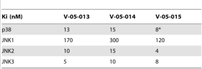
Table 1.Ki values for dual p38 and JNK MAPK inhibitors.
Ki (nM) V-05-013 V-05-014 V-05-015
p38 13 15 8*
JNK1 170 300 120
JNK2 10 15 4
JNK3 5 10 8
All compounds have a Ki greater than 1mM for all other kinases tested. *This one value is an IC50, not a Ki.
6.47 (m, 1H), 5.03 (s, H), 4.18-4.00 (m, 1H), 4.18-4.00 (m, 1H), 3.80-3.35 (m, 5H), 1.71 (m, 1H), 1.52-1.23 (m, 5H); LC-MS (method X) tR= 0.60 min., (M+H+) 452.37. Ki or IC50 was
determined to be.1mM for the following kinases: AKT3, AurA, CDK2, ERK-2, EphA, FLT3, IGF1R, IRAK, ITK, JAK2, JAK3, KDR, MAPKAP2, cMET, MKK4, MKK6, MKK7, PIM1, PKA, PLK1, PRAK, ROCK1, SRC, SYK, TIE2, ZAP70; GSK3b Ki = 0.73mM.
V-05-014:1H NMR (400 MHz, DMSO-d6) 11.04 (s, 0.3 H),
10.22 (s, 0.2 H), 8.33 (s, 1 H), 8.08 (s, 1 H), 7.38–7.79 (m, 4 H), 6.68 (s, 1 H), 6.20 (s, 1 H), 5.15–5.23 (m, 0.7 H), 4.86–4.91 (m, 1H), 3.78 (s, 3 H), 3.53 (s, 1 H), 3.33 (s, 1 H), 1.56–2.13 (m, 9 H), 1.09–1.28 (m, 6 H); LC-MS (method Y)tR = 2.47 min., (M+H+
) 452.25; LC-MS (method X) tR= 0.63 min., (M+H+) 452.37. Ki or
IC50 was determined to be .1mM for the following kinases: AKT3, AurA, COT, CDK2, ERK2, EphA, FAK, GSK3b, IGF1R, IRAK, JAK3, KDR, LCK, MAPKAP2, cMET, MKK4, MKK6, MKK7, NIK, PDK1, PIM1, PKA, PLK1, PRAK, ROCK1, SRC, SYK, TIE2, ZAP70.
V-05-015:1H NMR (300 MHz, MeOD) 8.20 (d, J = 5.1 Hz,
1H), 7.62 (dd, J = 5.3, 8.6 Hz, 2H), 7.29 (t, J = 8.7 Hz, 2H), 6.68-6.42 (m, 1H), 5.00 (s, 2H), 3.75-3.53 (m, 3H), 3.34-3.15 (m, 2H), 2.10-1.77 (m, 9H), 1.88 (m, 1H), 1.65-1.20 (m, 7H); LC-MS (method X) tR= 0.64 min., (M+H+) 436.38. Ki or IC50 was
determined to be.1mM for the following kinases: AKT3, AurA, CDK2, ERK2, FLT3, GSK3b, IGF1R, IRAK, JAK2, JAK3, KDR, LCK, MAPKAP2, cMET, MKK4, MKK6, MKK7, PDK1, PIM1, PKA, PLK1, PRAK, ROCK1, SRC, SYK, ZAP70.
Measurement of Pulmonary Inflation Pressure
One day after exposure to ozone, guinea pigs were anesthetized with 1.9 g/kg urethane i.p. (Sigma-Aldrich, St. Louis, MO). This dose produces a deep anesthesia lasting 8–10 hours [31] though no experiments lasted longer than 4 hours.
Physiological measurements were made as previously described [32]. The jugular veins were cannulated for intravenous admin-istration of drugs and the right carotid artery was cannulated to measure heart rate and blood pressure. Both vagus nerves were cut and distal ends placed on platinum electrodes submerged in liquid paraffin. Animals were tracheostomized, ventilated (1 ml/100 g body weight, 100 breaths per minute) and paralyzed with a constant infusion of succinylcholine (10mg/kg/min iv,
Sigma-Aldrich). Pulmonary inflation pressure was measured at the trachea and bronchoconstriction was measured as the increase in pressure over basal inflation pressure produced by the ventilator.
Measurement of Vagally Induced Bronchoconstriction
Electrical stimulation of both vagus nerves (10V, 0.2 ms pulse width, 1–25 Hz, 5 sec duration at 1 minute intervals) produced frequency dependent bronchoconstriction and bradycardia due to release of acetylcholine onto muscarinic receptors. To confirm vagally induced bronchoconstriction was cholinergic, atropine (1 mg/kg iv, Sigma-Aldrich) was given at the end of each experiment.
Measurement of Smooth Muscle M3Muscarinic Receptor Function
Recovery from vagal stimulation was confirmed by pulmonary inflation pressure and heart rate returning to baseline before measuring smooth muscle M3muscarinic receptor function (5–10
minutes after cessation of vagal stimulation). In vagotomized guinea pigs, M3 muscarinic receptor function on airway smooth
muscle was tested by measuring bronchoconstriction after administration of acetylcholine (1–10mg/kg iv, Sigma-Aldrich).
Measurement of Neuronal M2Muscarinic Receptor Function
Recovery from administration of intravenous acetylcholine was confirmed by pulmonary inflation pressure and heart rate returning to baseline before measuring M2 muscarinic receptor
function (5–10 minutes after the last dose of acetylcholine). To test the function of neuronal M2muscarinic receptors, vagally induced
bronchoconstriction was measured before and after administration of gallamine (0.1–10 mg/kg iv, Sigma-Aldrich) an M2 receptor
antagonist. Electrical stimulation of both vagus nerves (3–30V, 0.2 ms pulse width, 15 Hz, 5 sec duration at 1 minute intervals) produced reproducible, frequency dependent, bronchoconstric-tions. In the presence of normally functioning M2 receptors,
gallamine (0.1–10 mg/kg iv) will block them, resulting in increased vagally induced bronchoconstriction [33]; an effect that is suppressed if M2 receptors are not responding to endogenous
acetylcholine [10].
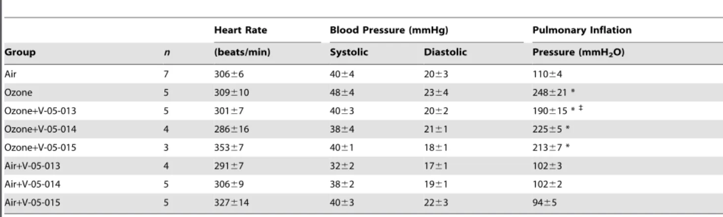
Table 2.Baseline cardiovascular and pulmonary parameters.
Heart Rate Blood Pressure (mmHg) Pulmonary Inflation
Group n (beats/min) Systolic Diastolic Pressure (mmH2O)
Air 7 30666 4064 2063 11064
Ozone 5 309610 4864 2364 248621 *
Ozone+V-05-013 5 30167 4063 2062 190615 *`
Ozone+V-05-014 4 286616 3864 2161 22565 *
Ozone+V-05-015 3 35367 4061 1861 21367 *
Air+V-05-013 4 29167 3262 1761 10263
Air+V-05-014 5 30669 3862 1961 10262
Air+V-05-015 5 327614 4063 2263 9465
Values are means6SEM. Baseline pulmonary inflation pressure significantly increased after ozone exposure. Treatment with dual p38/JNK MAPK inhibitor V-05-013 significantly reduced the ozone-induced increase in baseline pulmonary inflation pressure. *p,0.05 Significantly different from air exposure.`
p,0.05 Significantly different from ozone exposure.
Bronchoalveolar Lavage (BAL)
At the end of each experiment, the lungs were lavaged with five 10 ml aliquots of phosphate buffered saline (PBS) that contained 100mg isoproterenol (Sigma-Aldrich). Lavage fluid was centri-fuged (400g, 10 min) and the pellets were resuspended in PBS. Cells were counted using a hemocytometer and slides made from centrifuged lavaged cells were stained with Hemacolor (EMD
Chemicals, Gibbstown, NJ) and used to determine cell differen-tials.
Drugs
Acetylcholine, succinylcholine, and urethane were purchased from Sigma (St. Louis, MO) and were dissolved and diluted in PBS.
Figure 2. Blocking p38 and JNK MAPK completely prevented ozone-induced airway hyperreactivity mediated by the vagus nerves.In anesthetized and vagotomized guinea pigs, stimulation of the vagus nerves (10V, 0.2 ms pulse width, 1–25 Hz, 5 sec duration at 1 minute intervals) caused frequency dependent bronchoconstriction (A open circles; measured as an increase in inflation pressure in mmH2O)
that is significantly potentiated one day post-ozone exposure (A closed circles). Pretreatment with dual MAPK inhibitors V-05-013 (A closed squares), V-05-014 (B closed triangles), or V-05-015 (C closed inverted triangles) completely prevented ozone-induced airway hyperreactivity. All three dual MAPK inhibitors suppressed parasympathetic nerve activity (A open squares, B open triangles, C open inverted triangles). Ozone and air exposed control data are the same in A-C. *p,0.05, **p,0.01 Significantly different from air exposed controls. Data are mean6SEM. n = 4–7.
doi:10.1371/journal.pone.0075351.g002
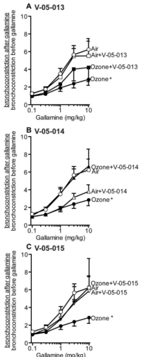
Figure 3. In control (air exposed) guinea pigs electrical stimulation of the vagus nerves (3–30V, 0.2 ms pulse width, 15 Hz, 5 sec duration at 1 minute intervals) resulted in vagally induced bronchoconstriction (measured as an increase in pulmonary inflation pressure; 1661 mmH2O). An M2 receptor
antagonist, gallamine, potentiated vagally induced bronchoconstriction up to 6-fold in air exposed animals (open circles) demonstrating that functional M2 receptors were limiting acetylcholine release. The
potentiation by gallamine was decreased in ozone-exposed animals, demonstrating M2receptors were dysfunctional after ozone exposure
(closed circles). V-05-013 partially prevented M2receptor dysfunction (C
closed squares), while V-05-014 (B closed triangles) and V-05-015 (C closed inverted triangles) completely protected M2receptor function.
Vagally induced bronchoconstriction in the absence of gallamine was not different from control among all groups. Ozone and air exposed controls are the same in A–C. *p,0.05, **p,0.01 Significantly different from air exposed controls. Data are mean6SEM. n = 4–7.
Data Analysis and Statistics
All data are expressed as means 6 SE. In vivo frequency response and dose response curves were compared using two-way ANOVA for repeated measures. Baseline data were analyzed by one-way ANOVA with Bonferroni’s correction. APvalue of less than 0.05 was considered significant. Analyses were made with GraphPad Prism (version 5.0; GraphPad Software, La Jolla, CA).
Results
Baselines
One day after ozone exposure, baseline pulmonary inflation pressure was significantly increased compared to air-exposed controls (Table 2). All the dual p38 and JNK inhibitors partially attenuated the ozone induced increase in baseline airway inflation pressure, although the attenuation only reached statistical signif-icance in the group treated with V-05-013. None of the MAPK inhibitors affected baseline inflation pressure in air-exposed controls. Neither ozone nor the MAPK inhibitors affected baseline heart rate or blood pressure.
Airway Physiology
Ozone significantly potentiated bronchoconstriction in response to electrical stimulation of the vagus nerves compared to air-exposed controls as previously reported (Figure 2). Treatment with any of the dual MAPK inhibitors prevented ozone induced airway hyperreactivity (Figures 2A–C). Vehicle treatment had no effect on vagally mediated bronchoconstriction in either air or ozone exposed animals (data not shown). M2muscarinic receptors were
dysfunctional in ozone treated animals as gallamine, an M2
selective inhibitor, potentiated bronchoconstriction in response to vagal stimulation in air-exposed animals but not in ozone-exposed animals (Figure 3); an effect that is consistent with decreased function of neuronal M2 muscarinic receptors [34]. Ozone
induced M2 receptor dysfunction was prevented by treatment
with V-05-014 and V-05-015 (Figure 3B–C), and attenuated by treatment withV-05-013 (Figure 3A). Airway smooth muscle responses to intravenous acetylcholine were potentiated by ozone (Figure 4). This was not prevented by any of the MAPK inhibitors, but was partially attenuated by V-05-015 (Figure 4C). V-05-015 also produced a paradoxical increase in airway response to intravenous acetylcholine in air-exposed animals.
Ozone exposure potentiated falls in heart rate in response to vagal stimulation compared to air-exposed controls (Figure 5A–C). Ozone and air-exposed controls are the same in figure 5A–C. Separate pretreatment with all three dual MAPK inhibitors prevented the ozone-induced potentiation in falls in heart rate and had no effect in air-exposed animals (Figure 5A–C). Falls in heart rate in response to intravenous acetylcholine were not affected by either ozone or the MAPK inhibitor (Figure 5D–F). Ozone and air exposed controls are the same in figure 5D–F.
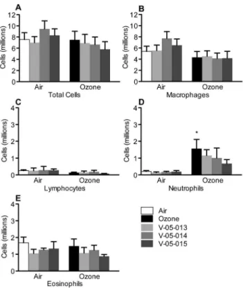
Bronchoalveolar Lavage and Peripheral Blood
One day after ozone exposure neutrophils were increased in bronchoalveolar lavage (Figure 6D). All the MAPK inhibitors slightly, though not significantly, attenuated the ozone induced increase in neutrophils (Figure 6D). None of the other inflamma-tory cell types were affected by either ozone or the MAPK inhibitors (Figure 6).
There were no significant differences between inflammatory cells in peripheral blood after either ozone exposure, or treatment with the dual MAPK inhibitors (Figure 7).
Discussion
Ozone induces airway hyperreactivity, measured as potentiation of vagally induced bronchoconstriction, in guinea pigs one day after exposure confirming previous studies [35,36]. Ozone also significantly potentiated bronchoconstriction in response to intravenous acetylcholine; an effect that has also been previously reported [10]. Blocking both p38 and JNK MAPK with three different, but related, inhibitors prevented vagally mediated
Figure 4. Bronchoconstriction (measured as an increase in inflation pressure in mmH2O) in response to intravenous
hyperreactivity in ozone-exposed animals but had no effect on inflammatory cell numbers in bronchoalveolar lavage. The prevention of vagally mediated hyperreactivity was associated with prevention of ozone induced M2 receptor dysfunction that
was complete in animals treated with V-05-014 and V-05-015, and partial in animals treated with V-05-013. Ozone induced hyperreactivity to intravenous acetylcholine was partially attenu-ated by treatment with the MAPK inhibitors.
All three MAPK inhibitors were administered at a dose of 30 mg/kg i.p. one hour before ozone. While the compounds are active with submicromolar potencies, preliminary studies suggested there is a significant shift in in vivo potency from the tens of nanomolar to hundreds of nanomolar IC50 s presum-ably the result of plasma protein binding (unpublished data). Nonetheless, the compounds were chosen because they exhibit adequate pharmacokinetic profiles (table 1) to test our hypoth-esis in vivo. The relatively high in vivo clearances and half lives are somewhat limiting, leading to the need for a sufficient dose to demonstrate a role for MAPKs in ozone induced hyperre-activity. However, as with most compounds, different effects could occur at lower doses.
Treatment of air-exposed guinea pigs with any of the three MAPK inhibitors decreased the airway response to vagal stimulation slightly. This effect was most pronounced at high frequency stimulation, but could not be explained by changes in M2 receptor function, as the effects of gallamine were not
potentiated by the MAPK inhibitors in air exposed animals. This effect was also not due to decreased smooth muscle responsiveness, as the effects of intravenous acetylcholine were
not decreased by the MAPK inhibitors. Although response to the MAPK inhibitors was variable in air-exposed animals the overall effect with ozone exposure was prevention of ozone-induced airway hyperreactivity. These minor differences may be due to off target effects of the inhibitors, or to the dose of inhibitors used in this study. Thus, in air exposed guinea pigs, p38 and JNK MAPK inhibitors inhibit vagally induced bronchoconstriction by suppressing release of acetylcholine from airway parasympathetic nerves.
The mechanism for this decreased acetylcholine release is unknown. p38 and JNK are involved in nerve regeneration and development [37,38] but whether they inhibit ganglionic trans-mission, action potentials or transmitter release (by a mechanism separate from M2 receptors, since there was no change in the
response to gallamine) is not well studied. InAplysia, activation of p38 by the peptide neurotransmitter FMRFa leads to long-term depression in sensory neurons in the pleural ganglia [39], although the mechanism is not known. In Drosophila motor neurons, expression of constitutively active JNK decreases neurotransmitter release [40] while in primary cultures of rat cortical neurons, IL-1b signaling activates p38, decreasing synaptophysin, a protein involved in synaptic transmission [41]. These varied and sometimes contradictory effects of MAPKs on neural function and transmitter release may be involved in the effects we observed. In neutrophils, activation of p38 MAPK is required for granule exocytosis after stimulation by CXCR1/2 ligands [42]; if neurotransmitter exocytosis were similarly mediated by MAPK, kinase inhibitors would block secretion. Thus, the role of MAPK is cell type dependent and additionally may differ between central
Figure 5. Ozone potentiated vagally mediated falls in heart rate (measured as beats/minute; A–C closed circles) compared to air exposed animals (A–C open circles).Separate pretreatment with all three dual MAPK inhibitors (V-05-013: A closed squares; V-05-014: B closed squares; V-05-015: C closed squares) prevented the ozone-induced potentiation of frequency induced falls in heart rate. Fall in heart rate following intravenous acetylcholine administration was not changed by either ozone, or MAPK inhibitors (D–E). Ozone and air exposed controls are the same for A–C, and are the same for D–F. **p,0.01 Significantly different from air exposed controls. Data are mean6SEM. n = 3–7.
neurons where kinases inhibit neurotransmission and peripheral neurons, where they have not been well studied. The data in this paper suggest p38 or JNK MAPK may additionally play a previously unrecognized role in release of acetylcholine from lung parasympathetic nerves.
None of the MAPK inhibitors completely reversed the ozone-induced increase in baseline pulmonary inflation pressure, which is commonly due to airway edema and not increased vagal tone. Ozone also significantly increased the numbers of neutrophils in bronchoalveolar lavage compared to air exposed controls confirming previously published data [35,36]. However, blocking both p38 and JNK MAPK did not prevent the neutrophil influx. No other inflammatory cell population in the lavage was effected by ozone or by the p38 and JNK MAPK inhibitors. Thus, prevention of ozone-induced airway hyperreactivity did not occur via a decrease in airway inflammatory cells.
Previously we have shown major basic protein, released from eosinophils, inhibits neuronal M2 muscarinic receptor function,
thereby increasing acetylcholine release and subsequently leading to increased bronchoconstriction and airway hyperreac-tivity after ozone exposure [43,44]. Depletion of eosinophils with an antibody to IL-5, or blocking major basic protein with heparin, prevents M2 receptor dysfunction and ozone-induced
airway hyperreactivity one day post-ozone exposure [11]. Thus, although neutrophils are the cells that increase in the bronchoalveolar lavage after ozone, it is tissue eosinophils around airway nerves that mediate ozone-induced hyperreac-tivity. In eosinophils, eotaxin and IL-5 signal through both
ERK and p38 MAPK activation [45,46]. Inhibition of p38 reduces eosinophil degranulation as measured by decreased eosinophil cationic protein release [45]. Major basic protein has also been shown to alter smooth muscle contractility [47]. Thus, while not tested directly in this study, blocking eosinophil degranulation with MAPK inhibitors could also contribute to preventing smooth muscle hyperreactivity.
Thus, p38 and JNK MAPK inhibitors inhibit ozone-induced hyperreactivity by multiple mechanisms. Exposure to high levels of environmental ozone increases hospitalizations from asthma exacerbations. Over 4 million children and 10 million adults with asthma live in counties with unhealthy levels of ozone, and those with asthma are an especially susceptible population to the adverse health effects of ozone [1]. Our data show that treatment with p38 and JNK inhibitors, immediately prior to ozone exposure prevented subsequent development of airway hyperreactivity. Currently there is no specific therapy for ozone related asthma complications and our data suggest both p38 and JNK are potential targets for additional therapeutic candidates; and that inhibitors could be tested as prophylactic treatment for asthma exacerbations on days with anticipated high ozone.
Author Contributions
Conceived and designed the experiments: KCV FGS MWL ADF DBJ. Performed the experiments: KCV. Analyzed the data: KCV. Contributed reagents/materials/analysis tools: FGS MWL. Wrote the paper: KCV ADF DBJ MWL. Designed and characterized the molecules used in this study: FGS MWL.
Figure 6. Ozone exposure increased neutrophils in bronchoal-veolar lavage (D closed bar). No other inflammatory cell type number was affected by either ozone or the dual p38/JNK MAPK inhibitors. *p,0.05 Significantly different from air exposed controls. Data are mean6SEM. n = 3–6.
doi:10.1371/journal.pone.0075351.g006
Figure 7. Neither ozone nor the dual p38/JNK MAPK inhibitors affected inflammatory cell numbers in peripheral blood.Data are mean6SEM. n = 3–6.
References
1. Association AL (2009) State of the Air Report.
2. Hiltermann JT, Lapperre TS, van Bree L, Steerenberg PA, Brahim JJ, et al. (1999) Ozone-induced inflammation assessed in sputum and bronchial lavage fluid from asthmatics: a new noninvasive tool in epidemiologic studies on air pollution and asthma. Free Radic Biol Med 27: 1448–1454.
3. Bell ML, McDermott A, Zeger SL, Samet JM, Dominici F (2004) Ozone and short-term mortality in 95 US urban communities, 1987–2000. Jama 292: 2372– 2378.
4. Lewis TC, Robins TG, Dvonch JT, Keeler GJ, Yip FY, et al. (2005) Air pollution-associated changes in lung function among asthmatic children in Detroit. Environ Health Perspect 113: 1068–1075.
5. Kreit JW, Gross KB, Moore TB, Lorenzen TJ, D’Arcy J, et al. (1989) Ozone-induced changes in pulmonary function and bronchial responsiveness in asthmatics. J Appl Physiol 66: 217–222.
6. Wagner EM, Jacoby DB (1999) Methacholine causes reflex bronchoconstriction. J Appl Physiol 86: 294–297.
7. Foster WM, Brown RH, Macri K, Mitchell CS (2000) Bronchial reactivity of healthy subjects: 18–20 h postexposure to ozone. J Appl Physiol 89: 1804–1810. 8. Lee LY, Bleecker ER, Nadel JA (1977) Effect of ozone on bronchomotor
response to inhaled histamine aerosol in dogs. J Appl Physiol 43: 626–631. 9. Mitchell HW, Adcock J (1988) Vagal mechanisms and the effect of
indomethacin on bronchoconstrictor stimuli in the guinea-pig. Br J Pharmacol 94: 522–527.
10. Schultheis AH, Bassett DJ, Fryer AD (1994) Ozone-induced airway hyperre-sponsiveness and loss of neuronal M2 muscarinic receptor function. J Appl Physiol 76: 1088–1097.
11. Yost BL, Gleich GJ, Fryer AD (1999) Ozone-induced hyperresponsiveness and blockade of M2 muscarinic receptors by eosinophil major basic protein. J Appl Physiol 87: 1272–1278.
12. Pryor WA (1992) How far does ozone penetrate into the pulmonary air/tissue boundary before it reacts? Free Radic Biol Med 12: 83–88.
13. Hamilton RF Jr, Hazbun ME, Jumper CA, Eschenbacher WL, Holian A (1996) 4-Hydroxynonenal mimics ozone-induced modulation of macrophage function ex vivo. Am J Respir Cell Mol Biol 15: 275–282.
14. Kirichenko A, Li L, Morandi MT, Holian A (1996) 4-hydroxy-2-nonenal-protein adducts and apoptosis in murine lung cells after acute ozone exposure. Toxicol Appl Pharmacol 141: 416–424.
15. Kumagai T, Nakamura Y, Osawa T, Uchida K (2002) Role of p38 mitogen-activated protein kinase in the 4-hydroxy-2-nonenal-induced cyclooxygenase-2 expression. Arch Biochem Biophys 397: 240–245.
16. Cui CH, Adachi T, Oyamada H, Kamada Y, Kuwasaki T, et al. (2002) The role of mitogen-activated protein kinases in eotaxin-induced cytokine production from bronchial epithelial cells. Am J Respir Cell Mol Biol 27: 329–335. 17. Atherton HC, Jones G, Danahay H (2003) IL-13-induced changes in the goblet
cell density of human bronchial epithelial cell cultures: MAP kinase and phosphatidylinositol 3-kinase regulation. Am J Physiol Lung Cell Mol Physiol 285: L730–739.
18. Nath P, Leung SY, Williams A, Noble A, Chakravarty SD, et al. (2006) Importance of p38 mitogen-activated protein kinase pathway in allergic airway remodelling and bronchial hyperresponsiveness. Eur J Pharmacol 544: 160–167. 19. Denhardt DT (1996) Signal-transducing protein phosphorylation cascades mediated by Ras/Rho proteins in the mammalian cell: the potential for multiplex signalling. Biochem J 318 (Pt 3): 729–747.
20. Kyriakis JM, Avruch J (1996) Protein kinase cascades activated by stress and inflammatory cytokines. Bioessays 18: 567–577.
21. Kalesnikoff J, Huber M, Lam V, Damen JE, Zhang J, et al. (2001) Monomeric IgE stimulates signaling pathways in mast cells that lead to cytokine production and cell survival. Immunity 14: 801–811.
22. Peng Q, Matsuda T, Hirst SJ (2004) Signaling pathways regulating interleukin-13-stimulated chemokine release from airway smooth muscle. Am J Respir Crit Care Med 169: 596–603.
23. Liu W, Liang Q, Balzar S, Wenzel S, Gorska M, et al. (2008) Cell-specific activation profile of extracellular signal-regulated kinase 1/2, Jun N-terminal kinase, and p38 mitogen-activated protein kinases in asthmatic airways. J Allergy Clin Immunol 121: 893–902 e892.
24. Duan W, Chan JH, McKay K, Crosby JR, Choo HH, et al. (2005) Inhaled p38alpha mitogen-activated protein kinase antisense oligonucleotide attenuates asthma in mice. Am J Respir Crit Care Med 171: 571–578.
25. Underwood DC, Osborn RR, Kotzer CJ, Adams JL, Lee JC, et al. (2000) SB 239063, a potent p38 MAP kinase inhibitor, reduces inflammatory cytokine production, airways eosinophil infiltration, and persistence. J Pharmacol Exp Ther 293: 281–288.
26. Fujisawa T, Ide K, Holtzman MJ, Suda T, Suzuki K, et al. (2008) Involvement of the p38 MAPK pathway in IL-13-induced mucous cell metaplasia in mouse tracheal epithelial cells. Respirology 13: 191–202.
27. Williams AS, Issa R, Leung SY, Nath P, Ferguson GD, et al. (2007) Attenuation of ozone-induced airway inflammation and hyper-responsiveness by c-Jun NH2 terminal kinase inhibitor SP600125. J Pharmacol Exp Ther 322: 351–359. 28. Williams AS, Issa R, Durham A, Leung SY, Kapoun A, et al. (2008) Role of p38
mitogen-activated protein kinase in ozone-induced airway hyperresponsiveness and inflammation. Eur J Pharmacol 600: 117–122.
29. Cho HY, Morgan DL, Bauer AK, Kleeberger SR (2007) Signal transduction pathways of tumor necrosis factor–mediated lung injury induced by ozone in mice. Am J Respir Crit Care Med 175: 829–839.
30. Fox T, Coll JT, Xie X, Ford PJ, Germann UA, et al. (1998) A single amino acid substitution makes ERK2 susceptible to pyridinyl imidazole inhibitors of p38 MAP kinase. Protein Sci 7: 2249–2255.
31. Green C (1982) Animal Anesthesia. London: Elsevier.
32. Fryer AD, Stein LH, Nie Z, Curtis DE, Evans CM, et al. (2006) Neuronal eotaxin and the effects of CCR3 antagonist on airway hyperreactivity and M2 receptor dysfunction. J Clin Invest 116: 228–236.
33. Fryer AD, Maclagan J (1984) Muscarinic inhibitory receptors in pulmonary parasympathetic nerves in the guinea-pig. Br J Pharmacol 83: 973–978. 34. Fryer AD, Wills-Karp M (1991) Dysfunction of M2-muscarinic receptors in
pulmonary parasympathetic nerves after antigen challenge. J Appl Physiol 71: 2255–2261.
35. Yost BL, Gleich GJ, Jacoby DB, Fryer AD (2005) The changing role of eosinophils in long-term hyperreactivity following a single ozone exposure. Am J Physiol Lung Cell Mol Physiol 289: L627–635.
36. Verhein KC, Jacoby DB, Fryer AD (2008) IL-1 receptors mediate persistent, but not acute, airway hyperreactivity to ozone in guinea pigs. Am J Respir Cell Mol Biol 39: 730–738.
37. Hirai S, Kawaguchi A, Suenaga J, Ono M, Cui DF, et al. (2005) Expression of MUK/DLK/ZPK, an activator of the JNK pathway, in the nervous systems of the developing mouse embryo. Gene Expr Patterns 5: 517–523.
38. Agthong S, Koonam J, Kaewsema A, Chentanez V (2009) Inhibition of MAPK ERK impairs axonal regeneration without an effect on neuronal loss after nerve injury. Neurol Res.
39. Guan Z, Kim JH, Lomvardas S, Holick K, Xu S, et al. (2003) p38 MAP kinase mediates both short-term and long-term synaptic depression in aplysia. J Neurosci 23: 7317–7325.
40. Etter PD, Narayanan R, Navratilova Z, Patel C, Bohmann D, et al. (2005) Synaptic and genomic responses to JNK and AP-1 signaling in Drosophila neurons. BMC Neurosci 6: 39.
41. Li Y, Liu L, Barger SW, Griffin WS (2003) Interleukin-1 mediates pathological effects of microglia on tau phosphorylation and on synaptophysin synthesis in cortical neurons through a p38-MAPK pathway. J Neurosci 23: 1605–1611. 42. Rittner HL, Labuz D, Richter JF, Brack A, Schafer M, et al. (2007) CXCR1/2
ligands induce p38 MAPK-dependent translocation and release of opioid peptides from primary granules in vitro and in vivo. Brain Behav Immun 21: 1021–1032.
43. Fryer AD, Jacoby DB (1992) Function of pulmonary M2 muscarinic receptors in antigen-challenged guinea pigs is restored by heparin and poly-L-glutamate. J Clin Invest 90: 2292–2298.
44. Evans CM, Fryer AD, Jacoby DB, Gleich GJ, Costello RW (1997) Pretreatment with antibody to eosinophil major basic protein prevents hyperresponsiveness by protecting neuronal M2 muscarinic receptors in antigen-challenged guinea pigs. J Clin Invest 100: 2254–2262.
45. Kampen GT, Stafford S, Adachi T, Jinquan T, Quan S, et al. (2000) Eotaxin induces degranulation and chemotaxis of eosinophils through the activation of ERK2 and p38 mitogen-activated protein kinases. Blood 95: 1911–1917. 46. Adachi T, Choudhury BK, Stafford S, Sur S, Alam R (2000) The differential
role of extracellular signal-regulated kinases and p38 mitogen-activated protein kinase in eosinophil functions. J Immunol 165: 2198–2204.