Determination of lytic enzyme activities of indigenous
Trichoderma
isolates from Pakistan
Saeed Ahmad Asad
1, Ayesha Tabassum
2, Abdul Hameed
2, Fayyaz ul Hassan
3,
Aftab Afzal
4, Sabaz Ali Khan
5, Rafiq Ahmed
5, Muhammad Shahzad
5 1Centre for Climate Research and Development, COMSATS University, Islamabad, Pakistan. 2
Department of Microbiology, Quaid-i-Azam University, Islamabad, Pakistan. 3
Department of Biosciences, COMSATS University, Islamabad, Pakistan. 4
Department of Botany, Hazara University, Mansehra, Pakistan. 5
Department of Environmental Sciences, COMSATS University, Abbottabad, Pakistan.
Submitted: September 18, 2014; Approved: February 2, 2015.
Abstract
This study investigated lytic enzyme activities in three indigenous Trichoderma strains namely, Trichoderma asperellum, Trichoderma harzianumandTrichodermasp. NativeTrichodermastrains and a virulent strain ofRhizoctonia solaniisolated from infected bean plants were also included in the study. Enzyme activities were determined by measuring sugar reduction by dinitrosalicylic acid (DNS) method using suitable substrates. The antagonists were cultured in minimal salt medium with the following modifications: medium A (1 g of glucose), medium B (0.5 g of glucose + 0.5 g of deac-tivatedR. solanimycelia), medium C (1.0 g of deactivated respective antagonist mycelium) and me-dium D (1 g of deactivatedR. solanimycelia).T. asperellumshowed presence of higher amounts of chitinases,b-1, 3-glucanases and xylanases in extracellular protein extracts from medium D as com-pared to medium A. While, the higher activities of glucosidases and endoglucanses were shown in medium D extracts byT. harzianum.b-glucosidase activities were lower compared with other en-zymes; however, activities of the extracts of medium D were significantly different.T. asperellum exhibited maximum inhibition (97.7%). On the other hand,Trichodermasp. did not show any effect on mycelia growth ofR. solanion crude extract.
Key words:lytic enzymes, mycoparasitic activity,Rhizoctonia solani,Trichoderma.
Introduction
Soil-borne pathogens cause diseases that result in sig-nificant loss of quality and yield of the valuable crops around the globe. Fungi, being the most aggressive soil-borne pathogens, have been intensely investigated due to inflicting damage to major crops. The main pathogenic fun-gal genera involved in such damage are:Phythium, Botry-tis, Rhizoctonia and Fusarium (Djonovic et al., 2007). Pesticides have been widely used to control these patho-gens (Gerhardson, 2002) but their applications have been increasingly criticized due to environmental and human health concerns (Punja and Utkhede, 2003; Bues et al., 2004).
In Pakistan, being an agricultural country, almost all of the economically important crops are affected by phyto-pathogenic fungal species such as R. solani, Fusarium moniliforme, andFusarium solani(Ahmedet al., 1997).R. solanilives in subterranean forms, which makes it the most resistant pathogen. Hence, chemical control is ineffective, unless highly selective fungicides are used. The search for novel bio-control agents, therefore, is a prime target of many plant pathologists (Harman et al., 2004); and Trichodermaspecies, such asT. harzianum,Trichoderma hamatum,Trichoderma reeseiorTrichoderma virens,are recommended by many researchers as preferred choice for controlling phytopathogens (Punja and Utkhede 2003;
DOI: http://dx.doi.org/10.1590/S1517-838246420140787
Send correspondence to S.A. Asad. Centre for Climate Research and Development COMSATS University, Park Road, Chak Shahzad, 45550 Islamabad, Pakistan. E-mail: [email protected].
Steyaertet al., 2003; Harmanet al., 2004; Montealegreet al., 2010; Castroet al., 2014). .
The genus Trichoderma includes cosmopolitan saprophytic fungi found in soil and have long been known to be effective against plant pathogen, R. solani (Wein-dling, 1932).Trichodermaacts through direct fungal pene-tration (Asadet al., 2014) and/or by secreting antifungal compounds, such as hydrolytic enzymes, in order to inhibit the growth of phytopathogens. For instance,T. harzianum releases hydrolytic enzymes againstCrinipellis perniciosa, the causative agent of cocoa (Theobroma cacao) disease (De Marco et al., 2003). Due to the significant role of hydrolytic enzymes in the biocontrol activity of Trichoderma species against R. solani,the present study was undertaken to assess the activities of hydrolytic en-zymes from threeTrichoderma species. Furthermore, the antagonistic potential (antibiosis) of metabolites, obtained from crude extracts of selected isolates, was studied.
Materials and Methods
Microorganisms
ThreeTrichodermastrains (Trichoderma asperellum, Trichoderma harzianum and Trichoderma sp.) isolated from native agricultural soils were obtained from Fungal Culture Bank of the University of the Punjab, Lahore, Paki-stan. A virulent strain ofR. solanipreviously isolated from the infected bean plants was courteously supplied by Agro-innova culture bank, University of Torino, Italy (Minutoet al., 2008; Asadet al., 2014). All microbial cultures were grown and maintained on potato dextrose agar (Difco, Becton, Dickinson, Sparks, MD) at 4 °C. The antagonists were cultured in the minimal salt medium according to the method of Lilly and Barnett (1951) for chitinase, xylanase,
b-1, 3-glucanase, endoglucanase (CMCase) andb -gluco-sidase activities. Minimal salt medium (Tsenget al., 2008) was used with minor modifications as medium A (1 g of glucose), medium B (0.5 g of glucose + 0.5 g of deactivated R. solanimycelia), medium C (1.0 g of deactivated respec-tive antagonist mycelium) and medium D (1 g of deacti-vated R. solani mycelia). Fifty milliliters of each of the mentioned media was incubated at 25 °C on a rotary shaker at 150 rpm for 0, 24, 48, 72, 96 and 120 h. Filtrates were collected by centrifugation at 3000 xgfor 10 min at 4 °C and the filtrate was used for enzyme activity assays.
Deactivation of mycelium
Mycelia from seven-day old cultures ofTrichoderma andR. solaniwere collected by centrifugation (3000 xg) for 10 min and subsequently washed twice with 50 mL of sterile and deionized water. The collected mycelia were boiled twice for 20 min to obtain the deactivated mycelia and stored at -20 °C until used.
Enzyme activity assays
Enzyme activities ofTrichodermastrains were deter-mined by using dinitro-salicylic acid (DNS) reagent (Mil-ler, 1959) and sugar reduction in the respective substrates was measured.
Chitinase activity
Chitinase activity was determined according to Tseng et al. (2008), an artificial substrate containing 10 mL of 0.5% 4-nitrophenylN,N‘-diacetyl-b-D-chitobioside (Sig-ma-Aldrich, USA) and 250mL of the enzyme samples were mixed in 250mL of 100 mM acetate buffer having pH 5. Af-ter 30 min, 50mL of 0.4 M Na2CO3was added to terminate the reaction and turbidity (OD) was measured at 415 nm. One unit of the enzyme activity was defined as the amount of enzyme, required to produce 1 mmol of the product per milligram of the protein per hour.
Beta-1, 3-glucanase activity
Enzyme activity was quantified according to Masih and Paul (2002) by incubating 250mL of the enzyme sam-ples and 250mL of 1% laminarin dissolved in 0.2 M acetate buffer (pH 5) at 50 °C for 40 min. The reaction was termi-nated by 500mL of DNS reagent and kept for 10 min in a boiling water bath. After boiling, the solution was diluted by adding 4 mL of distilled water and after cooling, the amount of the reducing sugars was measured at 540 nm us-ing D-glucose as benchmark. Specific activity of the en-zyme was manifested as mmol of glucose released per milligram of the protein per hour.
Beta-glucosidase activity
Beta-Glucosidase activity was determined by the method of Tokaoet al.(1985). The reaction mixtures were prepared by adding 250mL enzyme sample and 400mL of 17 mmol L-1salicin solution dissolved in 0.2 mol L-1 so-dium acetate buffer (pH 4.6). 250mL distilled water was added to the solution and incubated at 50 °C for 40 min. Op-tical density was recorded at 540 nm using glucose as stan-dard. Specific activity of the enzyme was expressed as mmol of glucose released per milligram of the protein per hour.
Xylanase activity
For xylanase activity, 100mL enzyme sample was mixed with 500 mL of 1% oat spelt xylan dissolved in 0.1 mol L-1phosphate buffer (pH 7), and then 400mL of 0.1 mol L-1phosphate buffer (pH 7) was added (Baileyet al., 1992). The resultant mixture was incubated at 30 °C for 20 min, cooled, and turbidity was recorded at 540 nm with D-xylose as standard. One unit of enzyme activity was de-fined as 1 mmol of xylose released per milligram of the pro-tein per hour.
Endoglucanase activity
Endoglucanase activity was determined as described by Koet al.(2005). Briefly, 250mL enzyme sample was added to 250mL of 1% carboxymethyl cellulose dissolved in 0.2 mol L-1sodium acetate buffer (pH 5). The enzyme containing mixture was incubated at 50 °C for 40 min and terminated by adding 1.5 mL of DNS reagent. Turbidity was recorded at 540 nm using glucose as a standard. En-zyme activity was defined as mmol of xylose released per milligram of the protein per hour.
Total protein concentration in enzyme solution was estimated using bovine serum albumin as standard (Brad-ford, 1976). The specific activity of the enzyme was ex-pressed as the amount of enzyme that catalyzed formation of 1 mmol of the product per hour under the assay condi-tions. The experiments were performed twice with three replicates for each treatment.
Effect of crude extract onR. solanimycelial inhibition (Antibiosis)
Three discs of mycelial agar plugs (5 mm diameter) were inoculated in 100 mL potato dextrose broth (PDB). The samples were incubated for 7 days at 25±1 °C on a ro-tary shaker at 100 rpm and then filtered through a Millipore filter (0.2 mm). The samples were sterilized by passing through a biological membrane filter (0.2 mm). Filtered supernatant (500mL) from each of theTrichodermaspecies was spread over the surface of PDA plates and a 5 mm disc ofR. solaniwas inoculated at the center of each plate. The plates were incubated at 25 °C until the colony spread over the surface of PDA plates in the control treatment (Dennis and Webster 1971). Radial growth of the pathogens was re-corded on a daily basis. The percent inhibition of the aver-age growth of mycelia in relation to the growth of the controls was calculated by using Equation 1 (Edingtonet al., 1971).
Mycelial inhibition (%)=é
-ë ê ù û ú ´ C C C 2 1 2 100 (1)
where C1= radial mycelial growth ofR. solaniin the pres-ence ofTrichoderma, and C2= radial mycelial growth ofR. solaniin control.
Experimental design and statistical analyses
The experiments were repeated twice in a complete randomized block design with four replicates for each treat-ment. The data were analyzed using SPSS software (ver-sion 17.0 Chicago IL, USA). Analysis of variance (ANOVA) was carried out with a significance defined at p < 0.05. Duncan’s HSD multiple range test was used as a post-hoc analysis to compare means.
Results
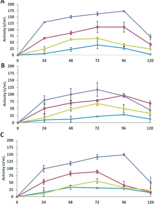
Chitinases
Chitinase activity assays showed that all the antago-nists cultured in medium D, (containing deactivated myce-lium of R. solani) expressed the highest enzyme activity compared to other three media (Figure 1). The maximum activity (173 U mL-1) was detected byT. asperellumin the extracts of medium D after 96 h of incubation, which was the highest among all the isolates. The medium B, contain-ing glucose and deactivated mycelium ofR. solani, also showed high activities (111 U mL-1) as compared to me-dium A (glucose) and C (deactivated antagonist myce-lium).
The maximum chitinase activity byT. harzianumwas detected in medium D after 72 h of incubation (117 U mL-1). The medium B showed maximum activity of 97 U mL-1after 96 h of incubation. While the medium A and C expressed very low chitinases activities (Figure 1).
Trichodermasp. also showed an increased chitinase activity (149 U mL-1) until 96 h of incubation in the extracts of medium D. While in medium B, the activity increased (89 U mL-1) until 72 h of incubation and then decreased. Significantly low chitinase activities were expressed in me-dium A and C (Figure 1).
b- 1, 3-glucanase
The assays forb-1,3-glucanase showed that all the antagonists were able to express much higher enzyme ac-tivity in extracts of medium D than the extracts of other me-dia. After 48 h of incubation, Trichoderma sp. showed maximum activity of 149 U mL-1, whileT. asperellumand T. harzianumexhibited activity of 146 and 137 U mL-1, re-spectively.T. harzianummaintained maximum activity till 120 h, while the activities decreased in the extracts ofT. asperellumandTrichodermasp. after reaching the maxi-mum level (Figure 2). Very high activities were observed in the extracts of medium B. The activities dropped to mini-mum after 72 and 96 h of incubation, in the case ofT. asperellum and T. harzianum respectively, while for Trichodermasp., the activity retained at 102 U mL-1until 120 h of incubation. The results showed that the glucanase activities of all the strains in extracts of medium A and C were non-significant (Figure 2).
b- glucosidase
maxi-mum activities were 51 U mL-1in medium A after 72 h of incubation and 50 and 55 U mL-1after 24 h of incubation in medium B and C, respectively. T. harzianum and Trichodermasp. exhibited very low enzyme activities in the extracts of all the three media. These activities were also lower than the activities expressed byT. asperellumin the respective media (Figure 3).
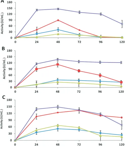
Xylanase
All antagonists showed a higher xylanase activity in medium D compared to other media.T. asperellum exhib-ited maximum xylanase activity of 162 U mL-1in the ex-tracts of medium D after 72 h of incubation, which
decreased to 81 U mL-1 after 120 h of the incubation (Figure 4). The extracts from medium B exhibited activity of 140 U mL-1at 72 h of the incubation and then decreased thereafter to 45 U mL-1.
In the extracts of medium A and C, some activity was shown byT. asperellumafter 72 h, which then diminished at the end of the incubation period. The xylanase activities byT. harzianumwere also comparatively high in extracts of medium D. The maximum activity was 152 U mL-1after 72 h of incubation. A significant activity was expressed by the extracts of medium B, while the extracts of medium A and C showed very low activities (Figure 4).
1056 Asadet al.
Trichodermasp. showed highest maximum xylanase activity of 175 U mL-1in the extracts of medium D after 72 h of the incubation. The medium B also showed signifi-cantly higher activities upon induction by deactivated pathogen mycelium, while in the extracts of other media, the enzyme activities were comparable to other isolates (Figure 4).
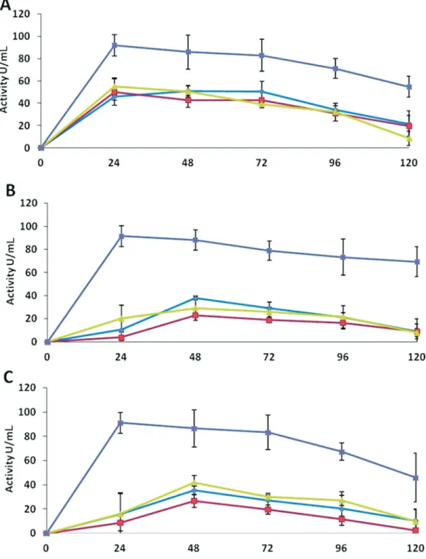
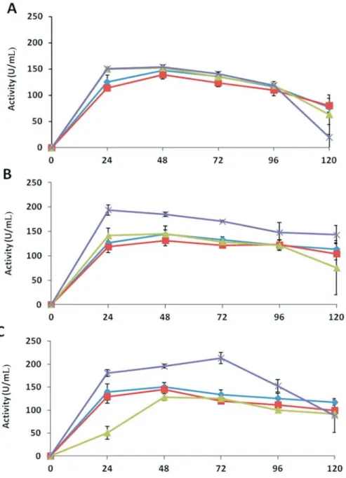
Endoglucanase
As shown in Figure 5,T. asperellumshowed much higher endoglucanase activity than other enzymes in the extracts of all media. There was no effect of the presence of pathogen mycelium in medium D compared with the ex-tracts of medium A, containing only glucose, the same level of enzyme activity was exhibited.
The maximum activities in medium A and B were 147 and 139 U mL-1, respectively; while in media C and D both showed about 153 U mL-1activity after 48 h of
incuba-tion. At the end of the time course, the activities signifi-cantly decreased to 77, 80, 63 and 20 U mL-1in medium A, B, C, and D, respectively (Figure 5).
T. harzianumshowed higher activities in extracts of medium D. The maximum enzyme activity (194 U mL-1) was expressed within first 24 h of the incubation and then decreased to 143 U mL-1after 120 h. In other media, the ac-tivities increased after 48 h of incubation to 144, 131 and 145 U mL-1in medium A, B, and C, respectively; these ac-tivities were retained as 114, 104 and 75 U mL-1for the three media, respectively, after 120 h (Figure 5). Trichodermasp. also showed high endoglucanase activity (214 U mL-1) in medium D extracts after 72 h of incubation. In medium A, B and C maximum endoglucanase activities were 151, 145 and 128 U mL-1after 48 h of incubation, which decreased to 116, 99 and 92 U mL-1, respectively, by the end of the time course (Figure 5).
Effect of crude extracts of different species of
Trichodermaon the growth inhibition ofR. solani
The effect of the metabolites present in the crude ex-tracts of the media was statistically significant for all the
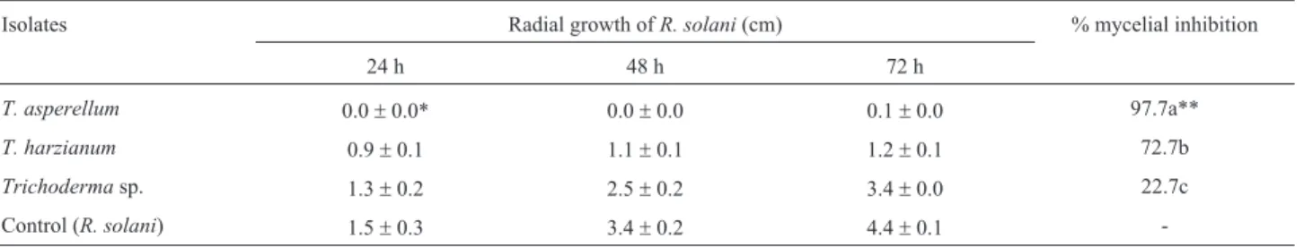
strains (p < 0.05).T. asperellumwas most effective to in-hibit the mycelial growth ofR. solaniby 97.7%, whileT. harzianum was effective with 72.7% of inhibition, and Trichoderma sp. was failed to show any effect on the mycelial growth (Table 1).
1058 Asadet al.
Table 1- Effect of crude extracts of differentTrichodermaspecies on growth inhibition ofR. solani.
Isolates Radial growth ofR. solani(cm) % mycelial inhibition
24 h 48 h 72 h
T. asperellum 0.0±0.0* 0.0±0.0 0.1±0.0 97.7a**
T. harzianum 0.9±0.1 1.1±0.1 1.2±0.1 72.7b
Trichodermasp. 1.3±0.2 2.5±0.2 3.4±0.0 22.7c
Control (R. solani) 1.5±0.3 3.4±0.2 4.4±0.1
-*The results are the mean value (±SE) of two independent experiments, each with four replicates.
**Values followed by the different letters are statistically different by Duncan’s multiple range test (p < 0.05).
Discussion
Apart from other mechanisms, antagonistic ability of Trichodermaalso includes production of lytic enzymes that hydrolyze the cell wall of the host fungus (Woo et al., 2006). In most fungi, chitin and glucans are the most abun-dant microfibrilar components in the cell wall, while pro-teins and glucans act as cementing matrix (Peberdy, 1990). In the current study, deactivatedR. solanimycelium was used rather than cell walls of the pathogen to simulate pro-duction of natural metabolites byTrichodermawhich re-sulted in significant activities of chitinase, xylanase and
b-1, 3-glucanase. The higher activities of extracellular en-zymes in medium D and B as compared to medium A (containing only glucose) illustrate that these enzymes were induced by the presence of deactivated mycelium of R. Solani, this observation concords with the study of Tsenget al.(2008). This behavior can be correlated to the reduction ofR. solanimycelium after seven days of incuba-tion, when co-inoculatedin vitro. Chitin is the major com-ponent of fungal cell walls (Free, 2013). Hence, the chiti-nases were considered to be involved in the degradation of cell wall ofR. solanibyT. virens(Baeket al., 1999).
Simi-1060 Asadet al.
lar observations were recorded by Viterbo et al. (2002), where Chitinase catalyzed cleavage ofb-1,4 linkages be-tween N-acetyl-b-D-glucosamine units. Our results dem-onstrated thatT. asperellum produced higher amount of chitinases compared with other species, andT. harzianum produced lowest one. The enzyme activities were observed until 96 h and 72 h of incubation byT. asperellumandT. harzianum, respectively, which decreased significantly af-ter 120 h (Figure 1). De Marcoet al.(2003) also reported increase in chitinase activity by Trichoderma isolates within 72 h of incubation. The chitinolytic system of Trichoderma comprises many enzymes, including 1,4-b -acetylglucosaminidase, endochitinases, and exochitinases (Brunneret al., 2003). The chitinases are thought to be the major enzyme reported participating in mycoparasitic in-teraction. In current study, increased production of the en-zymes upon induction by pathogen’s deactivated mycelium indicated their possible role in antagonistic activity in dual culture assays. Harighiet al.(2007) purified chitinase-42 (Chi42) from Trichoderma atroviride, which showed a strong potential to lyse R. solani cell wall and inhibit mycelial growth.T. virensmutants over-expressing Chi42 showed an enhanced biocontrol activity againstR. solaniin cotton seedlings compared to the wild types (Howell, 2003). Haran et al. (1996) detected the presence of GlcNAcases (1,4-b-acetylglucosaminidase), Chit73, and Chit102 inT. harzianumTM andT. asperellum. However, Kullniget al.(2000) reported that endochitinases were reg-ulated by chit33, chit36 and chit42 genes during stress. Danaet al. (2001) suggested thatchit33 is not expressed during overgrowth ofR. solani, but expressed only during the contact phase; whereas,chit36Yexpression does not re-quire direct contact with the pathogen. Two proteins, 1,3-b-glucosidase and a 42 kDa endochitinase, were identi-fied by Grinyeret al.(2004) in the culture supernatant ofT. Atroviride, grown in the medium containing cell walls ofR. solanias the sole carbon source.
The major component ofR. solanicell wall has been identified asb-glucan in addition to chitin (Lahsenet al., 2001); therefore, glucanases can play important role in an-tagonistic activity.b-1,3-glucanases cleaveb-1,3 linkages between two molecules of glucose (Viterboet al., 2002). In the present study, medium D, containing deactivated myce-lium of the pathogen, showed highest activity ofb -1,3-glucanase for all the three isolates; and, in addition, T. harzianum retained the activity to a maximum level throughout the incubation period (Figure 2). In contrast to this, De Marcoet al.(2003) reported increase in glucanase activity byTrichodermaisolates within 72 h of incubation. De La Curzet al.(1995) and Vázquez-Garcidueñaset al. (1998) also showed an increased activity of glucanases by Trichodermasp. within 48 h of induction, in the presence of different substrates having hydrolytic ability in combina-tion with other enzymes. However, Loritoet al.(1994)
pu-rified an endo-b-1,3-glucosidase involved in the inhibition of spore germination of B. cinerea, establishing role of glucanase in mycoparasitism. El-Katatnyet al.(2001) also purified a glucanase having a potential to inhibit the growth ofS. rolfsiifromT. harzianumisolate T-24. Therefore, the activities of glucanases by allTrichodermaspecies can be speculated to be involved in the antagonistic potential in dual culture assays.
In the present study, all testedTrichodermaspecies showed some activity in medium A containing only glu-cose until 48 h of incubation and then decreased (Figure 2). However these findings are contrary to Ramotet al.(2000), who concluded that glucose inhibited the secretion of
b-1,3-glucanases. Similarly, Tsenget al.(2008) hypothe-sized thatTrichodermasecretsb-1,3-glucanases at a very low level to detect long-chainb-1,3-glucans. The enzymes are secreted for a short period in a glucose-rich medium and degrade if no polysaccharides withb-1,3-linkages are pres-ent. However, in ab-1,3-glucan-rich medium, oligosaccha-rides with b-1,3-linkages are generated that induce
b-1,3-glucanases to cleave long chains ofb-1,3-glucan pro-ducing glucose, which disintegrates cell wall of the patho-gen. Xylanase catalyzes hydrolysis of xylan to xylose, and thus xylan is used as a nutrient source but its role in bio-control is not yet established.Trichoderma reesei pro-duces two specific, xylan-inducible xylanases encoded by xyn1andxyn2 to degrade theb-1,4-D-xylan backbone of hemicelluloses (Zeilingeret al., 1996; Machet al., 1996). The role of the enzymes in bio-control has not been studied possibly due to the fact thatR. solanicell wall lacks xylan. All the tested strains ofTrichodermashowed a high activity of xylanase, while grown in a medium containing deacti-vated pathogen mycelium (Figure 4). Therefore, b- 1,3-glucanase activities byTrichodermaspecies were assumed to exhibit xylanase activity, which was in agreement with Tsenget al.(2008). It might have contributed to the com-bined antagonistic activities ofTrichodermastrains against R. solaniin dual culture assays.
Trichodermaspecies are known to produce cellulases and b-glucosidase which hydrolyze b-1,4-glucans (De Marcoet al., 2003). Thecre1gene in the filamentous fungi Trichoderma reeseiandT. harzianumregulates cellulase expression (Ilménet al., 1996). The mycoparasitic interac-tion relieves binding of the Cre1 carboncatabolite repressor protein to promoter sequences of ech42 (endochitinase-encoding) gene inT. harzianum(Strausset al., 1995; Lorito et al., 1996; Portnoyet al., 2011; Silva-Rochaet al., 2014).
mycelium on the activity of cellulytic enzymes, suggesting that the major enzymes involved in the mycoparasitism by T. asperellumdo not include cellulases. De Marco et al. (2003) reported increased activities of cellulytic enzymes within 24 h of incubation in the presence of specific sub-strates;Trichodermamay be involved in the hydrolysis of cell wall of pathogen, which differs from Tseng et al. (2008), who reported an insignificant cellulose activity by T. harzianum.
Mycoparasitism is associated with the production of cell wall-degrading enzymes such as chitinase and gluco-sidase and their induction as a response to infection by pathogen (Zhanget al., 2010); and these two classes of the hydrolytic enzymes show synergistic activity against sev-eral pathogenic fungi (Qin et al., 2003). The cell wall-degrading enzymes ofTrichodermaare of special impor-tance to induce the defense mechanisms of plants (Jayalakshmiet al., 2009). Abd-El-Khairet al.(2010) also reported the induction of enzymes such as chitinase and peroxidase byTrichoderma, which play an important role in the defense mechanisms of plants against pathogens.
The production of antibiotics as well as secondary metabolites was determined by applying crude cell free ex-tract of Trichoderma indigenous isolates on pathogen growth to establish antagonistic potential. The inhibition of mycelia growth ofR. solaniby crude extracts produced in liquid medium was significantly different among Trichodermaspecies.T. asperellumshowed maximum in-hibition of 97%, whileTrichodermasp. did not show any effect on mycelia growth of the pathogen (Table 1), which indicated that the secondary metabolites/antibiotics pro-duction can be related to the combined antagonistic activity byT. asperellumagainstR. solani. These results are in ac-cordance with Castilloet al.(2011), who observed 100% mycelial growth inhibition of the pathogen by T. asperellum. Similar findings were reported by Etebarian (2006), showing the antifungal effect of metabolites se-creted byT.harzianumandT.virensisolates causing 100% inhibition of mycelia growth of Macrophomina phaseolina. However, Vinale et al. (2006) isolated and characterized secondary metabolites from culture filtrates of two commercialT. harzianumisolates (T22 and T39) and reported their production in relation to mycoparasitic interaction withR. solani.
Conclusions
T. asperellumproduced higher amounts of chitinases,
b-1,3-glucanases and xylanases in a medium containing de-activatedR. solanimycelium than the medium containing only glucose.T. asperellumshowed maximum inhibition (97.7%) of mycelial growth of R. solani, while Trichodermasp. did not show any effect. This study estab-lished antagonistic potential ofTrichoderma using crude protein extract.
Acknowledgments
The authors thank the University of Torino, Italy for partial financial support and Mas-simo Pugliese, Maria Lodovica Gullino for providing the technical assistance in sample analysis. We are also grateful to the anonymous ref-erees who made valuable comments and suggestions. We also thank Professor James Peters from the University of Manitoba, Canada for improving English quality of the manuscript.
References
Abd-El-Khair H, Khalifa RM, Hagga KE (2010) Effect of Trichoderma Species on damping off diseases incidence, some plant enzymes activity and nutritional status of bean plants. J American Sci 6:486-497.
Ahmad S, Iqbal SH, Khalid AN (1997)Fungi of Pakistan. Myco-logical Society of Pakistan. Uni. Punjab, 248 pp.
Ait-Lahsen H, Soler A, Rey Met al.(2001) An antifungal exo-b -1,3-glucanase (AGN13.1) from the bio control fungus Trichoderma harzianum. Appl Environ Microbiol 67:5833-5839.
Asad SA, Ali N, Hameed Aet al.(2014) Biocontrol efficacy of different isolates ofTrichodermaagainst soil borne patho-genRhizoctonia solani. Pol J Microbiol 63:95-103. Baek JM, Howell CR, Kenerley CM (1999) The role of
extra-cellular chitinase fromTrichoderma virensGv29-8 in the bio control ofRhizoctonia solani. Curr Genet 35:41-50. Bailey MJ, Bailey P, Poutanen K (1992) Inter laboratory testing of
methods for assay of xylanase activity. J Biotechnol 23:257-270.
Bradford MM (1976) A rapid and sensitive method for the quanti-tation of microgram quantities of protein utilizing the princi-ple of protein-dye binding. Anal Biochem 72:1105-1112. Brunner K, Montero M, Mach RLet al.(2003) Expression of the
ech42(endochitinase) gene ofTrichoderma atroviride un-der carbon starvation is antagonized via a BrlA-like cis-act-ing element. FEMS Microbiol Lett 218:259-264.
Bues R, Bussieres P, Dadomo Met al.(2004) Assessing the envi-ronmental impacts of pesticides used on processing tomato crops. Agri Ecosys Environ 102:155-116.
Castillo FDH, Padilla AMB, Morales GGet al.(2011)In vitro an-tagonist action of Trichodermastrains against sclerotinia sclerotiumandsclerotium cepivorum.Am J Agri Biol Sci 6:410-417.
Castro Ldos S, Antoniêto ACC, Pedersoli WRet al.(2014) Ex-pression pattern of cellulolytic and xylanolytic genes regu-lated by transcriptional factors XYR1 and CRE1 are af-fected by carbon source inTrichoderma reesei. Gene Expr Patterns 14:88-95.
Dana MM, Limón MC, Mejías R (2001) Regulation of chitinase 33 (chit33) gene expressions in Trichoderma harzianum. Curr Genet 38:335-342.
De la Cruz J, Pintor-Toro JA, Benitez T (1995) A novel endo-b-1, 3-glucanase, BGN13.1 involved in the mycoparasitism of Trichoderma harzianum. J Bacteriol 177:6937-6945. De Marco JL, Valadares MC, Felix CR (2003) Production of
hydrolytic enzymes byTrichoderma isolates with antago-nistic activity against Crinipellis perniciosa, the causal
agent of witches’ broom of cocoa. Braz J Microbiol 34:33-38.
Dennis C, Webster J (1971) Antagonistic properties of species groups ofTrichodermaI, production of non-volatile antibi-otics. Trans Br Mycol Soc 57:25-39.
Djonovic S, Vargas WA, Kolomiets MV (2007) A proteinaceous elicitor Sm1 from the beneficial fungusTrichoderma virens is required for induced systemic resistance in maize. Plant Physiol 145:875-889.
Edington LV, Khew KL, Barron GL (1971) Fungi toxic spectrum of benzimidazole compounds. Phytopathol 61:42-44. El-Katatny MH, Gudelj M, Robra KH (2001) Characterization of
a chitinase and an endo-beta-1,3-glucanase from Trichoderma harzianumRifai T24 involved in control of the phytopathogen Sclerotium rolfsii. Appl Microbiol Bio-technol 56:137-143.
Etebarian HR (2006) Evaluation ofTrichodermaisolates for bio-logical control of charcoal stem rot in melon caused by Macrophomina phaseolina. J Agri Sci Technol 8:243-250. Free SJ (2013) Fungal cell wall organization and biosynthesis.
Adv Genet 81:33-82.
Gerhardson B (2002) Biological substitutes for pesticides. Trends Biotechnol 20:338-343.
Grinyer J, McKay M, Herbert B (2004) Fungal proteomics: map-ping the mitochondrial proteins of a Trichoderma harzianumstrain applied for biological control. Curr Genet 45:170-175.
Haran S, Schickler H, Oppenheim A, Chet I (1996) Differential expression of Trichoderma harzianum chitinases during mycoparasitism. Phytopathol 86:980-985.
Harman GE, Howell CR, Viterbo A (2004)Trichoderma spe-cies-opportunistic, a virulent plant symbionts. Nat Rev Microbiol 2:43-56.
Harighi MJ, Zamani MR, Motallebi M (2007) Evaluation of antifungal activity of purified chitinase 42 from Trichoderma atroviridePTCC5220. Biotechnol 6:28-33. Howell CR (2003) Mechanisms employed byTrichoderma
spe-cies in the biological control of plant diseases; the history and evolution of current concepts. Plant Dis 87:4-10. Ilmén M, Thrane C, Penttilä M (1996) The glucose repressor gene
cre1 of Trichoderma: isolation and expression of a full-length and a truncated mutant form. Mol Gen Genet 251:451-460.
Jayalakshmi SK, Rajus S, Benagi VI (2009) Trichoderma harzianumL1 as a potential source for lytic enzymes and elicitor of defense responses in chickpea (Cicer arietinum L.) against wilt disease caused byFusarium oxysporum. sp. ciceri. Aus J Crop Sci 3:44-52.
Ko HG, Parka SH, Kimb SH (2005) Detection and recovery of hydrolytic enzymes from spent compost of four mushroom species. Foliar Microbiol 50:103-106.
Kullnig C, Mach RL, Lorito Met al.(2000) Enzyme diffusion from Trichoderma atroviride (T. harzianum P1) to Rhizoctonia solani is a prerequisite for triggering of Trichoderma ech42gene expression before mycoparasitic contact. Appl Environ Microbiol 66:2232-2234.
Lilly VG, Bernett HL (1951) Physiology of the Fungi. McGraw-Hill, New York.
Lorito M, Harman CK, Di Pietro Aet al.(1994) Purification, char-acterization and synergistic activity of a glucans 1, 3-beta
glucosidase and an N-acetylglucosaminidase from Trichoderma harzianum. Phytopathol 84:398-405. Lorito M, Mach RL, Sposato Pet al.(1996) Mycoparasitic
inter-action relieves binding of the Cre1 carbon catabolite repres-sor protein to promoter sequence of ech-42 (endochitinase-encoding) gene ofTrichoderma harzianum. Proc Natl Acad Sci 93:14868-14872.
Mach RL, Strauss J, Zeilinger Set al.(1996) Carbon catabolite re-pression of xylanase I (xyn1) gene exre-pression in Trichoderma reesei. Mol Microbiol 21:1273-1281. Masih EI, Paul B (2002) Secretion ofb-1, 3-glucanase by the
yeastPichia membranefaciensand its possible role in the bio control ofBotrytis cinereacausing grey mold of grape-vine. Curr Microbiol 44:391-395.
Miller GL (1959) Use of dinitrosalicylic acid reagent for determi-nation of reducing sugar. Anal Chem 31:426-428.
Minuto A, Gaggero L, Gullino ML (2008) Influence of pH, nutri-ent solution disinfestation and antagonists application in a closed soilless system on severity of Fusarium wilts of gerbera. Phytopara 36:294-303.
Montealegre J, Valderrama L, Sánchez S (2010) Biological con-trol of Rhizoctonia solani in tomatoes withTrichoderma harzianummutants. Electron J Biotechnol 13:1-11 Peberdy JF, (1990) Fungal cell walls. A review. In: Kuhn PJ,
Trinci APJ, Jung MJet al.(ed) Biochemistry of Cell Walls and Membranes in Fungi. Springer-Verlag, London, pp 5-30.
Portnoy T, Margeot A, Linke Ret al.(2011)The CRE1 carbon catabolite repressor of the fungus Trichoderma reesei: a master regulator of carbon assimilation. BMC Genomics 12:269.
Punja ZK, Utkhede RS (2003) Using fungi and yeasts to manage vegetable crop diseases. Trend Biotechnol 21:400-407. Qin GZ, Tian SP, Xu Y (2003) Enhancement of bio control
effi-cacy of antagonistic yeasts by salicylic acid in sweet cherry fruit. Physiol Mol Plant Pathol 62:147-154.
Ramot O, Cohen-Kupiec R, Chet I (2000) Regulation ofb -1,3-glucanase by carbon starvation in the mycoparasite Trichoderma harzianum. Mycol Res 104:415-420. Silva-Rocha R, Castro Ldos S, Antoniêto ACCet al.(2014)
Deci-phering theCis-Regulatory Elements for XYR1 and CRE1 Regulators inTrichoderma reesei. PLoS One 9:1-10. Steyaert JM, Ridgway HJ, Elad Y (2003) Genetic basis of
myco-parasitism; a mechanism of biological control by species of Trichoderma. New Zea J Crop Hort Sci 31:281-291. Strauss J, Mach RL, Zeilinger Set al.(1995) Cre l, the carbon
catabolite repressor protein fromTrichoderma reesei. FEBS Letters 376:103-107.
Tokao S, Kamagata Y, Sasaki T (1985) Cellulase production by Penicillium purpurogenum. J Agri Sci 93:217-222. Tseng SC, Liu SY, Yang HH (2008) Proteomic Study of Bio
con-trol mechanisms ofTrichoderma harzianumETS 323 in re-sponse toRhizoctonia solani.J Agri Food Chem 56:6914-6922.
Vázquez-Garcidueñas S, Leal-Morales C, Herrera-Estrella A (1998) Analysis of theb-1, 3-glucanolytic system of the bio control agent Trichoderma harzianum. Appl Environ Microbiol 64:1442-1446.
ac-tive against different phytopathogens. Letts Appl Microbiol 43:143-148.
Viterbo A, Ramot O, Chemin LYet al.(2002) Significance of lytic enzymes fromTrichodermaspp. in the bio control of fungal plant pathogens. Ant van Leeuw 81:549-556.
Weindling R (1932) Trichoderma lignorumas a parasite of other fungi. Phytopathol 22:837-845.
Woo SL, Scala F, Ruocco Met al.(2006) The molecular biology of the interactions between Trichoderma spp. phytopa-thogenic fungi and plants. Phytopathol 96:181-185.
Zeilinger S, Mach RL, Schindler Met al.(1996) Different indu-cibility of expression of the two xylanase genes xyn1 and xyn2 inTrichoderma reesei. J Biol Chem 271:25624-25629. Zhang D, Spadaro D, Garibaldi Aet al.(2010) Selection and eval-uation of new antagonists for their efficacy against postharvest brown rot of peaches. Postharvest Biol Technol 55:174-181.
Associate Editor: Miguel Juan Beltran-Garcia
All the content of the journal, except where otherwise noted, is licensed under a Creative Commons License CC BY-NC.